/
Myocardial Infarction Myocardial Infarction

Myocardial Infarction - PowerPoint Presentation

trish-goza
trish-goza . @trish-goza
Follow
724 views
Uploaded On 2016-11-06

Myocardial Infarction - PPT Presentation

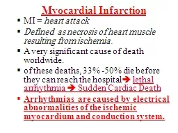
MI heart attack Defined as necrosis of heart muscle resulting from ischemia A very significant cause of death worldwide of these deaths 33 50 die before they can reach the hospital ID: 485192

cardiac myocardial ventricular heart myocardial cardiac heart ventricular infarct infarction rupture muscle acute death microscopic repair tissue days features rate necrotic myocardium

Share:

Link:

Embed:

Download Presentation from below link

Download Presentation The PPT/PDF document "Myocardial Infarction" is the property of its rightful owner. Permission is granted to download and print the materials on this web site for personal, non-commercial use only, and to display it on your personal computer provided you do not modify the materials and that you retain all copyright notices contained in the materials. By downloading content from our website, you accept the terms of this agreement.


Presentation Transcript

Slide1

Myocardial Infarction

MI =

heart attack

Defined as necrosis of heart muscle resulting from ischemia.

A very significant cause of death worldwide.

of these deaths, 33% -50% die before they can reach the hospital

lethal arrhythmia

Sudden Cardiac Death

Arrhythmias are caused by electrical abnormalities of the ischemic myocardium and conduction system. Slide2

Acute occlusion of the proximal left anterior descending (LAD) artery is the cause of

40% to 50% of all MIs

and typically results in infarction of the anterior wall of the left ventricle, the anterior two thirds of the ventricular septum, and most of the heart apexSlide3

The frequency of MIs rises progressively with increasing age

and presence of other risk factors such as hypertension, smoking, and diabetes

Approximately only 10% of MIs occur in people younger than 40 years. Slide4
Slide5

Evaluation of MI

Clinical signs and symptoms

Electrocardiographic(ECG) abnormalities

Laboratory evaluation:

is based on measuring the blood levels of intracellular macromolecules that leak out of injured myocardial cells through damaged cell membranes.

these molecules include :

1-myoglobin.

2-cardiac

troponins

T and I (

TnT

,

TnI

)

3-creatine

kinase

(CK, and more specifically the myocardial-specific

isoform

, CK-MB)

4- lactate

dehydrogenaseSlide6

Cardiac enzymes in MI

Cardiac

troponins

T and I (

TnT

,

TnI

), are

the

best

markers for acute MI.

persistence of elevated

troponin

levels for approximately 10 days allows the diagnosis of acute MI long after CK-MB levels have returned to normal.

creatine

kinase

CK-

MB

is the

second best

marker after the cardiac-specific

troponins

.

Slide7

Since various forms of creatine

kinase

(

CK)

are found in brain, myocardium, and skeletal muscle, total CK activity is not a reliable marker of cardiac injury (i.e. it could come from skeletal muscle injury). Thus, the

CK-MB

isoform

-principally derived from myocardium is the more specific indicator of heart damage.

CK-MB activity begins to rise within 2-4 hours of MI, peaks at 24-48 hours, and returns to normal within approximately 72 hours.Slide8

Microscopic changes of MI and its repair.

(<24 hr)

coagulative

necrosis

and

wavy

fibers

. Necrotic cells are separated by edema fluid.

2- to 3-day old - infarct

Dense

neutrophil

infiltrate

(7 to 10 days)

complete removal of necrotic

myocytes

by

phagocytic

macrophages

up to 14 days

Granulation

tissue

characterized by loose connective tissue and abundant capillaries.

several weeks

Healed myocardial infarct consisting of a dense

collagenous

scar

. Slide9

Microscopic features of myocardial infarction and its repair.

(<24 hr)

coagulative

necrosis

and

wavy

fibers

Necrotic cells are separated by edema fluidSlide10

2- to 3-day old - infarct Dense

neutrophil

infiltrate

in case of reperfusion

 contraction bands

Microscopic features of myocardial infarction and its repair. Slide11

7 to 10 days) complete removal of necrotic

myocytes

by

phagocytic

macrophages

Microscopic features of myocardial infarction and its repair. Slide12

up to 14 days Granulation

tissue

characterized by loose connective tissue

(blue) and

abundant

capillaries (red)

Microscopic features of myocardial infarction and its repair. Slide13

several weeks Healed myocardial infarct consisting of a dense

collagenous

scar

Microscopic features of myocardial infarction and its repair. Slide14

Consequences and Complications of MI

1-

Death

:

Unfortunately,

50% of the deaths

associated with acute MI occur in individuals who never reach the hospital

(within 1 hour of symptom onset-usually as a result of arrhythmias)

Extraordinary progress has been made in patient outcomes subsequent to acute MI (

the

in

-hospital)

death

rate

has declined from approximately 30% to an overall rate of between 10% and 13%).Slide15

Consequences and Complications of MI

2-

cardiogenic

shock.

(10% to 15%) of patients after acute MI

with a large infarct ( >40% of the

Lef

t

ventricle).

- 70% mortality

rate; 2/3

of in-hospital deaths.

3-Myocardial rupture

4-

Pericarditis.

5-

Infarct expansion

6-

Ventricular aneurysm

7-

Progressive late heart failureSlide16

Complications of myocardial rupture include:

(1) rupture of the ventricular free wall

hemopericardium

and cardiac

tamponade

(usually fatal)

(2) rupture of the ventricular septum

VSD and left-to-right shunt

(3) papillary muscle rupture

severe mitral regurgitation Slide17

myocardial ruptureSlide18

4-Pericarditis.

-

fibrinous

or hemorrhagic

pericarditis

- usually

2

to 3 days of a

transmural

MI

- typically spontaneously resolves with time (immunologic mechanism).

5-Infarct expansion

.

Because of the weakening of necrotic muscle, there may be disproportionate stretching, thinning, and dilation of the infarct region (especially with

anteroseptal

infarcts)Slide19

6-Mural thrombus

.

-the combination of a local loss of contractility (causing stasis) +

endocardial

damage (causing a

thrombogenic

surface

)

thromboembolism

7-Ventricular aneurysm

.

- A late complication

- most commonly result from a large transmural

anteroseptal

infarct that heals with the formation of thin scar tissue

Slide20

Ventricular aneurysm

Complications of ventricular aneurysms

include:

1-mural thrombus

2-arrhythmias

3-heart failureSlide21

8-Papillary muscle dysfunction

(

post-infarct

mitral regurgitation )

dysfunction of a papillary muscle after MI occurs due to:

1- rupture.

2- ischemic dysfunction

3- fibrosis and shortening

4- ventricular dilation.

9-Progressive late heart failureSlide22

Long-term prognosis after MI

depends on many factors, the most important of which are left ventricular function and the severity of atherosclerotic narrowing of vessels

perfusing

the remaining viable myocardium.

Mortality rate within the first year =30%

Thereafter, the annual mortality rate is 3% to 4%. Slide23

Chronic Ischemic Heart Disease

Chronic IHD usually results from

post-infarction

cardiac decompensation that follows exhaustion of the hypertrophic viable myocardium.

progressive

heart failure

as a consequence of ischemic myocardial damage; sometimes punctuated by episodes of angina or MI.

Arrhythmias

are common along with

CHFSlide24

Sudden Cardiac Death (SCD)

Affecting some 300,000 to 400,000 individuals annually in the United States

SCD is most commonly defined as

unexpected death from cardiac causes either without symptoms or within 1 to 24 hours of symptom onset

Coronary artery disease is the most common underlying cause

In many adults SCD is the first clinical manifestation of IHD.

With

younger

victims, other

non-atherosclerotic

causes are more common: Slide25

Other non-atherosclerotic

causes of SCD

Congenital coronary arterial abnormalities

Aortic valve

stenosis

Mitral valve

prolapse

Myocarditis

Dilated or hypertrophic

cardiomyopathy

Pulmonary hypertension

Hereditary or acquired abnormalities of the cardiac conduction system.

Isolated myocardial hypertrophy.

unknown causes.