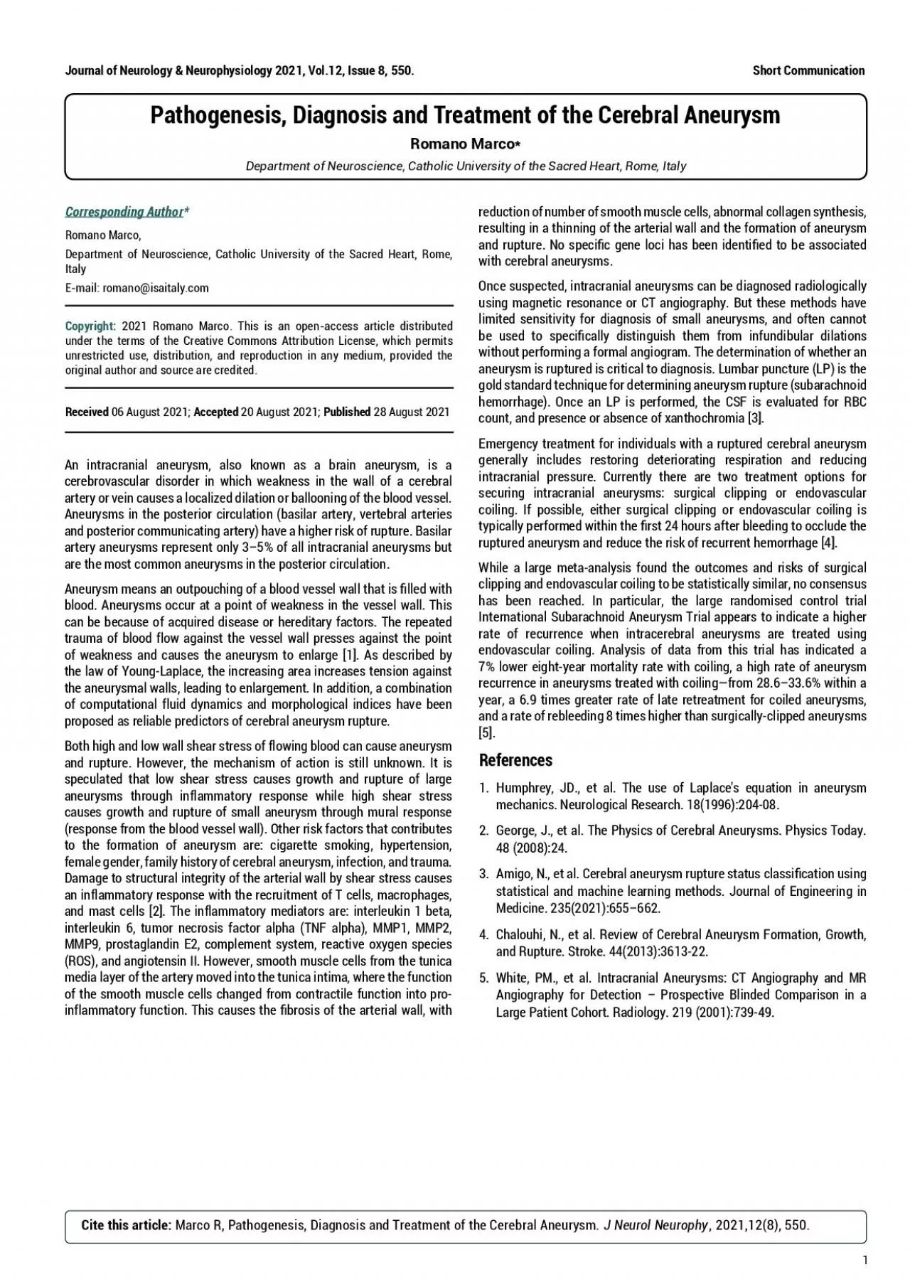
Corresponding AuthorRomano Marco Department of Neuroscience Catholic University of the Sacred Heart Rome Italy Email romanoisaitalycom Copyright 2021 Romano Marco This is an openaccess ar ID: 938182
Download Pdf The PPT/PDF document "Journal of Neurology Neurophysiology 20..." is the property of its rightful owner. Permission is granted to download and print the materials on this web site for personal, non-commercial use only, and to display it on your personal computer provided you do not modify the materials and that you retain all copyright notices contained in the materials. By downloading content from our website, you accept the terms of this agreement.
Journal of Neurology & Neurophysiology 2021, Vol.12, Issue 8, 550.1Short CommunicationPathogenesis, Diagnosis and Treatment of the Cerebral AneurysmRomano Marco*Department of Neuroscience, Catholic University of the Sacred Heart, Rome, Italy Corresponding Author*Romano Marco, Department of Neuroscience, Catholic University of the Sacred Heart, Rome, Italy E-mail: romano@isaitaly.com Copyright: 2021 Romano Marco. This is an open-access article distributed under the terms of the Creative Commons Attribution License, which permits unrestricted use, distribution, and reproduction in any medium, provided the original author and source are credited. Received 06 August 2021; Accepted 20 August 2021; Published 28 August 2021 An intracranial aneurysm, also known as a brain aneurysm, is a cerebrovascular disorder in which weakness in the wall of a cerebral artery or vein causes a localized dilation or ballooning of the blood vessel. Aneurysms in the posterior circulation (basilar artery, vertebral arteries and posterior communicating artery) have a higher risk of rupture. Basilar artery aneurysms represent only 35% of all intracranial aneurysms but are the most common aneurysms in the posterior circulation. # P G W T [ U O O G C P U C P Q W V R Q W E J K P I Q H C D N Q Q F X G U U G N Y C N N V J C V K U N N G F Y K V J blood. Aneurysms occur at a point of weakness in the vessel wall. This can be because of acquired disease or hereditary factors. The repeated trauma of blood flow against the vessel wall presses against the point of weakness and causes the aneurysm to enlarge [1]. As described by the law of Young-Laplace, the increasing area increases tension against the aneurysmal walls, leading to enlargement. In addition, a combination of computational fluid dynamics and morphological indices have been proposed as reliable predictors of cerebral aneurysm rupture.Both high and low wall shear stress of flowing blood can cause aneurysm and rupture. However, the mechanism of action is still unknown. It is speculated that low shear stress causes growth and rupture of large aneurysms through inflammatory response while high shear stress causes growth and rupture of small aneurysm through mural response (response from the blood vessel wall). Other risk factors that contributes to the formation of aneurysm are: cigarette smoking, hypertension, female gender, family history of cerebral aneurysm, infection, and trauma. Damage to structural integrity of the arterial wall by shear stress causes an inflammatory response with the recruitment of T cells, macrophages, and mast cells [2]. The inflammatory mediators are: interleukin 1 beta, interleukin 6, tumor necrosis factor alpha (TNF alpha), MMP1, MMP2, MMP9, prostaglandin E2, complement system, reactive oxygen species (ROS), and angiotensin II. However, smooth muscle cells from the tunica media layer of the artery moved into the tunica intima, where the function of the smooth muscle cells changed from contractile function into pro- K P H N C O O C V Q T [ H W P E V K Q P 6 J K U E C W U G U V J G D T Q U K U Q H V J G C T V G T K C N Y C N N Y K V J reduction of number of smooth muscle cells, abnormal collagen synthesis, resulting in a thinning of the arterial wall and the formation of aneurysm C P F T W R V W T G 0 Q U R G E K E I G P G N Q E K J C U D G G P K F G P V K G F V Q D G C U U Q E K C V G F with cerebral aneurysms.Once suspected, intracranial aneurysms can be diagnosed radiologically using magnetic resonance or CT angiography. But these methods have limited sensitivity for diagnosis of small aneurysms, and often cannot D G W U G F V Q U R G E K E C N N [ F K U V K P I W K U J V J G O H T Q O K P H W P F K D W N C T F K N C V K Q P U without performing a formal angiogram. The determination of whether an aneurysm is ruptured is critical to diagnosis. Lumbar puncture (LP) is the gold standard technique for determining aneurysm rupture (subarachnoid hemorrhage). Once an LP is performed, the CSF is evaluated for RBC count, and presence or absence of xanthochromia [3].Emergency treatment for individuals with a ruptured cerebral aneurysm generally includes restoring deteriorating respiration and reducing intracranial pressure. Currently there are two treatment options for securing intracranial aneurysms: surgical clipping or endovascular coiling. If possible, either surgical clipping or endovascular coiling is V [ R K E C N N [ R G T H Q T O G F Y K V J K P V J G T U V J Q W T U C H V G T D N G G F K P I V Q Q E E N W F G V J G T W R V W T G F C P G W T [ U O C P F T G F W E G V J G T K U M Q H T G E W T T G P V J G O Q T T J C I G = ? While a large meta-analysis found the outcomes and risks of surgical clipping and endovascular coiling to be statistically similar, no consensus has been reached. In particular, the large randomised control trial International Subarachnoid Aneurysm Trial appears to indicate a higher rate of recurrence when intracerebral aneurysms are treated using endovascular coiling. Analysis of data from this trial has indicated a 7% lower eight-year mortality rate with coiling, a high rate of aneurysm recurrence in aneurysms treated with coilingfrom 28.633.6% within a year, a 6.9 times greater rate of late retreatment for coiled aneurysms, and a rate of rebleeding 8 times higher than surgically-clipped aneurysms [5].References1. Humphrey, JD., et al. The use of Laplace's equation in aneurysm O G E J C P K E U 0 G W T Q N Q I K E C N 4 G U G C T E J \n 2. George, J., et al. The Physics of Cerebral Aneurysms. Physics Today. \n 3. # O K I Q 0 G V C N % G T G D T C N C P G W T [ U O T W R V W T G U V C V W U E N C U U K E C V K Q P W U K P I statistical and machine learning methods. Journal of Engineering in Medicine. 235(2021):655662. Chalouhi, N., et al. Review of Cerebral Aneurysm Formation, Growth, C P F 4 W R V W T G 5 V T Q M G \n 5. White, PM., et al. Intracranial Aneurysms: CT Angiography and MR Angiography for Detection Prospective Blinded Comparison in a Large Patient C Q J Q T V 4 C F K Q N Q I [ \n Cite this article: Marco R, Pathogenesis, Diagnosis and Treatment of the Cerebral Aneurysm. J Neurol Neurophy, 2021,12(8), 550. Journal of Neurology & Neurophysiology 2021, Vol.12, Issue 8, 550. Short Communication