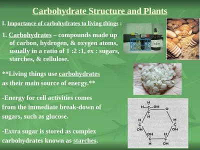
a linear polymer of approximately 2800 Dglucose units per molecule joined by b 14glycosidic bonds fully extended conformation with alternating 180 flips of glucose units extensive intra and intermolecular hydrogen bonding between chains ID: 779428
Download The PPT/PDF document "Cellulose : the major structural compon..." is the property of its rightful owner. Permission is granted to download and print the materials on this web site for personal, non-commercial use only, and to display it on your personal computer provided you do not modify the materials and that you retain all copyright notices contained in the materials. By downloading content from our website, you accept the terms of this agreement.
Slide1
Slide2Cellulose
:
the major structural component of plants, especially wood and plant fibers
a linear polymer of approximately 2800 D-glucose units per molecule joined by b-1,4-glycosidic bondsfully extended conformation with alternating 180° flips of glucose unitsextensive intra- and intermolecular hydrogen bonding between chains
Polysaccharides
Slide3Cellulose is important source of energy for animals
And it is very important for our digestive system .
It contains glucose but different in linkage beta (1-4)
Slide4Cellulose
also decrease absorption of lipids (fat) and cholesterol , so it decrease cancer risk , because cancer increase by increasing fat and
consumption of protein
decrease cholesterol , decrease artery thersosis , decrease cardiovascular disease , weight decreaseCellulose is digestive by animals because they have cellulase
enzyme in bacteria which located in their digestive system
Slide5Carbohydrates – Complex (Polysaccharides)
Cellulose = polysaccharide
found in plant
cell walls
Cellulose fibers
Macrofibril
Microfibril
Chains of
cellulose
Slide6Starch
A
polymers of
a-D-glucose units and used for energy storage in plants, 2 formsamylose: continuous, helical unbranched chains of up to 4000 a-D-glucose units joined by
-1,4-glycosidic bonds
amylopectin: a highly branched helical polymer consisting of 24-30 units of D-glucose joined
by
_
1,4-glycosidic
bonds and branches created by -1,6-glycosidic bondsamylases catalyze hydrolysis of -1,4-glycosidic bondsdebranching enzymes catalyze the hydrolysis of -1,6-glycosidic bonds
Slide7Amylase found in saliva
And there are two type:-
Salivary amylase and pancreatic amylase .
both are the same but pancreatic amylase continue to digest what salivary amylase that begin to digest it .
Slide8Starch
Slide9Polysaccharides
Branching in
amylopectin and glycogen
Slide10Glycogen is the energy storage of glucose in animals , and we find it in muscles and liver .
It is highly branched and globular .
It is highly branched to decrease the storage space , and it is easy to dissociated and to synthesis.
Our body use the storage of glycogen when we do exercise ( in muscles ) or fasting(in liver) .In case of hypocalcemia? When blood sugar level decrease the glycogen will give blood to body to rest of body .*Blood glucose necessary for brain
Cellulose
, chitin and starch are all called
homo
polysaccharides
Slide15Polysaccharides
Chitin:
the major structural component of the exoskeletons of invertebrates, such as insects and crustaceans; also occurs in cell walls of algae, fungi, and yeasts
composed of units of N-acetyl--D-glucosamine joined by -1,4-glycosidic bonds
Slide16Polysaccharides
Bacterial cell walls:
prokaryotic cell walls are constructed on the framework of the repeating unit NAM-NAG joined by
b-1,4-glycosidic bonds
Slide17Bacterial Cell Walls
The N-acetyl-D-
glucoseamine
and N-acetylmuramic acid polysaccharide is in turn cross-linked by small peptidesin Staphylococcus aureus, the cross link is a
tetrapeptidethis tetrapeptide
is unusual in that it contains two amino acids of the D-series, namely D-Ala and D-Glneach tetrapeptide is cross linked to an adjacent
tetrapeptide
by a
pentapeptide
of five
glycine
unitsPeptidoglycan: is the resulted cross linking of polysaccharides by peptides
Slide18Bacterial
Cell Walls
Slide19Bacterial Cell Walls
The
peptidoglycan of a bacterial cell wall
Staphylococcus
aureus
Slide20End