/
CHANGES AFTER DEATH MUDr. Kateřina CHANGES AFTER DEATH MUDr. Kateřina

CHANGES AFTER DEATH MUDr. Kateřina - PowerPoint Presentation

uoutfeature
uoutfeature . @uoutfeature
Follow
343 views
Uploaded On 2020-06-16

CHANGES AFTER DEATH MUDr. Kateřina - PPT Presentation

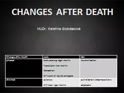
Stoklásková Changes after death early late physical body cooling algor mortis h ypostasis livor mortis desiccation diffusion of liquids and gases mummification chemical ID: 779275

body death rigor tissues death body tissues rigor hours temperature mortis hypostasis decomposition brain cooling minutes blood skin activity

Share:

Link:

Embed:

Download Presentation from below link

Download The PPT/PDF document "CHANGES AFTER DEATH MUDr. Kateřina" is the property of its rightful owner. Permission is granted to download and print the materials on this web site for personal, non-commercial use only, and to display it on your personal computer provided you do not modify the materials and that you retain all copyright notices contained in the materials. By downloading content from our website, you accept the terms of this agreement.


Presentation Transcript

Slide1

CHANGES AFTER DEATH

MUDr. Kateřina Stoklásková

Changes after death

early

late

physical

body

cooling

-

algor

mortis

h

ypostasis

-

livor

mortis

desiccation

diffusion

of liquids and

gases

mummification

chemical

autolysis

stiffness

-

rigor

mortis

p

utrefaction

(

decomposition

)

a

dipoc

e

re

Slide2

Cessation

of

heartbeat

and respiration

traditionally

,

both

the

legal

and

medical

communities

determined

death

through

the

end

of

respiration

and

heartbeat

but

with

the

increasing

ability

of

medicine

to

resuscitate

patients

, a

need

for

better

definition

of

death

became

obvious

life

support

equipment

can

manintain

body

functions

for

a long

time

a

demand

for

organ

transplantation

is

rising

Slide3

Patophysiology

of death

I

t is conventional to describe two types of death

:

S

omatic

death

– the person is unconscious, unable to be aware of the environment, to communicate, unable to initiate any voluntary movement

reflex nervous activity may persist, circulatory and respiratory functions continue either spontaneously or with artificial

support

C

ellular

death

– cells no longer function and have no metabolic activity

it is a

process

- not

an

event

-

different

tissues die at different rates, the cerebral cortex being vulnerable to only few

minutes’

anoxia, connective tissues

surviving

for many hours

Slide4

B

rain death

equals somatic

deaththe number of minutes for which total anoxia will cause cortical damage is estimated 8 minutes

hypothermia (

e

.g

.

immersion under water) reduces the oxygen needs of the tissues – even 40 minutes of hypoxia has been claimed as not leading to brain damage

when the higher levels of cerebral activity are selectively lost (hypoxia, trauma, toxic insult), the victim will exist in

vegetative state

(coma

vigile

,

apalic

syndrome)

the

vicitim

can remain in deep coma certainly for years, is able to sustain circulation and respiration, control body temperature,

excerete

waists, heal wounds, gestate

featus

Slide5

http://news.bbc.co.uk/onthisday/hi/dates/stories/november/19/newsid_2520000/2520581.stm

https

://en.wikipedia.org/wiki/Terri_Schiavo_case

Slide6

http://brno.idnes.cz/zena-porodila-v-komatu-0be-/brno-zpravy.aspx?c=A160209_164845_brno-zpravy_zde

Slide7

B

rainstem death

it is another matter when the brain death spreads below the tentorium (midbrain, pons, upper medulla) – the victim is not only irreversibly comatose, but also incapable of spontaneous breathing

without medical intervention, hypoxic cardiac arrest inevitably follows within minutes and then the usual progression of cellular death ensues

Diagnosis

of

brain

death

panagiography

,

cerebral

blood

flow

and echo-

doppler

-

can

prove

the

death

of

higher

brain”, but not

the

brain stem

irreversible

brain stem

dysfunction

is diagnosed using

ABR

(auditory brain stem

responses

) and

SEP

(

short

latency

somatosensory

potentials

)

EEG-

in

the

United

States

a

flat

EEG test

is

not

required

to

certify

death

,

it

is

considered

to

have

confirmatory

value

. In

the

UK

it

is

not

considered

to

be

of

value

.

Slide8

dead

…or not?

vita minima

- apparent

death

-

hypnotics

overdose

,

electricity

injury

,

lightning

strike,

coma

,

drowning

agony-

vita

reducta

-

the

struggle

that

precedes

death

Slide9

Uncertain

signs of

death- paleness, no pulse, no

blood

pressure

,

areflexia

Certain

signs

of

death

-

livores

mortis

,

rigor

mortis

,

putrefaction

, …

Slide10

Body Cooling – Algor

Mortis

the rate

of

cooling

is

proportional

to

the

difference

in

temperature

between

the

body

surface

and

its

surroundings

the

ideal

cooling

curve

would

be

exponential

, but

it

is

not

in the real human body

estimation

of

the

time

of

death

is

complicated

,

there

are many

factors

affecting

cooling

of

the

body:

initial

body

temperature

(

fever

,

hypotermia

,

diurnal

variation

,

physical

activity

, …)

body

size

(obesity,

children

)

body

posture

clothing

and

coverings

ambient

temperature

air

movement

and humidity

air x

water

Slide11

Body

Coolingroughly the

decrease in temperature is 1°C/

hour

, but

without

considering

all

the

factors

affecting

cooling

,

this

is

highly

inaccurate

Slide12

Hypostasis

hypostasis occurs when the circulation ceases and the gravity acts upon the stagnant blood and pulls it down to the lowest accessible areas

bluish red

discoloration begins as blotchy patches which soon

coalesce

,

the color

depend

ing

on the state of oxygenation at death

darker tint

is a result of reduced

haemoglobin

in skin vessels – asphyxia

pink or dark red

in hypothermia

cherry pink

color of

carboxyhaemoglobin

in carbon monoxide poisoning

pattern

of hypostasis depends on the posture of the body after death

pale

areas are the result of pressure against a hard supporting surface

hypostasis

may

start

forming

after

30 minutes

,

reaches

maximum

within

8-12

hours

does

not

have

much

value

for

estimation of the time since death – in some cases may not be visible at all (infants, old people,

anaemia

, bleeding out…), in some cases it can be observed in the living if the heart action is failing

Slide13

move

completely to the newly lowest areas – can be examined by thumb pressure

be

partly relocated and partly fixed

remain

fixed due to staining of the tissues by

haemolysis

H

ypostasis

O

nce

hypostastis

is established, it can undergo subsequent gravitational shift –

if

the body is moved into a different posture, the primary hypostasis may either

:

Slide14

H

ypostasisblood also settles in the

internal tissues and organsdifferentiation between hypostasis and bruising – the classic test is to incise the suspect area to see if the underlying blood is intravascular (hypostasis) or infiltrating the tissues outside the vessels (contusion), histological examination may be necessary

Slide15

Desiccation

caused by evaporation

of fluids from the

body

’s naturally wet

regions

(

eyes

,

lips

,

genitals

)

or

wounds

Autolysis

cellular

lysozomes

disintegrate

and

hydrolases

are

released

, which break down cells and cellular membranes

starts

in

pancreas

,

stomach

mucosa

Diffusion

of

liquids

and

gases

result

of

autolysis

natural

barriers

do not

function

anymore

and

liquids

and

gases

spread

freely

though

the

tissues

Slide16

Rigor

mortisimmediately after death there is general muscular flaccidity, usually followed by a period of partial or total rigidity, which in turn passes off as the signs of decomposition appear

rigor is initiated when the ATP concentration falls to 85 % of normal

stiffness may

start

develop

ing

within

30 minutes

or may be postponed almost indefinitely (physical activity before death, electrocution X freezing)

reach

es

maximum

within 6-12 hours

duration of full rigor may be 18-36 hours

rigor

is first apparent in the smaller muscle groups – jaw, facial muscles, neck

fades in the same order of muscle groups as it appeared

rigor can be “broken” during manipulation with the body

rigor mortis in other tissues – iris, heart,

dartos

muscle, seminal vesicles, prostate, erector

pili

muscles

poorly

developed in infants, elderly, cachecticcadaveric spasm

– a rare form of virtually instantaneous rigor that develops at the time of death with no period of post-mortem flaccidity (soldiers,

fall

ing

from

height

)

Slide17

A rough

estimation of the post-

mortem

interval in average condition (18°C):

the

body

is

:

warm

and

flaccid

less

than

3

hours

warm

and

stiff

– 3-8

hours

cold

and

stiff

– 8-36

hours

cold and

flaccid

– more

than

36

hours

Slide18

death

putrefaction

skeletization

adipocere

complete dissolution

(unless fossilized)

mummification

cremation

Slide19

Putrefaction (

decomposition)onset

depends on the environment, but in

average

conditions

the

putrefaction

starts

after

approximately

3

days

first

external

naked-eye

sign

is

discoloration of the lower abdominal wall (caecum)1

week

the

color

of

the

skin

is

green/

black

/

reddish

putrefactive

bacteria

spread

through

the venous system, hemolyze the

blood

and

stain

the

vessels

and

adjacent

tissues

greenish-black

the

face and

other

soft

tissues

become

swolen

and

pervaded

by fluid and

gas

2-3

weeks

skin

blisters

appear

,

filled

with

clear

, pink

or

red

fluid

later

the

skin

starts

peeling

off

,

the

hair

and

nails

detach

the

scrotum

and

breasts

are

swolen

bloody

fluid (

tissue

liquefaction

stained

by

haemolysis

)

leaks

from

body

orifices

several

months

soft

tissues

disintegrate

,

leaving

ligamentous

and

tendineous

tissues

attached

to skeleton

Slide20

Forensic Enthomology

animal, especially

insect

infestation depends on

the

ambient

temperature

and

environment

Slide21

Decomposition in

immersed bodies

the

rate of decomposition

is

slower

-

approximately

twice

compared

to

decopmosition

in air

water

slows

down

putrefaction

mainly

because of the lower ambient temperature and protection from insect

the

epidermis

becomes

macerated

and

eventually

detached

gas

formation

is

the

reason

for

flotation

of an unweighted body

the

usual

posture

of

a

freely

floating

body

is

face

down

Slide22

Decomposition in

buried bodies

the

rate of decay

of

bodies

buried

in

earth

is

much

slower

than

those

in

either

air (

approximately

eight

times) or water- due to lower temperature, exclusion of animal and insect

predators

and

lack

of

oxygen

after

10

years

only

bones

should

be

found

in

the

grave

unless

Slide23

Adipocere

the body tissues

transform

into a waxy substance

derived

from

the

body fat

caused

by

hydrolysis

and

hydrogenation

of

adipose

tissue

by

anaerobic

bacteria

(

fatty

acids + K, Na, Ca, Mg ions)the tissues are greasy or waxy,

later

brittle

and

chalky

,

white

,

pinkish

,

greenish

or

grey

requires

MOISTURE

(

usually

wet

soil or water), usually

warmth

and

alcalic

pH

adipocere

starts

forming

after

approximately

3

months

Slide24

Mummification

dried out body,

usually

only some body parts

(

fingers

,

toes

,

ears

, nose)

the

skin

is

dry, hard,

brown

secondary

colonization

by

moulds

causes

white

, green

or

black patches on the skincan only occur in a

DRY

environment

,

preferably

with

moving

air

current

,

which

is

usually

also

warm

mummification can

take weeks or months,

depending

on

the

environment

Slide25

Damage

caused by animals

after

death

there

is

no

bleeding

,

oedema

or

reddening

of

the

edges

of

the

wound

, no other vital signs of injuries

Slide26

References

:

Knight´s

Forensic

Pathology

,

2004