/
DNA  Reccombination Barbara DNA  Reccombination Barbara

DNA Reccombination Barbara - PowerPoint Presentation

valerie
valerie . @valerie
Follow
342 views
Uploaded On 2022-06-11

DNA Reccombination Barbara - PPT Presentation

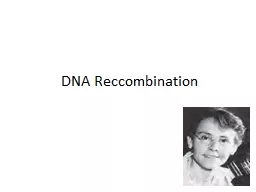
McClintock 19021992 An American scientist and  cytogeneticist  who was awarded the 1983  Nobel Prize in Physiology She received her PhD from   Cornell University  in 1927 She studied ID: 916529

gene dna recombination chromosome dna gene chromosome recombination genetic chromosomes genes coding sequences meiosis homologous cell sequence genome pairs

Share:

Link:

Embed:

Download Presentation from below link

Download Presentation The PPT/PDF document "DNA Reccombination Barbara" is the property of its rightful owner. Permission is granted to download and print the materials on this web site for personal, non-commercial use only, and to display it on your personal computer provided you do not modify the materials and that you retain all copyright notices contained in the materials. By downloading content from our website, you accept the terms of this agreement.


Presentation Transcript

Slide1

DNA Reccombination

Slide2

Barbara

McClintock, (1902–1992)

An

American scientist and 

cytogeneticist

 who was awarded the 1983 

Nobel Prize in

Physiology.

She received her

Ph.D

from


Cornell University

 in

1927. She studied

chromosomes


and

how they change during reproduction in maize.

One

of those ideas was the notion of 

genetic recombination

 by 

crossing-over

 during 

meiosis

—a mechanism by which chromosomes exchange information. She produced the first 

genetic map

 for maize, linking regions of the chromosome to physical traits. She demonstrated the role of the 

telomere

 and 

centromere

, regions of the chromosome that are important in the conservation of 

genetic information

. She was recognized among the best in the field, awarded prestigious fellowships, and elected a member of the 

National Academy of Sciences

 in 1944.

Slide3

Robin Holliday

(1932-2014)

A

British 

molecular

biologist

.

 Holliday described a mechanism of DNA-strand exchange that attempted to explain gene-conversion events that occur during meiosis in fungi. That model first proposed in 1964 and is now known as the 

Holliday

Junction

.

In 1975 he suggested that DNA methylation could be an important mechanism for the control of gene expression in higher organisms, and this has now become documented as a basic 

epigenetic

 mechanism in normal and also cancer

cells.

Slide4

A

Holliday intermediate formed between

two bacterial

plasmids in vivo, as seen with

the electron

microscope

.

Note that such intermediates can form only when the nucleotide sequences of the two parental duplexes are very similar or identical in the region of recombination because specific base pairs must form between the bases of the two parental duplexes.

Slide5

Recombination

Two DNA molecules can recombine with each other to form new DNA molecules that have segments from both parental molecules.

Slide6

Classes Of Recombination

1

.

Homologous genetic recombination

(also called general recombination)

This involves genetic exchanges between any two DNA molecules (or segments of the same molecule) that share an extended region of nearly identical sequence. The actual sequence of bases is irrelevant, as long as it is similar in the two DNAs.

Slide7

2.

S

ite-specific recombination :

The exchanges occur only at a particular DNA sequence.

3

.

DNA transposition :It is usually involves a short segment of DNAwith the remarkable capacity to move from

one location in a chromosome to another.

These

“jumping genes”

were first observed

in maize in the 1940s by Barbara McClintock.

Slide8

Functions of genetic recombination

1. They include roles in specialized DNA repair systems.

2. Specialized activities in DNA replication.

3. Regulation of expression of certain genes.

4. Facilitation of proper chromosome segregation

during eukaryotic cell division.

5. Maintenance of genetic diversity in a population.

6. Implementation of programmed genetic

rearrangements during embryonic development.

Slide9

Recombination during meiosis

Model of double-strand break repair

for homologous

genetic recombination. The

two homologous

chromosomes involved in this recombination event have similar sequences.Each of the two genes shown has different

alleles on

the two chromosomes. The DNA strands

and alleles are colored differently so that their fate

is evident

.

Slide10

Slide11

Slide12

Replication

Prophase I

separation of

Homologous pairs

first

Meiotic division

second

Meiotic division

Haploid gametes

Meiosis in eukaryotic germ-line cells.

Diploid

germ-line cell

Slide13

DNA recombination

In

meiosis homologous pair of chromosomes are arranged in pairs, so

that each

chromosome with two parental sister chromatids are facing each others

in this

arrangement to give a special tetrad arrangement of 4 chromatids .In this arrangement, the opposite chromatids which come from each chromosome will undergo a process

of crossover

. That is opposite

chromatids exchange pieces of DNA between each

others

.

Slide14

DNA recombination

Exchange

of DNA

which allows mixing of genetic information between gametes

that originate from father and mother and produces new combinations of genes. Without this phenomenon, the new gametes will have exactly the same genetic information as the original parents and no genetic variations occur.

Slide15

Recombination involves the breakage and rejoining of two chromosomes (M and F) to produce two re-arranged chromosomes (C1 and C2).

Slide16

Crossing

over. (a)

Crossing over often produces an

exchange of genetic material.

(b)

The homologous chromosomes of

a grasshopper are shown during prophase I of meiosis. Many points of joining (

chiasmata

) are evident between the two homologous pairs

of chromatids. These

chiasmata

are the physical manifestation of

prior homologous

recombination (crossing over) events.

Slide17

Recombination of the V and J

gene segments of the human IgG

kappa light

chain.

Slide18

The light chain can combine

with any of 5,000 possible

heavy

chains to produce an

antibody molecule

Slide19

Slide20

Slide21

Slide22

Chromosome

is highly folded form of the interphase chromatin (extended or relaxed threads of the same nucleoprotein structure).The chromosome form appears during cell division, particularly in the metaphase stage.

Slide23

Chromatin present in two forms

Euchromatin

: the part of chromatin that, during

interphase

, are uncoiled (decondensed) and non-

stained

but containing high concentrations of

transcribed genes (

transcriptionally active)

2. Heterochromatin: The part of the chromatin that

remains

tightly coiled (condensed) and intensely

stained

during interphase, but inactive in gene

expression

.

Slide24

Yeast is known as

Saccharomyces cerevisiae

, which is the single-celled

fungus used to make bread. Yeast is haploids with a genome of 16 chromosomes single

set

.

The gene for mating has a locus on one chromosome which makes the yeast exists either in dominant mating type (G) or recessive mating

type

(g

).

Slide25

The yeast has two types of cell division,

asexual in which the cell divide to produce

two haploid cells with the same mating type

. A

second type of

cell division

called sexual in which two cells conjugate together during mating toproduce new hybrid cell .The hybrid cell is a diploid having pair of two alleles, similar to the pair of alleles arrangements in diploid system of human

cells.

Slide26

Slide27

In diploid system

The

gene is responsible for certain trait in phenotype expression(color of

eyes, color

of skin

).

The allele is a variant of the gene (in eye color expressed as black or

brown

or

blue

) so that certain people have a specific allele of

that gene

, which results in the trait variant. The genes code for proteins,

which might

result in different traits, but it is the

gene

, not the trait, which is inherited

Slide28

Behavior of 2 different genes at different positions on the same chromosome

When

chromosomes go through meiosis, there are two possible situations:

If

no cross-over between the two gene loci

occurs

(if they are present in short distance from

each

other' on the same chromosome):

-

Alleles segregate

together

on the same

chromosome

- A and B together and a and b

together

2. If there is a cross-over between the two gene

loci (

when they are present far distance from each other's on the

chromosome).

Slide29

Alleles

segregate from each other in Meiosis II

Two recombinant products:

- A and b now together in one meiotic product

- a and B now together in one meiotic product

Two parental products

the other two meiotic products are still AB and ab

Slide30

Slide31

Slide32

Gene

density

Not

all the DNA genome encodes protein, but usually it contains coding

and non-coding

sequences. The percentage of coding sequence in total genome

is called gene density.In bacteria the gene density is about 90 % .

In human this density is only about 2% .

Slide33

Therefore, majority of eukaryotes DNA contain non-coding DNA, or regions of DNA that serve no obvious function. Simple single-celled eukaryotes have relatively small amounts of such DNA, whereas the genomes of complex multicellular organisms, including humans contain an absolute majority of DNA without an identified function.

Slide34

This non-coding sequences include repetitive DNA sequence (satellite DNA) , introns and regulatory regions which occupies 98% of total human genomic DNA.

The genomic non-coding DNA sequences are components of an organism's DNA that do not encode protein sequences. Some noncoding DNA is transcribed into functional noncoding RNA molecules (e.g. transfer RNA, ribosomal RNA, and regulatory RNAs), while others are not transcribed or give rise to RNA transcripts of unknown function.

Slide35

Functions of noncoding DNA

1

. Protection of the genome:

Noncoding DNA separate genes from each other with long gaps, so alteration in one gene or part of a chromosome does not extend to the whole chromosome. In high genomic complexity like in case of human genome, not only different genes, but also inside one gene there are gaps of introns to protect the entire coding segment to minimize the changes caused by changing part of the

gene.

Slide36

2. Genetic switches:

Some noncoding DNA sequences function as genetic switches that decide when and where genes are expressed.

3. Regulation of gene expression:

Some noncoding DNA sequences quantitatively determine the expression levels of various genes.

Slide37

4. Act as regulatory sites for gene actions:

Some non-coding sequence can be promoters, operators, and enhancers ….etc.

Repeated sequences are patterns of DNA that occur in multiple copies throughout the genome.

Slide38

Satellite

DNA

Very

large sequence consists of tandem repeating, non-coding DNA. It is the main component of functional centromeres, and forms the main structural constituent of heterochromatin. A repeated pattern can be between 1 base pair long (a mononucleotide repeat) to several thousand base pairs long, and the total size of a satellite DNA block can be several

mega bases

(Unit of length for DNA fragments equal to 1 million nucleotides and roughly equal to 1

cm)

without interruption.

Slide39

Most satellite DNA is localized to the

telomeric or the

centromeric

region of the chromosome. The nucleotide sequence of the repeats is fairly well conserved across a species. However, variation in the length of the repeat is common. The difference in how many of the repeats are present in the region (length of the region) is the basis for DNA fingerprinting in forensic medicine.

Slide40

Tandem repeats

: copies which lie adjacent to each other, either directly or inverted

Minisatellite

- repeat units from about 10 to 70 base pairs, found in many places in the genome, including the centromeres.Microsatellite

- repeat units of less than 10 base pairs (typically have 6 to 8 base

repeat units) mainly found in telomeres

Slide41

Some types of satellite DNA in humans are

Type

Size of repeat unit (

bp

)

Location

α (

alphoid

DNA)

171

All chromosomes

β

68

Centromeres of chromosomes 1, 9, 13, 14, 15, 21, 22 and Y

Mini-satellite

25-48

Centromeres and other regions in heterochromatin of most chromosomes

Micro-

satalite

5

Most chromosomes

telomers