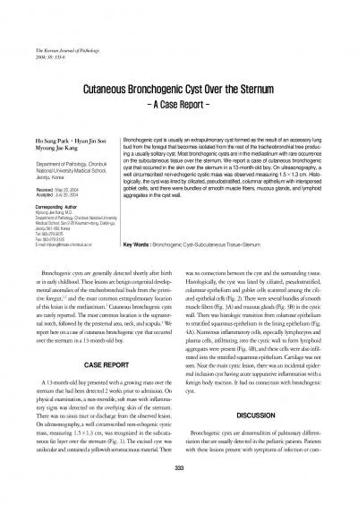
/
STUDY OF APPLICATION OF FLUORESCENCE BRONCHOSCOPY TO BRONCHOGENIC CARCINOMA DIAGNOSIS STUDY OF APPLICATION OF FLUORESCENCE BRONCHOSCOPY TO BRONCHOGENIC CARCINOMA DIAGNOSIS

STUDY OF APPLICATION OF FLUORESCENCE BRONCHOSCOPY TO BRONCHOGENIC CARCINOMA DIAGNOSIS - PowerPoint Presentation

vestibulephilips
vestibulephilips . @vestibulephilips
Follow
343 views
Uploaded On 2020-06-17

STUDY OF APPLICATION OF FLUORESCENCE BRONCHOSCOPY TO BRONCHOGENIC CARCINOMA DIAGNOSIS - PPT Presentation

NATIONAL LUNG HOSPITAL In the world amp Vietnam Prevalence 1 st in male 2 nd in female Mortality 1 st in both genders LUNG CANCER Common malignant disease INTRODUCTION Bronchogenic Carcinoma ID: 780506

carcinoma bronchoscopy bronchogenic amp bronchoscopy carcinoma amp bronchogenic lung cancer fluorescene tumor lesions features results light white fluorescence study

Share:

Link:

Embed:

Download Presentation from below link

Download The PPT/PDF document "STUDY OF APPLICATION OF FLUORESCENCE BRO..." is the property of its rightful owner. Permission is granted to download and print the materials on this web site for personal, non-commercial use only, and to display it on your personal computer provided you do not modify the materials and that you retain all copyright notices contained in the materials. By downloading content from our website, you accept the terms of this agreement.


Presentation Transcript

Slide1

STUDY OF APPLICATION OF FLUORESCENCE BRONCHOSCOPY TO BRONCHOGENIC CARCINOMA DIAGNOSIS

NATIONAL LUNG HOSPITAL

Slide2

In the world & Vietnam: Prevalence: 1st in male, 2nd in female Mortality: 1st in both genders.

LUNG CANCER

Common malignant disease

INTRODUCTION

Slide3

Bronchogenic Carcinoma: cancer of trachea, bronchi (main, lobar, segmental and subsegmental)Lung Cancer or

Bronchogenic Carcinoma(S.Ikeda 1974)

INTRODUCTION

Slide4

Diagnosis of Lung CancerChest X-ray, CT, MRI, PET…

Lung needle biopsy

Exploratory surgery

Bronchoscopy

INTRODUCTION

Slide5

1

Assessing the lesions of bronchogenic

carcinoma in

fluorescene

bronchoscopy

2

Describe some clinical,

paraclinical

features of bronchogenic carcinoma

Objectives

Slide6

Literature Review Causes and risk factors of lung cancer:Cigarette smokingSubstances not related to cigarette: Arsenic, Asbestos, Nickel…

Risk factors: + Age: peak in 55 – 65 + Gender: World: male/female: 6/1

Vietnam: male/female: 4/1

+ Other factors: family history, diet, chronic pulmonary diseases,

socioeconomic status.

Slide7

Literature ReviewClinical signs & symptoms:A. Local: Cough, chest pain, dyspneaB. Compression:Hoarseness, hiccup, dysphagia, superior vena cava syndrome…Pleural, pericardial effusionC. Paraneoplastic syndromes

D. General signs: Fever, loss of appetite, weight lossE. Symptoms of metastasis: headache (brain met), bone pain (bone met), abdominal pain (liver met)…

Slide8

Literature Review Paraclinial features:Diagnostic Imaging: Chest X-ray, CT Scanner, MRI…Tumor markers: CEA,

Cyfra 21-1, Pro-GRPBronchoscopyPathology: cytology, histopathology

EGFR mutation testing

Slide9

Lung cancer stages: AJCC 2010Stage group

T

N

M

0

T0

N0

M0

IA

IB

T1a

-b

T2a

N0

N0

M0

M0

IIA


IIB

T1a-b, T2a

T2b

T2b

T3

N1

N0

N1

N0

M0

M0

M0

M0

IIIA


IIIB

T3

T1-3

T4

T1-3

T4

N1

N2

N0-1

N3

N2-3

M0M0M0M0M0IVAny TAny NM1a-b

Literature Review

Slide10

Literature ReviewMain histopathologic types of lung cancer (WHO 2015): Adenocarcinoma Squamous Cell Carcinoma Carcinoid Tumor

Mixed Lung Cancer

Slide11

Fluorescence Bronchoscopy DevicesLiterature Review

Tumor with decrease in fluorescence

Slide12

Subjects

Patients were diagnosed primary lung cancer by histopathological evidence from bronchial biopsy specimens obtained at fluorescene

bronchoscopy in National Lung Hospital from December 2014 to August 2015

Slide13

Study design: Cross-sectional

Sampling technique: Convenient

Data

collection tool

:

Data collection form

Methods

Slide14

DATA ANALYSIS & MANAGEMENT Study variables Cilinical features: age, gender, history, time of detecting tumorClinical signs & symptoms: general, objective, subjective, performance status

Primary tumor location, metastatic sitesParaclinical

features:

- Imaging, laboratory tests

-

Fluorescene

bronchoscopy

- Histopathology

Methods

Slide15

Data was entered and analyzed using SPSS 16.0.Statistical issues:Descriptive: mean, standard deviation, max, min. Statistical tests: chi-square test was used. Statistically significant results (p < 0.05) were included. Fisher’s exact test was used when sample sizes are smaller than 5.DATA ANALYSIS & MANAGEMENT

Methods

Slide16

Bronchogenic Carcinoma

White light bronchoscopy

Fluorescence bronchoscopy

Images of lesions

Histopathology

T Classification

TNM Staging


Conclusion

Research Process Flowchart

Slide17

Chart 3.2: Gender distribution

1Clinical & paraclinical features

Study

Male/female

Hoàng

Đình

Chân (2004)

5.76

Thu

(2009)

4

RESULTS & DISCUSSION

Male

Female

Slide18

RESULTS & DISCUSSION1

Clinical &

paraclinical

features

< 40 40 – 49 50 – 59 60 – 69 > 70

Slide19

RESULTS & DISCUSSIONStudy

Smoking historyPha

n

Thắng

(2000)78.4%

Lê Thu Hà (2009)71

.

2%

1

Clinical &

paraclinical

features

Smoking

Non-smoking

%

< 20 20 – 30 > 30

5% 30% 45%

Slide20

1

RESULTS & DISCUSSION

Table 3.1.

Tumor location on chest

x-ray

Study

Tumor location

Nguyễn

Việt

Cồ

( 1994)

Right lung (64.2%), left

lung

(35.3%), upper lobe (52.4%), lower

lobe (

29.14%).

Tumor Location

Bronchogenic Carcinoma

n

%

Lung

Right

22

55

Left

14

35

Both

4

10

Total

40

100

Lobe

Upper

21

52,5

Middle

6

15

Lower

13

32,5

Total

40

100

Clinical &

paraclinical

features

Slide21

Chart 3.10. Histopathologic types

1RESULTS & DISCUSSION

Study

Histopathologic

types

Houwen

L (1994)

Central lung cancer: squamous cell

60%

Peripheral lung cancer:

adenocarcinoma 70%

Phan

Thắng

(2000)

Central lung cancer: squamous cell

38.5%

Peripheral lung cancer:

adenocarcinoma 74.5%

Clinical &

paraclinical

features

Small cell

Adenocarcinoma

Squamous cell

Large cell

Slide22

2Assessing the lesions of

bronchogenic carcinoma in white light bronchoscopy:

RESULTS & DISCUSSION

Study

Lesions

Ngô

Quý

Châu

(2002)

Papillary tumor

17%, infiltration 39%, compression 30%

Nguyễn

Đại

Bình

(1999)

Papillary tumor 29.2

%, infiltration 47.9%, compression 12.3%

Lesion

Number

Percentage

Papillary tumor

10

25

Infiltration

24

60

Compression

6

15

Total

40

100

Table 3.3. Lesions of

bronchogenic carcinoma in white light bronchoscopy

Slide23

RESULTS & DISCUSSIONTable 3.4. Lesions of bronchogenic carcinoma in fluorescence bronchoscopy Bronchoscopy

LesionWhite light

(

n = 40

)

Decrease in fluorescence

(n =

40)

Papillary

tumor

10

10

Infiltration

24

24

Compression

6

6

Decrease in fluorescence

significantly different from

papillary tumor

,

infiltration,

compression

0

40

Assessing the lesions of

bronchogenic carcinoma

in

fluorescene

bronchoscopy

:

2

Slide24

RESULTS & DISCUSSIONHistopathologic typeNumber

PercentageHyperplasia

4

10

Dysplasia

3

7.5

Metaplasia

3

7.5

Cis

0

0

Cancer

20

50

Chronic inflammation

10

25

Total

40

100

Table 3.5. Histopathologic types of 40 decrease in

fluorescene

lesions of

bronchogenic carcinoma

2

Assessing the lesions of

bronchogenic carcinoma

in

fluorescene

bronchoscopy

:

Slide25

RESULTS & DISCUSSIONTable 3.6. Comparison of T classification of bronchogenic carcinoma between white light bronchoscopy and fluorescene bronchoscopyT classification

White light bronchoscopy

Fluorescene

bronchoscopy

Number

Number

T1

0

0

T2

7

2

T3

4

3

T4

9

15

Total

20

20

Assessing the lesions of

bronchogenic carcinoma

in

fluorescene

bronchoscopy

:

2

Slide26

RESULTS & DISCUSSIONTable 3.7. Comparison of stage of bronchogenic carcinoma between white light bronchoscopy and fluorescene bronchoscopyStage group

White light bronchoscopyFluorescene

bronchoscopy

Number

Number

IIa

1

0

IIb

0

1

IIIa

5

1

IIIb

2

6

IV

1

1

Total

9

9

2

Assessing the lesions of

bronchogenic carcinoma

in

fluorescene

bronchoscopy

:

Slide27

CONCLUSION Clinical & paraclinical features

1

- Bronchogenic carcinoma has some features: male more common than female, common in smokers, squamous cell carcinoma has high percentage.

Assessing the lesions of bronchogenic

carcinoma in

fluorescene

bronchoscopy

-

Fluorescene

bronchoscopy helps to identify accurately the margin of primary tumor of bronchogenic carcinoma (50%).

-

Fluorescene

bronchoscopy helps to stage more accurately than white light bronchoscopy (9 in total of 40 patients changed stage).

2

Slide28

Lung cancer patients who are operable should be ordered a fluorescence bronchoscopy to operate scientifically and rationally.Recommendation

Slide29

Thanks for listening