12 Carbohydrate metabolism Chemistry 176

Published  . 0 views
↓ Download
12 Carbohydrate metabolism Chemistry 176
1 / 1
12 Carbohydrate metabolism Chemistry 176 - slide 1 of 21 12 Carbohydrate metabolism Chemistry 176 - slide 2 of 21 12 Carbohydrate metabolism Chemistry 176 - slide 3 of 21 12 Carbohydrate metabolism Chemistry 176 - slide 4 of 21 12 Carbohydrate metabolism Chemistry 176 - slide 5 of 21 12 Carbohydrate metabolism Chemistry 176 - slide 6 of 21 12 Carbohydrate metabolism Chemistry 176 - slide 7 of 21 12 Carbohydrate metabolism Chemistry 176 - slide 8 of 21 12 Carbohydrate metabolism Chemistry 176 - slide 9 of 21 12 Carbohydrate metabolism Chemistry 176 - slide 10 of 21 12 Carbohydrate metabolism Chemistry 176 - slide 11 of 21 12 Carbohydrate metabolism Chemistry 176 - slide 12 of 21 12 Carbohydrate metabolism Chemistry 176 - slide 13 of 21 12 Carbohydrate metabolism Chemistry 176 - slide 14 of 21 12 Carbohydrate metabolism Chemistry 176 - slide 15 of 21 12 Carbohydrate metabolism Chemistry 176 - slide 16 of 21 12 Carbohydrate metabolism Chemistry 176 - slide 17 of 21 12 Carbohydrate metabolism Chemistry 176 - slide 18 of 21 12 Carbohydrate metabolism Chemistry 176 - slide 19 of 21 12 Carbohydrate metabolism Chemistry 176 - slide 20 of 21 12 Carbohydrate metabolism Chemistry 176 - slide 21 of 21
Description: 12 Carbohydrate metabolism Chemistry 176 Hyperglycaemia and diabetes mellitus 183 Physiology 176 Hypoglycaemia 194 This chapter discusses carbohydrate metabolism and its synthesize glucose, abnormalities, with emphasis on diabetes mellitus

Related Topics

Download Presentation

"12 Carbohydrate metabolism Chemistry 176" is the property of its rightful owner. Permission is granted to download and print the materials on this website for personal, non-commercial use only, and to display it on your personal computer provided you do not modify the materials and that you retain all copyright notices contained in the materials. By downloading content from our website, you accept the terms of this agreement.

Presentation Transcript

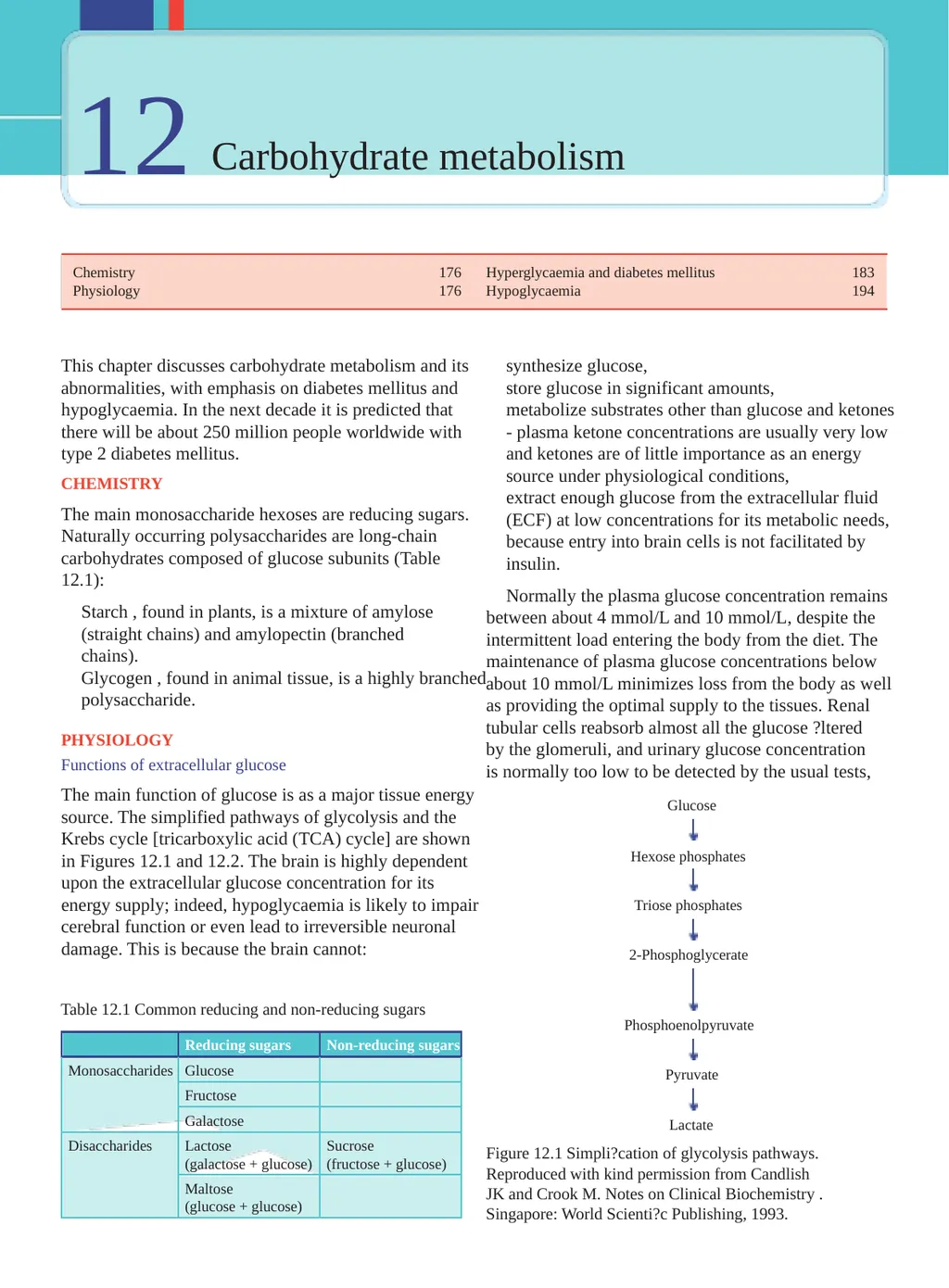
slide1. 12 Carbohydrate metabolism Chemistry 176 Hyperglycaemia and diabetes mellitus 183 Physiology 176 Hypoglycaemia 194 This chapter discusses carbohydrate metabolism and its synthesize glucose, abnormalities, with emphasis on diabetes mellitus and store glucose in significant amounts, hypoglycaemia. In the next decade it is predicted that metabolize substrates other than glucose and ketones there will be about 250 million people worldwide with - plasma ketone concentrations are usually very low type 2 diabetes mellitus. and ketones are of little importance as an energy source under physiological conditions, CHEMISTRY extract enough glucose from the extracellular fluid The main monosaccharide hexoses are reducing sugars. (ECF) at low concentrations for its metabolic needs, Naturally occurring polysaccharides are long-chain because entry into brain cells is not facilitated by carbohydrates composed of glucose subunits (Table insulin. 12.1): Normally the plasma glucose concentration remains Starch , found in plants, is a mixture of amylose between about 4 mmol/L and 10 mmol/L, despite the (straight chains) and amylopectin (branched intermittent load entering the body from the diet. The chains). maintenance of plasma glucose concentrations below Glycogen , found in animal tissue, is a highly branched about 10 mmol/L minimizes loss from the body as well polysaccharide. as providing the optimal supply to the tissues. Renal tubular cells reabsorb almost all the glucose ?ltered PHYSIOLOGY by the glomeruli, and urinary glucose concentration Functions of extracellular glucose is normally too low to be detected by the usual tests, The main function of glucose is as a major tissue energy Glucose source. The simplified pathways of glycolysis and the Krebs cycle [tricarboxylic acid (TCA) cycle] are shown Hexose phosphates in Figures 12.1 and 12.2. The brain is highly dependent upon the extracellular glucose concentration for its energy supply; indeed, hypoglycaemia is likely to impair Triose phosphates cerebral function or even lead to irreversible neuronal damage. This is because the brain cannot: 2-Phosphoglycerate Table 12.1 Common reducing and non-reducing sugars Phosphoenolpyruvate Reducing sugars Non-reducing sugars Monosaccharides Glucose Pyruvate Fructose Galactose Lactate Disaccharides Lactose Sucrose Figure 12.1 Simpli?cation of glycolysis pathways. (galactose + glucose) (fructose + glucose) Reproduced with kind permission from Candlish Maltose JK and Crook M. Notes on Clinical Biochemistry . (glucose + glucose) Singapore: World Scienti?c Publishing, 1993.<br>
slide2. Physiology 177 Insulin Glucose- Ribose- Glucose 6-phosphate 5-phosphate Insulin is the most important hormone controlling plasma glucose concentrations. A plasma glucose TPP Transketolase concentration of greater than about 5 mmol/L acting via the glucose transporter 2 stimulates insulin release from Sedoheptulose- Fructose- 7-phosphate the pancreas b -cell. These cells produce proinsulin, which 6-phosphate consists of the 51-amino-acid polypeptide insulin and a linking peptide (C-peptide, Fig. 12.3). Splitting of the peptide bonds by prohormone convertases releases via Fructose-1, intermediates (mostly 32-33 split proinsulin) equimolar 6-diphosphate amounts of insulin and C-peptide into the ECF. Insulin binds to speci?c cell surface receptors on muscle and adipose tissue, thus enhancing the rate Pyruvate Lactate of glucose entry into these cells. Insulin-induced Pyruvate activation of enzymes stimulates glucose incorporation TPP dehydrogenase into glycogen (glycogenesis) in liver and muscle (Fig 12.4). Insulin also inhibits the production of glucose Acetyl CoA (gluconeogenesis) from fats and amino acids, partly by inhibiting fat and protein breakdown (lipolysis and Oxaloacetate Citrate proteolysis). The transport of glucose into liver cells is insulin independent but, by reducing the intracellular glucose concentration, insulin does indirectly promote the a -Ketoglutarate Succinate passive diffusion of glucose into them. Insulin also a -Ketoglutarate directly increases the transport of amino acids, dehydrogenase TPP potassium and phosphate into cells, especially muscle; these processes are independent of glucose transport. Figure 12.2 Simpli?cation of the tricarboxylic acid (Krebs) cycle. CoA, coenzyme A; TPP, thiamine pyrophosphate. In the longer term, insulin regulates growth and Reproduced with kind permission from Candlish JK and development and the expression of certain genes. Crook M. Notes on Clinical Biochemistry . Singapore: Glucagon World Scienti?c Publishing, 1993. Glucagon is a single-chain polypeptide synthesized by the a -cells of the pancreatic islets. Its secretion is even after a carbohydrate meal. Signi?cant glycosuria stimulated by hypoglycaemia. Glucagon enhances usually occurs only if the plasma glucose concentration hepatic glycogenolysis (glycogen breakdown) and exceeds about 10 mmol/L - the renal threshold. gluconeogenesis. How the body maintains extracellular glucose concentrations Control of plasma glucose concentration C-PEPTIDE During normal metabolism, little glucose is lost unchanged from the body. Maintenance of plasma COO - glucose concentrations within the relatively narrow NH 3+ range of 4-10 mmol/L, despite the widely varying input S S from the diet, depends on the balance between the S S glucose entering cells from the ECF and that leaving S INSULIN them into this compartment. S Hormones concerned with glucose homeostasis Figure 12.3 Structure of proinsulin, indicating the Some of the more important effects of hormones on cleavage sites at which insulin and C-peptide are glucose homeostasis are summarized in Table 12.2. produced.<br>
slide3. Carbohydrate metabolism 178 Table 12.2 Action of hormones that affect intermediary metabolism Insulin Glucagon Growth hormone Glucocorticoids Adrenaline Carbohydrate metabolism In liver Glycolysis + Glycogenesis + Glycogenolysis + + Gluconeogenesis - + + In muscle Glucose uptake + - - Glycogenesis + Glycogenolysis + Protein metabolism Synthesis + + Breakdown - + Lipid metabolism Synthesis + Lipolysis - + + + Secretion Stimulated by Hyperglycaemia Hypoglycaemia Hypoglycaemia Hypoglycaemia Hypoglycaemia Amino acids Amino acids Stress Stress Stress Glucagon Fasting Sleep Gut hormones b -blockers Inhibited by Adrenaline Insulin Somatostatin Glucocorticoids Fasting IGF-1 Somatostatin Plasma NEFA concentrations Fall Rise Rise Rise Rise Plasma glucose concentrations Fall Rise Rise Rise Rise +, stimulates; -, inhibits; IGF-1, insulin-like growth factor 1; NEFA, non-esteri?ed fatty acid. Somatostatin in phaeochromocytoma (adrenaline and noradrenaline see Chapter 24) and thus oppose the normal action of This peptide hormone is released from the D cells of insulin. the pancreas and inhibits insulin and growth hormone release. The liver Other hormones The liver is the most important organ maintaining a When plasma insulin concentrations are low, for constant glucose supply for other tissues, including the example during fasting, the hyperglycaemic actions brain. It is also of importance in controlling the post- of hormones, such as growth hormone (GH), prandial plasma glucose concentration. glucocorticoids, adrenaline (epinephrine) and Portal venous blood leaving the absorptive area of the glucagon, become apparent, even if there is no increase intestinal wall reaches the liver ?rst, and consequently in secretion rates. Secretion of these so-called counter- the hepatic cells are in a key position to buffer the regulatory hormones may increase during stress and hyperglycaemic effect of a high-carbohydrate meal in patients with acromegaly (GH, see Chapter 6), (Fig. 12.5). Cushing's syndrome (glucocorticoids, see Chapter 8) or<br>
slide4. Physiology 179 lactate or the carbon chains resulting from deamination of certain amino acids (mainly alanine) (Table 12.3). The liver contains the enzyme glucose-6-phosphatase, which, by hydrolysing G6P derived from either glycogenolysis Outer or gluconeogenesis, releases glucose and helps to 1 1 1 1 branches 1 1 maintain extracellular fasting concentrations. Hepatic 1 glycogenolysis is stimulated by the hormone glucagon, 2 2 1 secreted by the a -cells of the pancreas in response 2 2 to a fall in the plasma glucose concentration, and by 3 catecholamines such as adrenaline or noradrenaline. 3 Inner branches During fasting, the liver converts fatty acids, 4 released from adipose tissue as a consequence of low insulin activity, to ketones. The carbon chains of some amino acids may also be converted to ketones (Table 12.3). Ketones can be used by other tissues, including R the brain, as an energy source when plasma glucose concentrations are low. Figure 12.4 Structure of glycogen. Open circles depict glucose moieties in a -1,4 linkage and the black Other organs circles those in a -1,6 linkages at branch points. R The renal cortex is the only other tissue capable of indicates the reducing end group. The outer branches gluconeogenesis, and of converting G6P to glucose. The terminate in non-reducing end groups. Reproduced with permission from Nyhan WL and Barshop BA. Atlas gluconeogenic capacity of the kidney is particularly of Inherited Metabolic Diseases, 3rd edition. London: important in hydrogen ion homeostasis and during Hodder Arnold, 2012. prolonged fasting. Other tissues, such as muscle, can store glycogen but, because they do not contain glucose-6-phosphatase, The entry of glucose into liver and cerebral cells is they cannot release glucose from cells and so can only not directly affected by insulin, but depends on the use it locally; this glycogen plays no part in maintaining extracellular glucose concentration. The conversion of the plasma glucose concentration. glucose to glucose-6-phosphate (G6P), the ?rst step in Systemic effects of glucose intake glucose metabolism in all cells, is catalysed in the liver by the enzyme glucokinase, which has a low af?nity for The liver modifies the potential hyperglycaemic effect glucose compared with that of hexokinase, which is found of a high-carbohydrate meal by extracting relatively in most other tissues. Glucokinase activity is induced by more glucose than in the fasting state from the portal insulin. Therefore, hepatic cells extract proportionally plasma. However, some glucose does pass through less glucose during fasting, when concentrations in the liver and the rise in the systemic concentration portal venous plasma are low, than after carbohydrate ingestion. This helps to maintain a fasting supply of Table 12.3 Metabolism of the carbon skeleton of some glucose to vulnerable tissues such as the brain. amino acids to either carbohydrate (glycogenic) or fat The liver cells can store some of the excess glucose as (ketogenic) glycogen. The rate of glycogen synthesis (glycogenesis) Glycogenic Glycogenic and ketogenic Ketogenic from G6P may be increased by insulin secreted by the b -cells of the pancreas in response to systemic Alanine Isoleucine Leucine hyperglycaemia. The liver can convert some of the excess Arginine Lysine glucose to fatty acids, which are ultimately transported Glycine Phenylalanine as triglyceride in very low-density lipoprotein (VLDL) Histidine Tyrosine and stored in adipose tissue. Methionine Under normal aerobic conditions, the liver can Serine synthesize glucose by gluconeogenesis using the Valine metabolic products from other tissues, such as glycerol,<br>
slide5. Carbohydrate metabolism 180 BRAIN MUSCLE G6P Insulin GLYCOGEN INTESTINE CO 2 + H 2 O G6P GLUCOSE Glucose Insulin Insulin G6P G6P GLYCOGEN Triose-P Triose-P Acetyl CoA Fatty acid + Glycerol-3-P Fatty acid + Glycerol-3-P Glycerol Triglyceride TRIGLYCERIDE VLDL LIVER ADIPOSE TISSUE Figure 12.5 Post-prandial metabolism of glucose. CoA, coenzyme A; G6P, glucose-6-phosphate; Glycerol-3-P, glycerol-3-phosphate; Triose-P, triose phosphate or glyceraldehyde 3-phosphate; VLDL, very low-density lipoprotein. stimulates the b -cells of the pancreas to secrete insulin. these actions are impaired. Both muscle and adipose tissue store the excess post-prandial glucose, but the Insulin may further enhance hepatic and muscle mode of storage and the function of the two types of glycogenesis. More importantly, entry of glucose into cell are very different, as will be shown later. adipose tissue and muscle cells, unlike that into liver and brain, is stimulated by insulin and, under physiological Ketosis conditions, the plasma glucose concentration falls to Adipose tissue and the liver near fasting levels. Conversion of intracellular glucose Adipose tissue triglyceride is the most important long- to G6P in adipose and muscle cells is catalysed by the term energy store in the body. Greatly increased use enzyme hexokinase, which, because its affinity for of fat stores, for example during prolonged fasting, glucose is greater than that of hepatic glucokinase, is associated with ketosis. Adipose tissue cells, acting ensures that glucose enters the metabolic pathways in in conjunction with the liver, convert excess glucose these tissues at lower extracellular concentrations than to triglyceride and store it in this form rather than those in the liver. The relatively high insulin activity as glycogen. The components are both derived from after a meal also inhibits the breakdown of triglyceride glucose, fatty acids from the glucose entering hepatic (lipolysis) and protein (proteolysis). If there is relative cells and glycerol from that entering adipose tissue cells. or absolute insulin deficiency, as in diabetes mellitus,<br>
slide6. Physiology 181 In the liver, triglycerides are formed from glycerol- Most tissues, other than the brain, can oxidize fatty 3-phosphate (from triose phosphate or glyceraldehyde- acids to acetyl CoA, which can then be used in the TCA 3-phosphate) and fatty acids [from acetyl coenzyme A cycle as an energy source. When the rate of synthesis (CoA)]. The triglycerides are transported to adipose exceeds its use, the hepatic cells produce acetoacetic acid tissue cells incorporated in VLDL, where they are by enzymatic condensation of two molecules of acetyl CoA; acetoacetic acid can be reduced to b -hydroxybutyric hydrolysed by lipoprotein lipase. The released fatty acids (of hepatic origin) are re-esteri?ed within these acid and decarboxylated to acetone. These ketones can cells with glycerol-3-phosphate, derived from glucose, be used as an energy source by brain and other tissues at which has entered this tissue under the in?uence of a time when glucose is in relatively short supply. insulin. The resultant triglyceride is stored and is far Ketosis occurs when fat stores are the main energy more energy dense than glycogen (see Chapter 13). source and may result from fasting or from reduced During fasting, when exogenous glucose is nutrient absorption, for example due to vomiting. Mild unavailable and the plasma insulin concentration ketosis may occur after as little as 12 h of fasting. After is therefore low, endogenous triglycerides are short fasts, metabolic acidosis is not usually detectable, reconverted to free non-esteri?ed fatty acids (NEFAs) but, after longer periods, more hydrogen ions may and glycerol by lipolysis (Fig. 12.6). Both are be produced than can be dealt with by homeostatic transported to the liver in plasma, the NEFA being buffering mechanisms, depleting the plasma bicarbonate protein bound, predominantly to albumin. Glycerol concentration, which therefore falls (see Chapter 4). enters the hepatic gluconeogenic pathway at the The plasma glucose concentration is maintained triose phosphate stage; the glucose synthesized can be principally by hepatic gluconeogenesis, but during released from these cells, thus minimizing the fall in prolonged starvation, such as that in anorexia nervosa or glucose concentrations. during childhood, ketotic hypoglycaemia may occur. The BRAIN G6P Acetyl CoA CO 2 + H 2 O Glucose KETONES + H + Triglyceride G6P GLYCOGEN Triose-P Glycerol KETONES + H + + FA NEFA Pyruvate Acetyl CoA LIVER ADIPOSE TISSUE Figure 12.6 Intermediary metabolism during fasting: ketosis. CoA, coenzyme A; FA, fatty acid; G6P, glucose-6-phosphate; NEFA, non-esteri?ed fatty acid.<br>
slide7. Carbohydrate metabolism 180 BRAIN MUSCLE G6P Insulin GLYCOGEN INTESTINE CO 2 + H 2 O G6P GLUCOSE Glucose Insulin Insulin G6P G6P GLYCOGEN Triose-P Triose-P Acetyl CoA Fatty acid + Glycerol-3-P Fatty acid + Glycerol-3-P Glycerol Triglyceride TRIGLYCERIDE VLDL LIVER ADIPOSE TISSUE Figure 12.5 Post-prandial metabolism of glucose. CoA, coenzyme A; G6P, glucose-6-phosphate; Glycerol-3-P, glycerol-3-phosphate; Triose-P, triose phosphate or glyceraldehyde 3-phosphate; VLDL, very low-density lipoprotein. stimulates the b -cells of the pancreas to secrete insulin. these actions are impaired. Both muscle and adipose tissue store the excess post-prandial glucose, but the Insulin may further enhance hepatic and muscle mode of storage and the function of the two types of glycogenesis. More importantly, entry of glucose into cell are very different, as will be shown later. adipose tissue and muscle cells, unlike that into liver and brain, is stimulated by insulin and, under physiological Ketosis conditions, the plasma glucose concentration falls to Adipose tissue and the liver near fasting levels. Conversion of intracellular glucose Adipose tissue triglyceride is the most important long- to G6P in adipose and muscle cells is catalysed by the term energy store in the body. Greatly increased use enzyme hexokinase, which, because its affinity for of fat stores, for example during prolonged fasting, glucose is greater than that of hepatic glucokinase, is associated with ketosis. Adipose tissue cells, acting ensures that glucose enters the metabolic pathways in in conjunction with the liver, convert excess glucose these tissues at lower extracellular concentrations than to triglyceride and store it in this form rather than those in the liver. The relatively high insulin activity as glycogen. The components are both derived from after a meal also inhibits the breakdown of triglyceride glucose, fatty acids from the glucose entering hepatic (lipolysis) and protein (proteolysis). If there is relative cells and glycerol from that entering adipose tissue cells. or absolute insulin deficiency, as in diabetes mellitus,<br>
slide8. Hyperglycaemia and diabetes mellitus 183 During gluconeogenesis, hydrogen ions are reused. hepatic and renal gluconeogenesis from lactate Under aerobic conditions, the liver consumes much cannot occur anaerobically, more lactate than it produces. anaerobic glycolysis is stimulated because the The physiological accumulation of lactic acid during falling adenosine triphosphate (ATP) levels cannot muscular contraction is a temporary phenomenon and be regenerated by the TCA cycle under anaerobic rapidly disappears at rest, when slowing of glycolysis conditions. allows aerobic processes to `catch up'. The combination of impaired gluconeogenesis and increased anaerobic glycolysis converts the liver from Pathological lactic acidosis an organ that consumes lactate and H + to one that Lactic acid, produced by anaerobic glycolysis, may either generates large amounts of lactic acid. Severe hypoxia, be oxidized to CO 2 and water in the TCA cycle or be for example following a cardiac arrest, causes marked reconverted to glucose by gluconeogenesis in the liver. lactic acidosis. If diabetic ketoacidosis is associated with Both the TCA cycle and gluconeogenesis need oxygen; signi?cant volume depletion, this hypoxic syndrome anaerobic glycolysis is a non-oxygen-requiring pathway. may aggravate the acidosis. (See Chapter 4 for a further Pathological accumulation of lactate may occur because: discussion of lactic acidosis.) The glycolytic pathway as well as the TCA cycle are production is increased by an increased rate of summarized in Figures 12.1 and 12.2. anaerobic glycolysis, use is decreased by impairment of the TCA cycle or impairment of gluconeogenesis. HYPERGL YCAEMIA AND DIABETES MELLITUS Tissue hypoxia (Fig. 12.8) due to the poor tissue Hyperglycaemia may be due to: perfusion of the `shock' syndrome is usually the most common cause of lactic acidosis. Hypoxia increases intravenous infusion of glucose-containing fluids, plasma lactate concentrations because: severe stress (usually a transient effect) such as trauma, myocardial infarction or cerebrovascular the TCA cycle cannot function anaerobically and accidents, oxidation of pyruvate and lactate to CO 2 and water diabetes mellitus or impaired glucose regulation. is impaired, GLYCOGEN G6P G6P GLYCOGEN Pyruvate LACTATE + H + LACTATE + H + Pyruvate LACTATE+ H + MUSCLE LIVER Figure 12.8 Metabolic pathways during tissue hypoxia. G6P, glucose-6-phosphate.<br>
slide9. Carbohydrate metabolism 184 Diabetes mellitus There is a spectrum of disorders ranging from mainly insulin resistance with relative insulin deficiency to a Diabetes mellitus is caused by an absolute or relative predominantly secretory defect with insulin resistance. insulin deficiency. It has been defined by the World Health Organization (WHO), on the basis of Other specific types of diabetes mellitus laboratory findings, as a fasting venous plasma glucose A variety of inherited disorders may be responsible for concentration of 7.0 mmol/L or more (on more than one the syndrome, either by reducing insulin secretion or by occasion or once in the presence of diabetes symptoms) causing relative insulin deficiency because of resistance or a random venous plasma glucose concentration to its action or of insulin receptor defects, despite high of 11.1 mmol/L or more. Sometimes an oral glucose plasma insulin concentrations. tolerance test (OGTT) may be required to establish the diagnosis in equivocal cases. The interpretation of this Genetic defects of b -cell function test is shown below, but, briefly, diabetes mellitus can be Maturity-onset diabetes of the young (MODY): diagnosed if the venous plasma glucose concentration - MODY 1: mutation of the hepatocyte nuclear is 7.0 mmol/L or more (fasting) and/or 11.1 mmol/L factor ( HNF4A ) gene, or more 2 h after the oral ingestion of the equivalent - MODY 2: mutation of the glucokinase gene, of 75 g of anhydrous glucose. Diabetes mellitus can be - MODY 3: mutation of the HNF1A gene. classified into the following categories. Some cases are thought to be point mutations in Type 1 diabetes mellitus mitochondrial deoxyribonucleic acid (DNA) associated Previously called insulin-dependent diabetes mellitus, with diabetes mellitus and deafness and are usually this is the term used to describe the condition in patients autosomal dominant. for whom insulin therapy is essential because they are Genetic defects of insulin action prone to develop ketoacidosis. It usually presents during Type A insulin resistance (insulin receptor defect), childhood or adolescence. Most of these cases are due to for example leprechaunism, lipoatrophy and immune-mediated processes and may be associated with Rabson-Mendenhall syndrome. other autoimmune disorders such as Addison's disease, vitiligo and Hashimoto's thyroiditis. It has been suggested Insulin deficiency due to pancreatic disease that many cases follow a viral infection that has damaged Chronic pancreatitis. the b -cells of the pancreatic islets. Individuals most Pancreatectomy. at risk are those with human leucocyte antigen (HLA) Haemochromatosis. types DR3 and DR4 of the major histocompatibility Cystic fibrosis. complex. Autoantibodies to islet cells, insulin, tyrosine Endocrinopathies phosphatases IA-2 and IA-2 b and glutamic decarboxylase (GAD) are found in about 90 per cent of cases. There Relative insulin deficiency, due to excessive GH is a form of type 1 diabetes called idiopathic diabetes (acromegaly), phaeochromocytoma, glucocorticoid mellitus that is not autoimmune mediated but is strongly secretion (Cushing's syndrome). inherited and more common in black and Asian people. Drugs The insulin requirement of affected people can fluctuate Thiazide diuretics. widely and the cause is unknown. There is also LADA Interferon- a . (latent autoimmune diabetes of adults), sometimes Glucocorticoids. called slow-onset type 1 diabetes. Infections Type 2 diabetes mellitus Septicaemia. Previously called non-insulin-dependent diabetes Congenital rubella. mellitus, this is the most common variety worldwide Cytomegalovirus. (about 90 per cent of all diabetes mellitus cases). Patients Rare forms of autoimmune-mediated diabetes are much less likely to develop ketoacidosis than those Anti-insulin receptor antibodies. with type 1 diabetes, although insulin may sometimes Stiff man syndrome, with high levels of GAD be needed. Onset is most usual during adult life; there autoantibodies. is a familial tendency and an association with obesity.<br>
slide10. Hyperglycaemia and diabetes mellitus 185 Genetic syndromes associated with diabetes homeostasis and diabetes mellitus. The definition is that the fasting venous plasma glucose is 6.1 mmol/L Down's syndrome. or more but less than 7.0 mmol/L, and less than Turner's syndrome. 7.8 mmol/L 2 h after an OGTT. Klinefelter's syndrome. Myotonic dystrophy. Subjects at risk of developing diabetes mellitus Gestational diabetes mellitus A strong family history of diabetes mellitus may suggest In the UK, about 4-5 per cent of pregnancies are that an individual is at risk of developing diabetes complicated by gestational diabetes mellitus (GDM). mellitus (particularly type 2), as may a family history It is associated with increased fetal abnormalities, of GDM, IGT or IFG. Those with predisposing HLA for example high birthweight, cardiac defects and types and autoimmune disease may be susceptible polyhydramnios. In addition, birth complications, to developing type 1 diabetes. Type 2 diabetes is maternal hypertension and the need for caesarean more common in certain racial groups, such as Afro- section may occur. If maternal diet/lifestyle factors fail Caribbeans, South Asians and Pacific Islanders. One of to restore glucose levels, insulin is usually required to the reasons why type 2 diabetes is on the increase is the try to reduce the risk of these complications. increasing tendency to obesity and central adiposity in Women at high risk for GDM include those who urbanized and more sedentary populations consuming have had GDM before, have previously given birth to a high-calorie diets. high-birthweight baby, are obese, have a family history of The thrifty phenotype (Barker-Hales) hypothesis diabetes mellitus and/or are from high-risk ethnic groups, proposes that nutritional de?ciency in fetal and early for example black or South Asian. These women should infancy associated with low birthweight increases be screened at the earliest opportunity and, if normal, the risk of developing type 2 diabetes and insulin retested at about 24-28 weeks, as glucose tolerance resistance. progressively deteriorates throughout pregnancy. In some units 50 g oral glucose is used and the blood glucose is Insulin resistance syndrome or metabolic syndrome sampled at 1 h - plasma glucose of more than or equal to It has been recognized that certain coronary heart disease 7.8 mmol/L being diagnostic (O'Sullivan's screening test risk factors occur together. There is an aggregation of for gestational diabetes). If fasting venous plasma glucose lipid and non-lipid risk factors of metabolic origin. is 7.0 mmol/L or more and/or the random measurement A particular cluster is known as the metabolic syndrome, gives a concentration of 11.1 mmol/L or more (some syndrome X or Reaven's syndrome and is closely linked doctors prefer to use a lower cut-off of about 9.0 mmol/L to insulin resistance. One definition is the presence of in pregnancy), the woman has GDM. In equivocal cases, three or more of the following features: an OGTT is indicated. Six weeks post partum, the woman Abdominal obesity (waist circumference): should be reclassi?ed with a repeat OGTT. - male more than 102 cm (40 in), Impaired glucose tolerance - female more than 88 cm (35 in). The WHO definition of impaired glucose tolerance Fasting plasma triglycerides more than 1.7 mmol/L. (IGT) is a fasting venous plasma glucose concentration Fasting plasma high-density lipoprotein (HDL) of less than 7.0 mmol/L and a plasma glucose cholesterol: concentration between 7.8 mmol/L and 11.1 mmol/L - male less than 1.0 mmol/L, 2 h after an OGTT. Some patients with IGT develop - female less than 1.3 mmol/L, diabetes mellitus later and may require an annual OGTT Blood pressure more than or equal to 130/85 mmHg. to monitor for this. However, because of the increased Fasting blood glucose more than 5.5 mmol/L. risk of vascular complications, secondary causes of IGT Plasma levels of insulin would be expected to be should be sought, dietary advice given, if necessary, and raised, that is, hyperinsulinaemia. Other associated the patient followed up. In pregnancy IGT is treated as features may include polycystic ovary syndrome, fatty GDM because of the risks to the fetus. liver, raised ?brinogen and plasminogen activator Impaired fasting glucose inhibitor 1 concentrations, renal sodium retention, hyperuricaemia and dense low-density lipoprotein Impaired fasting glucose (IFG), like IGT, refers to a (LDL) particles (see Chapter 13). metabolic stage intermediate between normal glucose<br>
slide11. Carbohydrate metabolism 186 Metabolic features of diabetes mellitus Long-term effects of diabetes mellitus Vascular disease is a common complication of diabetes Patients with type 1 diabetes tend to be diagnosed before mellitus. Macrovascular disease due to abnormalities of the age of 40 years, are usually lean and have experienced large vessels may present as coronary artery, cerebrovascular weight loss at the time of presentation. They may or peripheral vascular insufficiency. The condition is present with diabetic ketoacidosis. Conversely, patients probably related to alterations in lipid metabolism and with type 2 diabetes often present later, usually after the associated hypertension. The most common cause of death age of 40 years, and are often overweight or obese. The is cardiovascular disease, including myocardial infarction. presentation can be insidious and they may have had Microvascular disease due to abnormalities of small diabetes years before diagnosis. blood vessels particularly affects the retina (diabetic Hyperglycaemia retinopathy) and the kidney (nephropathy); both may If plasma glucose concentration exceeds about be related to inadequate glucose control. Diabetes is 10 mmol/L, glycosuria would be expected. High urinary one of the most common causes of patients requiring glucose concentrations produce an osmotic diuresis and renal dialysis. Microvascular disease of the kidney is therefore polyuria. Cerebral cellular dehydration due to associated with proteinuria. hyperosmolality, secondary to hyperglycaemia, causes Kidney disease is associated with several thirst (polydipsia). A prolonged osmotic diuresis may abnormalities, including proteinuria and progressive cause excessive urinary electrolyte loss. These `classic' renal failure. Diffuse nodular glomerulosclerosis symptoms are suggestive of diabetes mellitus. (Kimmelstiel-Wilson lesions) may cause the nephrotic Diabetic patients on insulin may show the syndrome. The presence of small amounts of albumin following conditions. The `dawn' phenomenon is the in the urine (microalbuminuria) is associated with an physiological response of the elevation of blood glucose increased risk of developing progressive renal disease, concentration in the early morning prior to breakfast which may sometimes be prevented by more stringent due to nocturnal spikes in GH concentration and a rise plasma glucose and blood pressure control. The renal in plasma cortisol concentration that increase hepatic complications may be partly due to the increased gluconeogenesis. Conversely, in some diabetic patients glycation of structural proteins in the arterial walls nocturnal hypoglycaemia may evoke a rebound supplying the glomerular basement membrane; similar counter-regulatory hyperglycaemia called the Somogyi vascular changes in the retina may account for the high phenomenon. Patient blood glucose checking at 02.00- incidence of diabetic retinopathy. Glycation of protein 04.00 h, or continuous glucose monitoring if available, in the lens may cause cataracts. may distinguish these conditions, as the Somogyi Infections are also more common in diabetic phenomenon reveals hypoglycaemia. It is sometimes patients, for example urinary tract or chest infections, possible to ameliorate these conditions by giving cellulitis and candida. Diabetic neuropathy can occur, intermediate-acting insulin before bedtime. which can be peripheral symmetric sensory, peripheral Abnormalities in lipid metabolism painful, acute mononeuropathies or autonomic. It These may be secondary to insulin deficiency. Lipolysis has been suggested that sorbitol is implicated in the is enhanced and plasma NEFA concentrations rise. aetiology of diabetic neuropathy through the action of In the liver, NEFAs are converted to acetyl CoA and aldolase reductase. Erectile dysfunction is also relatively ketones, or are re-esterified to form endogenous common and in some cases may be partly neurologically triglycerides and incorporated into VLDLs; the latter mediated. accumulate in plasma because lipoprotein lipase, which Diabetic ulcers, for example of the feet, can lead to is necessary for VLDL catabolism, requires insulin for gangrene and amputation. The ulcers can be ischaemic, optimal activity. High-density lipoprotein cholesterol neuropathic or infective. The joints can also be affected, concentration tends to be low in type 2 diabetes. If for example Charcot's joints. Other features of diabetes insulin deficiency is very severe, there may also be mellitus are skin disorders, such as necrobiosis lipoidica, chylomicronaemia. The rate of cholesterol synthesis is and abscesses. also increased, with an associated increase in plasma Principles of management of diabetes mellitus LDL concentrations. Consequently, patients with The management of diabetes mellitus is considered diabetes may show high plasma triglyceride, raised briefly, although consulting a specialist text is cholesterol and low HDL cholesterol concentrations.<br>
slide12. Hyperglycaemia and diabetes mellitus 187 recommended if further information is required. Insulin proximal tubular cells reabsorb most of the glucose requirements vary in patients with type 1 diabetes. For in the glomerular filtrate. Glycosuria, as defined example, the dose may need to be increased during above, occurs only when the plasma, and therefore any illness or during pregnancy and reduced if there is glomerular filtrate, concentrations exceed the tubular increased activity or meals are missed. reabsorptive capacity. This may be because the plasma In patients with type 2 diabetes, plasma glucose and glomerular filtrate concentrations are more than concentrations may be controlled by diet, associated about 10 mmol/L, and therefore the normal tubular with weight reduction, and increased physical activity, reabsorptive capacity is significantly exceeded. Very but insulin may be required during periods of stress rarely, if the glomerular filtration rate is much reduced, or pregnancy. In this group insulin secretion can be there may be no glycosuria despite plasma glucose stimulated by the sulphonylurea drugs, such as gliclazide, concentrations more than 10 mmol/L. A diagnosis of glipizide, glibenclamide or glimepiride. Biguanides, diabetes mellitus should never be made on the basis of usually metformin, can also be used and are particularly glycosuria. useful in obese patients. Metformin decreases intestinal Blood glucose glucose absorption and hepatic gluconeogenesis as well Blood glucose concentrations may be measured using as increasing tissue insulin sensitivity. Metformin can glucose testing reagent strips. The colour change of inhibit oxidative phosphorylation, which can, under the strip can be assessed visually or by using a portable certain circumstances, lead to lactic acid accumulation. glucose meter and the reaction often involves an enzyme Acarbose delays post-prandial absorption of glucose by determination of glucose, for example glucose oxidase. inhibiting a -glucosidase. Meters should ideally be overseen by laboratory staff Other oral agents are the thiazolidinediones or expert in point of care testing (see Chapter 30). Although `glitazones', for example rosiglitazone and pioglitazone, the measurement of blood glucose concentrations which activate g -peroxisome proliferator-activated involves the discomfort of several skin punctures, receptors and which can reduce insulin resistance many motivated patients are able to adjust their insulin by a number of metabolic pathways, some of which dose more accurately based on these results than on involve increasing the transcription of nuclear proteins those obtained by testing their urine. This method of that control free fatty acid and tissue glucose uptake. testing is also useful in the detection of hypoglycaemia. Repaglinide is a meglitinide that increases insulin release For patients who do not like blood testing, urinary from pancreatic b -cells and enhances tissue insulin glucose testing can be used, but of course cannot detect sensitivity. The incretins are gastrointestinal hormones hypoglycaemia and is dependent on the renal glucose that increase insulin release from the pancreas after threshold. eating, for example glucagon-like peptide (GLP-1) Glycated haemoglobin and gastric inhibitory peptide (GIP). They are rapidly Glycated haemoglobin (HbA 1c ) is formed by non- inactivated by the enzyme dipeptidyl peptidase-4 (DPP- enzymatic glycation of haemoglobin and is dependent 4). Incretin mimetics such as exenatide or liraglutide on the mean plasma glucose concentrations and on the or DPP-4 inhibitors such as sitagliptin, vildagliptin or lifespan of the red cell; falsely low values may be found saxagliptin are being used in type 2 diabetes mellitus.. in patients with haemolytic disease. Measurement of It is now recognized that diabetes mellitus is not blood HbA 1c may not reveal potentially dangerous just a glucose disorder. It is important also to optimize short-term swings and nor does HbA 1c detect abnormal plasma lipids (see Chapter 13) and correct hypoglycaemic episodes and thus plasma glucose hypertension, particularly if there is microalbuminuria estimations may also be useful. or proteinuria (see Chapter 19). This was expressed as a percentage of total blood Monitoring of diabetes mellitus haemoglobin concentration and gives a retrospective Glycosuria assessment of the mean plasma glucose concentration during the preceding 6-8 weeks. The higher the Glycosuria can be defined as a concentration of glycated haemoglobin, the poorer the mean diabetic or urinary glucose detectable using relatively insensitive, glycaemic control. but specific, screening tests. These tests often depend Glycated haemoglobin used to be expressed in on the action of an enzyme, such as glucose oxidase, percentage units but now is expressed as mmol/mol incorporated into a diagnostic strip. Usually, the<br>
slide13. Carbohydrate metabolism 188 and conversion between the units is by the following and less than 3.5 g/mol in females. An abnormal result equation: IFCC-HbA 1c (mmol/mol) = [DCCT-HbA 1c should be con?rmed in two out of three urine samples (%) - 2.15]  10.929. HbA 1c tests are certi?ed by the in the absence of other causes of proteinuria (see National Glycohemoglobin Standardization Program Chapter 19). Apart from being predictive of diabetic (NGSP) to standardize them against the results of renal complications, urinary albumin excretion is also the 1993 Diabetes Control and Complications Trial associated with increased vascular permeability and (DCCT) but now are expressed as IFCC (International enhanced risk of cardiovascular disease. Federation of Clinical Chemistry) units. Intervention Optimization of glycaemic control can slow the trials for type 1 and type 2 diabetes have shown that progression of microalbuminuria, as can treating trying to optimize glycaemic control, as judged by HbA 1c , hypertension. Some recommend a target blood to about 7 per cent (or above 53 mmol/mol) reduces pressure lower than 140/80 mmHg in type 2 diabetes, or the risk of microvascular diabetic complications. 135/75 mmHg or lower if microalbuminuria is present. The blood pressure targets are usually more aggressive Fructosamine in type 1 diabetes, partly as the lifetime risk of overt The measurement of plasma fructosamine concentrations nephropathy is greater. Angiotensin-converting may be used to assess glucose control over a shorter enzyme (ACE) inhibitor therapy, such as lisinopril time course than that of HbA 1c (about 2-4 weeks), but in type 1 diabetic patients with microalbuminuria, the assay has methodological limitations. Fructosamine can result in a decline in the albumin excretion rate; reflects glucose bound to plasma proteins, predominantly similar ?ndings have been shown with enalapril in albumin, which has a plasma half-life of about 20 days type 2 diabetes. This action of ACE inhibitors is only but is problematic in patients with hypoalbuminaemia, partially dependent on their blood pressure-lowering for example due to severe proteinuria. This assay may ability, and therefore they presumably also have other sometimes be useful in pregnancy and also if haemoglobin important renal protective actions. The angiotensin II variants, for example HbS or HbC, exist that may interfere receptor antagonists (ARAs), for example irbesartan with certain HbA 1c assays. and losartan, have also been shown to have renal protective actions. Blood ketones Monitoring of blood ketones may have a place Acute metabolic complications of diabetes mellitus in the home management of type 1 diabetes. A b -hydroxybutyrate below 0.60 mmol/L is normal, Patients with diabetes mellitus may develop various metabolic complications that require emergency treat- whereas values between 0.60 mmol/L and 1.0 mmol/L ment, including coma, and these include the following. may necessitate more insulin, and concentrations greater than 1.0 mmol/L a warning to seek medical Hypoglycaemia advice. This is probably the most common cause of coma seen Urinary albumin determination and diabetic nephropathy in diabetic patients. Hypoglycaemia is most commonly caused by accidental overadministration of insulin or One of the earliest signs of diabetic renal dysfunction sulphonylureas or meglitinides. Precipitating causes is the development of small amounts of albumin in the include too high a dose of insulin or hypoglycaemic urine, called microalbuminuria. Untreated, this can drug; conversely, the patient may have missed a meal or progress to overt albuminuria or proteinuria (more taken excessive exercise after the usual dose of insulin than 300 mg/day), impaired renal function and finally or oral hypoglycaemic drugs. end-stage renal failure. Hypoglycaemia is particularly dangerous, and Microalbuminuria is de?ned as a urinary albumin some patients lack awareness of this; that is to say, excretion of 30-300 mg/day or 20-200 æg/min. An they lose warning signs such as sweating, dizziness albumin concentration less than 30 mg/day or less than and headaches. Driving is a major hazard under such 20 æg/min is de?ned as normoalbuminuria. A random circumstances. Patients should monitor their own urine sample or timed overnight collection can be blood glucose closely, carry glucose preparations useful to assess urinary albumin excretion, although to abort severe hypoglycaemia and avoid high-risk the standard test is the urinary albumin to creatinine activities during which hypoglycaemic attacks could ratio (ACR), which avoids a timed urine collection. be dangerous. This should normally be less than 2.5 g/mol in males<br>
slide14. Hyperglycaemia and diabetes mellitus 189 CASE 1 CASE 2 A 34-year-old woman with known type 1 diabetes A 24-year-old woman presented to the casualty mellitus was admitted to hospital following a `black department in a coma. The relevant biochemical out' while driving. She had recently increased her results were as follows: insulin dose because she felt unwell with `flu' but Plasma unwisely had missed two meals during the day. The Sodium 130 mmol/L (135-145) results of some of her biochemistry tests were as Potassium 5.9 mmol/L (3.5-5.0) follows: Bicarbonate 10 mmol/L (24-32) Plasma Chloride 92 mmol/L (95-105) Sodium 135 mmol/L (135-145) Glucose 35 mmol/L (5.5-11.1) Potassium 4.0 mmol/L (3.5-5.0) pH 7.10 (7.35-7.45) Bicarbonate 23 mmol/L (24-32) P a CO 2 3.1 kPa (4.6-6.0) Urea 5.4 mmol/L (2.5-7.0) P a O 2 11.1 kPa (9.3-13.3) Creatinine 100 æmol/L (70-110) Urine was positive for ketones. Glucose 1.5 mmol/L (5.5-11.1) DISCUSSION pH 7.43 (7.35-7.45) The patient was shown to have type 1 diabetes P a CO 2 5.3 kPa (4.6-6.0) mellitus and had presented in diabetic ketoacidosis, P a O 2 12.1 kPa (9.3-13.3) with hyperglycaemia, hyponatraemia, hyperkalaemia and a metabolic acidosis. DISCUSSION The blood glucose shows hypoglycaemia, secondary to the patient having increased her insulin dose although euglycaemic diabetic ketoacidosis has been despite having missed meals. Hypoglycaemia can described when plasma glucose concentrations are only present with neurological impairment, including slightly elevated. impaired memory, loss of consciousness and coma. Hyperglycaemia causes glycosuria and hence an This can be treated in the emergency situation by osmotic diuresis. Water and electrolyte loss due to giving glucose intravenously to avoid irreversible vomiting, which is common in this syndrome, increases neurological damage. It is important for patients on ?uid depletion. There may be haemoconcentration and insulin to monitor their own blood glucose closely, reduction of the glomerular ?ltration rate enough to particularly if they wish to drive. cause uraemia due to renal circulatory insuf?ciency. The extracellular hyperosmolality causes a shift of water out of the cellular compartment and severe cellular Diabetic ketoacidosis dehydration occurs. Loss of water from cerebral cells is probably the reason for the confusion and coma. Thus Diabetic ketoacidosis may be precipitated by infection, there is both cellular and extracellular volume depletion. acute myocardial infarction or vomiting. The patient The rate of lipolysis is increased because of decreased who reasons `no food, therefore no insulin' could insulin activity; more free fatty acids are produced than mistakenly withhold insulin. In the absence of insulin, can be metabolized by peripheral tissues. The free fatty there is increased lipid and protein breakdown, acids are either converted to ketones by the liver or, enhanced hepatic gluconeogenesis and impaired of less immediate clinical importance, incorporated glucose entry into cells. as endogenous triglycerides into VLDL, sometimes The clinical consequences of diabetic ketoacidosis causing severe hypertriglyceridaemia (see Chapter 13). are due to: Hydrogen ions, produced with ketones other than hyperglycaemia causing plasma hyperosmolality, acetone, are buffered by plasma bicarbonate. However, metabolic acidosis, when their rate of production exceeds the rate of glycosuria. bicarbonate generation, the plasma bicarbonate falls. Plasma glucose concentrations are usually in the Hydrogen ion secretion causes a fall in urinary pH. range 20-40 mmol/L, but may be considerably higher, The deep, sighing respiration (Kussmaul's respiration)<br>
slide15. Carbohydrate metabolism 190 Table 12.4 Clinical and biochemical ?ndings in a and the odour of acetone on the breath are classic patient presenting with diabetic ketoacidosis features of diabetic ketoacidosis. Plasma potassium concentrations may be raised, Findings Underlying abnormality secondarily to the metabolic acidosis, before treatment Clinical is started. This is due to failure of glucose entry into Confusion and later coma Hyperosmolality cells in the absence of insulin and because of the low Hyperventilation (Kussmaul's respiration) Metabolic acidosis glomerular ?ltration rate. Despite hyperkalaemia, there is a total body de?cit due to increased urinary Signs of volume depletion Osmotic diuresis potassium loss in the presence of an osmotic diuresis. Biochemical During treatment, plasma potassium concentrations Plasma may fall as potassium re-enters cells, sometimes causing Hyperglycaemia Insulin de?ciency severe hypokalaemia unless potassium is prescribed. Low plasma bicarbonate Metabolic acidosis Plasma sodium concentrations may be low Initial hyperkalaemia Intracellular potassium (hyponatraemia) or low-normal at presentation, partly moves out because of the osmotic effect of the high extracellular Mild uraemia Decreased glomerular glucose concentration, which draws water from the cells and ?ltration rate dilutes the sodium. In the presence of a very high plasma Urine glucose concentration, a normal or raised plasma sodium Glycosuria Insulin de?ciency concentration is suggestive of signi?cant water depletion. Ketonuria Insulin de?ciency If there is severe hyperlipidaemia, the possibility of pseudohyponatraemia must be considered (see Chapter 2). When insulin is given, gluconeogenesis is inhibited, glucose enters cells and sodium-free water follows along of the symptoms, including those of confusion and the osmotic gradient. If plasma sodium concentrations coma, are related to it. However, the term `hyperosmolal' rise rapidly, the patient may remain confused or even coma or `pre-coma' is usually confined to a condition comatose as long as the plasma osmolality remains in which there is marked hyperglycaemia but no signi?cantly raised, despite a satisfactory fall in plasma detectable ketoacidosis. The reason for these different glucose concentration. This may also occur if isosmolar presentations is not clear. It has been suggested that or stronger saline solutions are given inappropriately. insulin activity is sufficient to suppress lipolysis but Hyperphosphataemia followed by hypophosphataemia insufficient to suppress hepatic gluconeogenesis or to as plasma phosphate concentrations parallel those of facilitate glucose transport into cells. potassium may persist for several days after recovery from Hyperosmolal non-ketotic (HONK) coma now diabetic coma. Similarly, hypermagnesaemia can result, may be referred to as hyperosmolar hyperglycaemic partly because of the acidosis. state (HHS) and may be of sudden onset. It is Plasma and urinary amylase activities may be more common in older patients. Plasma glucose markedly elevated and, even in the presence of abdominal concentrations may exceed 50 mmol/L. The effects of pain mimicking an `acute abdomen', do not necessarily glycosuria are as described above, but hypernatraemia indicate acute pancreatitis. In some patients the amylase due to predominant water loss is more commonly is of salivary rather than pancreatic origin. Some plasma found than in ketoacidosis and aggravates the plasma creatinine assays cross-react with ketones, resulting in a hyperosmolality. Cerebral cellular dehydration, which spurious plasma creatinine elevation. Sometimes severe contributes to the coma, may also cause hyperventilation, hypertriglyceridaemia and chylomicronaemia result, and a respiratory alkalosis, although sometimes plasma due to reduced lipoprotein lipase activity in the face lactic acid may rise, evoking a metabolic acidosis and of insulin de?ciency. A summary of the usual clinical thus a mixed acid-base disturbance may occur. There and biochemical ?ndings in a patient presenting with may also be an increased risk of thrombosis. diabetic ketoacidosis is shown in Table 12.4. Lactic acidosis Hyperosmolal non-ketotic coma Lactic acidosis can cause a high anion gap metabolic In diabetic ketoacidosis there is always plasma acidosis and coma. It may be due to the use of hyperosmolality due to the hyperglycaemia, and many metformin in certain situations, such as high doses in<br>
slide16. Hyperglycaemia and diabetes mellitus 191 the very elderly, those with renal, liver or cardiac failure next hour and then 2 h and repeated at 4 h. Monitoring or those dehydrated or undergoing imaging tests with central venous pressure may be useful to assess fluid contrast media (see Chapter 4). replacement. Dextrose-saline may be used when the plasma glucose concentration is less than 15 mmol/L. Other causes of coma in patients with diabetes mellitus If the plasma glucose concentration is more than In addition to the comas described above, a patient 20 mmol/L, 10 U soluble insulin should be given. A with diabetes mellitus may present with other comas: sliding insulin scale should be instigated. Insulin is given either by continuous intravenous infusion or Cerebrovascular accidents are relatively common in by intermittent intramuscular injections, as soon as diabetic patients because of the increased incidence the plasma glucose and potassium concentrations of vascular disease. are known. Once the patient is eating, subcutaneous Diabetic patients can, of course, have any other insulin can be given instead. coma, for example drug overdose. If the metabolic acidosis is very severe (pH less than Diabetic patients are also more at risk of diabetic 7.0), bicarbonate may be infused, but only until the nephropathy and renal failure and thus uraemic coma. blood pH rises to between about 7.15 and 7.20. It is The assessment of a diabetic patient presenting in unnecessary and often dangerous to correct the plasma coma or pre-coma is outlined in Table 12.5. bicarbonate concentration completely; it rapidly returns to normal following adequate ?uid and insulin therapy. Principles of treatment of diabetic coma Remember that 8.4 per cent sodium bicarbonate is Only the outline of treatment will be discussed. For very hyperosmolar and may cause hypernatraemia and details of management, the reader should consult a aggravate hyperosmolality. A rapid rise in the blood textbook of medical emergencies. pH may aggravate the hypokalaemia associated with treatment. Hypoglycaemia The plasma potassium concentration should be Hypoglycaemic coma needs prompt glucose replacement measured before insulin is given. It is almost always to avoid irreversible brain damage, for example 50 mL raised at presentation due to the metabolic acidosis and of 20 per cent glucose intravenously. If intravenous reduced glomerular ?ltration rate, although total body access is not an option, glucagon 1 mg can be given potassium may be decreased. The plasma potassium intramuscularly. Once the patient is awake, glucose- concentration may fall rapidly once treatment is containing drinks can be given. started, and therefore it should be monitored frequently Diabetic ketoacidosis and potassium given as soon as it starts to fall. Usually Repletion of fluid and electrolytes should be vigorous. 20 mmol/L potassium is given to each litre bag apart A 0.9 per cent normal saline solution should be from the ?rst litre and provided there is no oliguria or administered, usually 1 L initially and then 1 L over the hyperkalaemia. Diabetic ketoacidosis is severe if blood Table 12.5 Clinical and biochemical features of a diabetic patient presenting in coma Laboratory ?ndings Plasma Urine Diagnosis Clinical features Glucose Bicarbonate Lactate Creatinine Ketones Hypoglycaemia Sweaty, drowsy Low N N N Neg Ketoacidosis Volume depletion High Low N N or up Pos Hyperventilating Hyperosmolar coma Volume depletion Very high N or slightly low N or up N or up Neg May be hyperventilating Lactic acidosis Hyperventilating Variable Low Up N Neg Uraemia Hyperventilating Variable Low N or up Up Neg Cerebrovascular accident Neurological May be raised May be low N N Neg N, normal; Neg, negative; Pos, positive.<br>
slide17. Carbohydrate metabolism 192 Hyperosmolal non-ketotic coma ketones are greater than 6 mmol/L and the treatment aim is for these to be less than 0.30 mmol/L. The treatment of HONK coma is similar to that of Urinary volume should be monitored; if it fails to ketoacidosis. A sudden reduction of extracellular rise despite adequate rehydration, further ?uid and osmolality may be harmful, and it is important to potassium should be given only if clinically indicated, give small doses of insulin to reduce plasma glucose and then with care. The risk of deep vein thrombosis is concentrations slowly, for example 1 U/h. These patients increased, in part due to dehydration, and thus heparin are often very sensitive to the action of insulin. Hypo- 5000 U every 8 h subcutaneously can be given. osmolal solutions are often used to correct volume Clinical conditions such as infection that may have depletion, but these too should be given slowly. Heparin precipitated the coma should be sought and treated. is usually given, as there is an increased risk of venous Frequent monitoring of plasma glucose, potassium and thrombosis. sodium concentrations is essential to assess progress and to detect developing hypoglycaemia, hypokalaemia or Initial investigation of a diabetic patient presenting hypernatraemia. Acid-base balance should also be assessed. in coma A diabetic patient may be in coma due to hyperglycaemia, CASE 3 hypoglycaemia or any of the causes shown in Tables 12.4 and 12.5. After a thorough clinical assessment, A 77-year-old man with known type 2 diabetes proceed as follows: mellitus presented to the casualty department feeling drowsy. His home blood glucose monitoring had Notify the laboratory that specimens are being taken recently averaged about 25 mmol/L and a recent and ensure that they are delivered promptly. This glycated haemoglobin (HbA 1c ) result obtained by his minimizes delays. general practitioner was 12 per cent (108 mmol/mol). Take blood immediately for estimation of: The following blood results were returned in hospital: - glucose, - sodium and potassium, Plasma - urea and creatinine, Sodium 160 mmol/L (135-145) - bicarbonate, Potassium 5.0 mmol/L (3.5-5.0) - arterial blood gases. Bicarbonate 21 mmol/L (24-32) Do a drug screen for aspirin and paracetamol if Urea 15 mmol/L (2.5-7.0) concomitant drug overdose suspected. Creatinine 130 æmol/L (70-110) Determination of plasma lactate will help diagnose a Glucose 65 mmol/L (5.5-11.1) lactic acidosis (see Chapter 4). Osmolality 380 mmol/kg (285-295) Test a urine sample or blood for ketones. pH 7.38 (7.35-7.45) A rapid assessment of blood glucose concentration P a CO 2 5.2 kPa (4.6-6.0) may be obtained using a point-of-care (POCT) P a O 2 11.8 kPa (9.3-13.3) device, but results may be dangerously wrong so Urine was negative for ketones. these should always be checked against the results DISCUSSION obtained from the laboratory (see Chapter 30). The patient was found to be in a hyperosmolal non- If severe hypoglycaemia is suspected on clinical ketotic (HONK) diabetic coma. Note the severe grounds or because of the results obtained using hyperglycaemia, hypernatraemia and high plasma reagent strips, glucose should be given immediately osmolality and presentation in an elderly patient. while waiting for the laboratory results. It is HONK coma is associated with type 2 diabetes less dangerous to give glucose to a patient with mellitus. Ketoacidosis is usually absent, as there has hyperglycaemia than to give insulin to a patient with been no conversion to ketone metabolism. This is hypoglycaemia. more common in the elderly, and severe dehydration The results of point-of-care testing (see Chapter 30) is present and there is an increased risk of thrombotic must be interpreted with caution. events and focal neurological signs. Treatment is with Also look for precipitating causes such as acute careful intravenous rehydration, insulin and heparin. myocardial infarction or infection.<br>
slide18. Hyperglycaemia and diabetes mellitus 193 Investigation of suspected diabetes mellitus Oral glucose tolerance test Before starting this test, contact your laboratory: local In most cases a diagnosis can be established from either details may vary. fasting or random blood glucose determinations. In equivocal cases an OGTT may be required. Procedure The patient should be resting and should not smoke Initial investigations during the test. The patient fasts overnight (for at least 10 h but Blood for plasma glucose estimation should be taken if not more than 16 h). Water, but no other beverage, is a patient presents with symptoms of diabetes mellitus allowed. or glycosuria or if it is desirable to exclude the diagnosis, A venous sample is withdrawn for plasma glucose for example because of a strong family history. estimation. If the glucose concentration is measured Blood samples may be taken: in whole blood, the results will be approximately at least 10 h after a fast, 1.0 mmol/L lower. at random, A solution containing 75 g of anhydrous glucose as part of an oral glucose load test. in 300 mL of water is hyperosmolar, and not only may cause nausea and occasionally vomiting and Diabetes mellitus is con?rmed if one of the following diarrhoea, but also, because of delayed absorption, is present: may affect the results of the test. It is therefore more usual to give a solution of a mixture of glucose and a fasting venous plasma concentration of 7.0 mmol/L its oligosaccharides, because fewer molecules per unit or more on two occasions or once with symptoms, volume have less osmotic effect than the equivalent a random venous plasma concentration of amount of monosaccharide; the oligosaccharides are 11.1 mmol/L or more on two occasions or once with all hydrolysed at the brush border, and the glucose symptoms. immediately enters the cells. Diabetes mellitus is unlikely if the fasting venous A solution that contains the equivalent of 75 g of plasma glucose concentration is less than 5.5 mmol/L anhydrous glucose is: 113 mL of Polycal made up to on two occasions. Samples taken at random times after approximately 300 mL with water. meals are less reliable for excluding than for con?rming This solution should be drunk slowly over a few the diagnosis. minutes. Further blood is taken 2 h after the ingestion The indications for performing an OGTT to diagnose of glucose. diabetes mellitus may include: Note that in the investigation of acromegaly, sampling is half-hourly over the 2-h period (see Chapter 7). fasting venous plasma glucose concentration Interpretation of the OGTT is shown in Table between 5.5 mmol/L and less than 7.0 mmol/L 12.6. There is controversy as to how best to interpret - this is debatable as the WHO recommends an the OGTT in pregnancy because of the differences in OGTT only if fasting plasma glucose is greater than maternal glucose metabolism, as stated earlier. 6.0 mmol/L, The following factors may affect the result of the test: random venous plasma concentration between Previous diet No special restrictions are necessary if 7.0 mmol/L and less than 11.1 mmol/L, the patient has been on a normal diet for 3-4 days. a high index of clinical suspicion of diabetes mellitus, However, if the test is performed after a period of such as a patient at high risk of gestational diabetes carbohydrate restriction, for example as part of a with equivocal blood glucose results. weight-reducing diet, this may cause abnormal glucose The OGTT is sometimes also useful in the diagnosis tolerance, probably because metabolism is adjusted to of acromegaly (see Chapter 7). the `fasted state' and so favours gluconeogenesis. It has been suggested that an HbA 1c of greater than Time of day Most OGTTs are performed in the 6.5 per cent is diagnostic of diabetes mellitus, but morning and the reference values quoted are for this this is not universally agreed as other factors such as time of day. There is evidence that tests performed haemoglobin variants and abnormal erythrocyte in the afternoon yield higher plasma glucose lifespan may affect HbA 1c levels. concentrations and that the accepted `reference<br>
slide19. Carbohydrate metabolism 194 Table 12.6 Interpretation of the oral glucose tolerance test (glucose mmol/L); venous plasma preferred Venous plasma Capillary whole blood Venous whole blood Fasting 2h Fasting 2h Fasting 2h Diabetes mellitus unlikely < 6.1 < 7.8 < 5.6 < 7.8 < 5.6 < 6.7 Impaired glucose tolerance < 7.0 7.8-11.1 < 6.1 7.8-11.1 < 6.1 6.7-10.0 Impaired fasting glucose 6.1-6.9 < 7.8 5.6-6.0 < 7.8 5.6-6.0 < 6.7 ò 7.0 ò 11.1 ò 6.1 ò 11.1 ò 6.1 ò 10.0 Diabetes mellitus values' may not be applicable. This may be due to a Symptoms of hypoglycaemia may develop at higher circadian variation in islet cell responsiveness. concentrations if there has been a rapid fall from a Drug Steroids, oral contraceptives and thiazide previously raised value, when adrenaline secretion is diuretics may impair glucose tolerance. stimulated and may cause sweating, tachycardia and agitation. As discussed earlier, cerebral metabolism HYPOGL YCAEMIA (FIG. 12.9) depends on an adequate supply of glucose from ECF, and the symptoms of hypoglycaemia may resemble By definition, hypoglycaemia is present if the plasma those of cerebral hypoxia (neuroglycopenia). Faintness, glucose concentration is less than 2.5 mmol/L in a dizziness or lethargy may progress rapidly to coma and, specimen collected into a tube containing an inhibitor if untreated, permanent cerebral damage or death may of glycolysis, for example fluoride oxalate. Blood occur. Existing cerebral or cerebrovascular disease may cells continue to metabolize glucose in vitro, and low aggravate the clinical picture. Whipple's triad is de?ned concentrations found in a specimen collected without as hypoglycaemia, neuroglycopenic symptoms, and such an inhibitor can be dangerously misleading relief of these symptoms on raising the blood glucose. (pseudohypoglycaemia). Unexplained hypoglycaemia Related to meals? No Yes Measure plasma Reactive hypoglycaemia? insulin and C-peptide Consider mixed meal test Insulin O Insulin Insulin Exogenous insulin? C-peptide O C-peptide C-peptide O Insulinoma or Measure plasma sulphonylurea -hydroxybutyrate drug or (ketone body) insulin receptor antibodies Raised Low Endocrine cause Liver or kidney or inborn error failure or of metabolism non-pancreatic islet (Box 12.1) cell tumours Figure 12.9 Algorithm for the investigation of hypoglycaemia in adults.<br>
slide20. Hypoglycaemia 195 Hypoglycaemia is a disease manifestation and hypoglycaemia, sometimes called Doege-Potter not a diagnosis. There is no completely satisfactory syndrome. Hypoglycaemia may be the presenting classi?cation of its causes. However, one useful approach feature. The mechanism is not always clear, but may is to divide hypoglycaemia into (inappropriate) sometimes be due to the secretion of insulin-like hyperinsulinaemia, (appropriate) hypoinsulinaemia growth factor 2 (IGF-2) or abnormal glycosylated big and reactive hypoglycaemia (Box 12.1). IGF-2. The IGF-2 suppresses GH and IGF-1. Tumours secreting IGF-2 are characterized by an increased Hypoinsulinaemic hypoglycaemia plasma total IGF-2:IGF-1 ratio and low plasma insulin Non-pancreatic tumours (non-islet cell tumours) concentration. Although carcinomas (especially of the liver) and Endocrine causes sarcomas have been reported to cause hypoglycaemia, this occurs most commonly in association with Hypoglycaemia may occur in hypothyroidism, retroperitoneal tumours of mesenchymal origin, pituitary or adrenal insufficiency. However, it is rarely but also with lymphomas, haemangiopericytomas, the presenting manifestation of these conditions. liver carcinoma and leukaemia. Pleural spindle cell tumours can be associated with a paraneoplastic Impaired liver function The functional reserve of the liver is so great that, despite Box 12.1 Some causes of hypoglycaemia its central role in the maintenance of plasma glucose in adults concentrations, hypoglycaemia is a rare complication of liver disease. It may complicate very severe hepatitis, Hyperinsulinaemic hypoglycaemia hypoxic liver disease associated with congestive cardiac Inappropriately high insulin concentrations due to: failure or liver necrosis if the whole liver is affected. Pancreatic tumour - insulinoma Plasma IGF-1 concentration may be low. Hyperplasia of the pancreatic islet cells Insulin receptor antibodies Renal failure Autoimmune insulin syndrome Renal failure can result in hypoglycaemia as the kidney, Exogenous insulin Sulphonylureas, meglitinides like the liver, is a gluconeogenic organ. Hypoinsulinaemic hypoglycaemia Hyperinsulinaemic hypoglycaemia Endocrine Insulin or other drugs are probably the most common Glucocorticoid de?ciency/adrenal insuf?ciency causes. It is most important to take a careful drug Severe hypothyroidism Hypopituitarism history. Unless the facts are deliberately concealed by the Organ failure patient, the offending drug should be easily identifiable. Severe liver disease Hypoglycaemia in a diabetic patient may be caused End-stage renal disease by accidental insulin overdosage, by changing insulin Severe congestive cardiac failure requirements, or by failure to eat after insulin has been Malaria (particularly if taking quinine) given. Self-administration for suicidal purposes or to Some non-pancreatic islet cell tumours gain attention is not unknown, and homicidal use is a Insulin-like growth factor (IGF)-2-secreting tumours, remote possibility. Sulphonylureas or meglitinides may e.g. liver, adrenal, breast, also induce hypoglycaemia, especially in the elderly. mesenchymal, haemangiopericytomas Hypoglycaemia due to exogenous insulin suppresses Leukaemias, lymphomas, myeloma Widespread metastases insulin and C-peptide secretion. Measurement of plasma C-peptide concentrations may help to Reactive hypoglycaemia Idiopathic differentiate exogenous insulin administration, when Post-gastric surgery C-peptide secretion is inhibited, from endogenous Alcohol induced insulin secretion, when plasma C-peptide is raised, Miscellaneous causes whether it is from an insulinoma or following pancreatic Von Gierke's disease (type 1 glycogen storage disease) stimulation by sulphonylurea drugs. Drugs, e.g. salicylates, quinine, haloperidol, An insulinoma is usually a small, histologically pentamidine, sulphonamides benign primary tumour of the islet cells of the pancreas.<br>
slide21. Carbohydrate metabolism 196 Reactive (functional) hypoglycaemia CASE 4 Some people develop symptomatic hypoglycaemia between 2 and 4 h after a meal or a glucose load. Loss A 45-year-old woman was being investigated in the of consciousness is very rare. Similar symptoms may endocrine unit because of hypoglycaemic episodes, follow a gastrectomy or bariatric gastric banding, when which manifested as sweating and dizzy attacks and rapid passage of glucose into the intestine, and rapid which were relieved by sweet drinks. Her renal, liver absorption, may stimulate excessive insulin secretion and thyroid functions were all normal. Some of her (`late dumping syndrome'). Reactive hypoglycaemia is fasting biochemical results were as follows: uncommon. Plasma Glucose 2.1 mmol/L (5.5-11.1) Alcohol-induced hypoglycaemia Insulin 168 pmol/L (10-50) Hypoglycaemia may develop between 2 and 10 h Insulin C-peptide 998 pmol/L (200-650) after the ingestion of large amounts of alcohol. It is A urinary sulphonylurea screen was negative. found most often in undernourished subjects and DISCUSSION chronic alcoholics but may occur in young subjects This patient has raised plasma insulin concentrations when they first drink alcohol. Hypoglycaemia is in the presence of fasting hypoglycaemia. She was probably caused by the suppression of gluconeogenesis subsequently shown to have an insulinoma. Note the during the metabolism of alcohol. Differentiation of raised plasma insulin and C-peptide concentrations hypoglycaemia from alcoholic stupor may be impossible in the presence of hypoglycaemia, suggesting the unless the plasma glucose concentration is estimated. It presence of endogenous insulin secretion (exogenous may be necessary to infuse glucose frequently during insulin administration would not be expected to be treatment, until glycogen stores are repleted and plasma associated with raised C-peptide concentrations). glucose concentrations are stable. Her symptoms and their relief by glucose-containing See Chapter 26 for a discussion of hypoglycaemia in drinks are classic indicators of hypoglycaemic neonates and children. episodes. Insulinomas can be associated with Investigation of adult hypoglycaemia multiple endocrine neoplasia (MEN) syndrome, which should be excluded. Some of the causes of hypoglycaemia are shown in Box 12.1 and can be divided into hyperinsulinaemic and hypoinsulinaemic groups. The following scheme may It may present at any age. Multiple tumours may occur be useful in investigating hypoglycaemia. It is important and may be part of the syndrome of multiple endocrine to exclude pseudohypoglycaemia due to in vitro neoplasia (MEN). As with other functioning endocrine glucose metabolism, for example an old blood sample tumours, hormone secretion is inappropriate and or one not collected into fluoride oxalate anticoagulant. usually excessive. C-peptide and proinsulin are released Sometimes a cause may be evident from the medical in parallel with insulin, and plasma concentrations and drug histories and clinical examination. are therefore inappropriately high in the presence One of the most important tests in a patient with of hypoglycaemia. Some insulinomas secrete just proven hypoglycaemia is to measure the plasma insulin proinsulin. Attacks of hypoglycaemia occur typically at and C-peptide concentrations when the plasma glucose night and before breakfast, associated with hunger, and concentration is low. Plasma for these assays should may be precipitated by strenuous exercise. Personality be separated from cells immediately and the plasma or behavioural changes may be the ?rst feature; some stored at -20øC until hypoglycaemia has been proven. patients present initially to psychiatrists. These tests should differentiate exogenous insulin Insulin antibodies can form in response to exogenous administration and endogenous insulin production, insulin, probably less so for human insulin than for for example an insulinoma, from other causes of animal types. Sometimes insulin antibodies form despite hypoglycaemia. the patient never having been exposed to exogenous Raised plasma insulin concentrations and insulin - autoimmune insulin syndrome (AIS). suppressed plasma concentrations of C-peptide suggest Insulin receptor antibodies may cause hypoglycaemia, exogenous insulin administration (hyperinsulinaemic although they sometimes lead to insulin resistance and hypoglycaemia). Conversely, a high plasma insulin hyperglycaemia.<br>