
Acute pancreatitis defined as an acute condition
KL
Published · 36 slides · 0 views

1 / 1
Description
Acute pancreatitis defined as an acute condition presenting with abdominal pain and is usually associated with raised pancreatic enzyme levels in the blood or urine as a result of pancreatic inflammation. Acute pancreatitis may recur. Due
Related Topics
Download this presentation From Below
"Acute pancreatitis defined as an acute condition" is the property of its rightful owner. Permission is granted to download and print the materials on this website for personal, non-commercial use only, and to display it on your personal computer provided you do not modify the materials and that you retain all copyright notices contained in the materials. By downloading content from our website, you accept the terms of this agreement.
Share
Embed code
Presentation Transcript
01
Acute pancreatitis defined as an acute condition presenting with abdominal pain and is usually associated with raised pancreatic enzyme levels in the blood or urine as a result of pancreatic inflammation. Acute pancreatitis may recur.
Due to premature activation of pancreatic enzymes within the pancreas, leading to a process of autodigestion.
Anything that injures the acinar cell and impairs the secretion of zymogen granules, or damages the duct epithelium and thus delays enzymatic secretion, can trigger acute pancreatitis. Once cellular injury has been initiated, the inflammatory process can lead to pancreatic oedema, haemorrhage and, eventually, necrosis. As inflammatory mediators are released into the circulation, systemic complications can arise, such as haemodynamic instability, bacteraemia (due to translocation of gut flora), acute respiratory distress syndrome and pleural effusions, gastrointestinal haemorrhage, renal failure and disseminated intravascular coagulation (DIC).<br>
Due to premature activation of pancreatic enzymes within the pancreas, leading to a process of autodigestion.
Anything that injures the acinar cell and impairs the secretion of zymogen granules, or damages the duct epithelium and thus delays enzymatic secretion, can trigger acute pancreatitis. Once cellular injury has been initiated, the inflammatory process can lead to pancreatic oedema, haemorrhage and, eventually, necrosis. As inflammatory mediators are released into the circulation, systemic complications can arise, such as haemodynamic instability, bacteraemia (due to translocation of gut flora), acute respiratory distress syndrome and pleural effusions, gastrointestinal haemorrhage, renal failure and disseminated intravascular coagulation (DIC).<br>
02
classification Mild acute pancreatitis is characterised by interstitial oedema of the gland and minimal organ dysfunction. 80% . Mortality 1%.
Severe acute pancreatitis is characterised by pancreatic necrosis, a severe systemic inflammatory response and often multi-organ failure. In those who have a severe attack of pancreatitis, the mortality varies from 20 to 50 per cent. About one-third of deaths occur in the early phase of the attack, from multiple organ failure, while deaths occurring after the first week of onset are due to septic complications<br>
Severe acute pancreatitis is characterised by pancreatic necrosis, a severe systemic inflammatory response and often multi-organ failure. In those who have a severe attack of pancreatitis, the mortality varies from 20 to 50 per cent. About one-third of deaths occur in the early phase of the attack, from multiple organ failure, while deaths occurring after the first week of onset are due to septic complications<br>
03
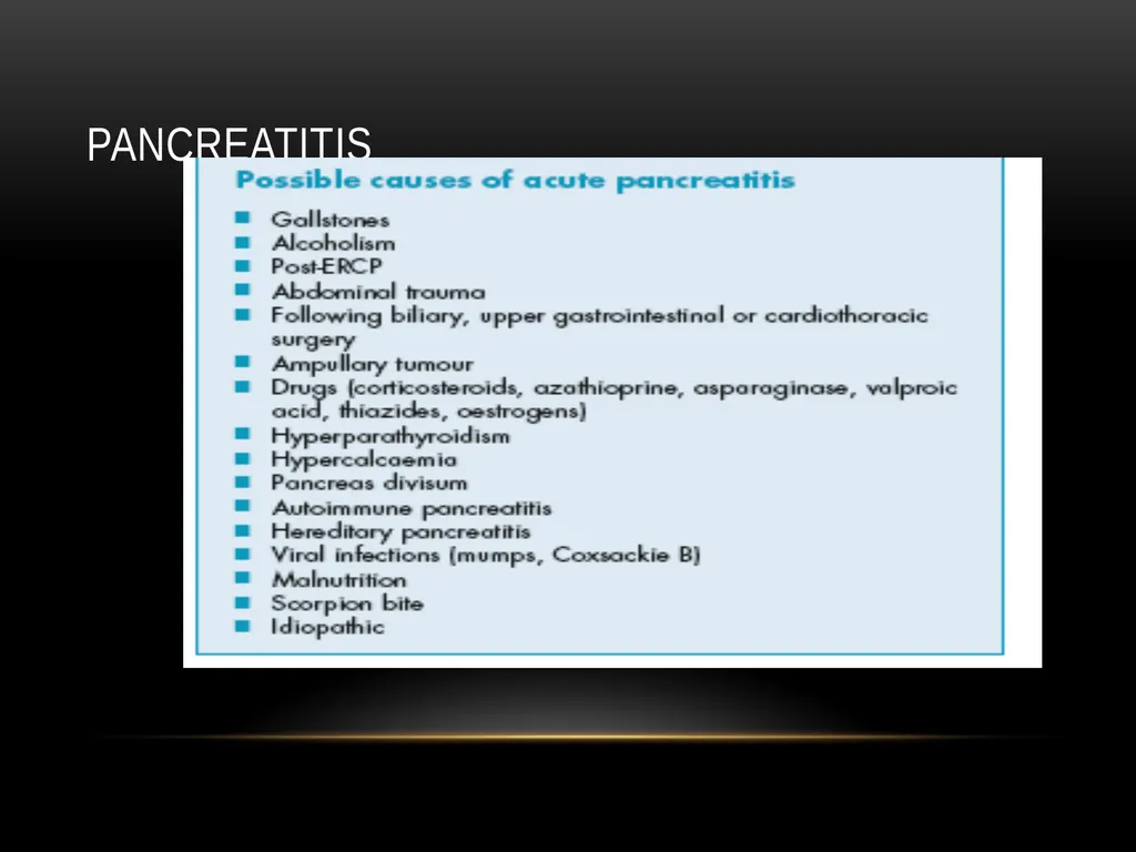
pancreatitis<br>
04
Clinical presentation Pain is the cardinal symptom.
develops quickly, reaching maximum intensity within minutes and persists for hours or even days.
The pain is frequently severe, constant and refractory to the usual doses of analgesics. Pain is usually experienced first in the epigastrium but may be localized to either upper quadrant or felt diffusely throughout the abdomen.
There is radiation to the back in about 50 per cent of patients, and some patients may gain relief by sitting or leaning forwards.
In fact, acute pancreatitis can mimic most causes of the acute abdomen<br>
develops quickly, reaching maximum intensity within minutes and persists for hours or even days.
The pain is frequently severe, constant and refractory to the usual doses of analgesics. Pain is usually experienced first in the epigastrium but may be localized to either upper quadrant or felt diffusely throughout the abdomen.
There is radiation to the back in about 50 per cent of patients, and some patients may gain relief by sitting or leaning forwards.
In fact, acute pancreatitis can mimic most causes of the acute abdomen<br>
05
Cont- Nausea, repeated vomiting and retching
Why ? Hiccough.
on examination: the appearance may be that of a patient who is well or, at the other extreme, one who is gravely ill with profound shock, toxicity and confusion.
Tachypnea is common, tachycardia is usual, and hypotension may be present. The body temperature is often normal or even subnormal, but frequently rises as inflammation develops<br>
Why ? Hiccough.
on examination: the appearance may be that of a patient who is well or, at the other extreme, one who is gravely ill with profound shock, toxicity and confusion.
Tachypnea is common, tachycardia is usual, and hypotension may be present. The body temperature is often normal or even subnormal, but frequently rises as inflammation develops<br>
06
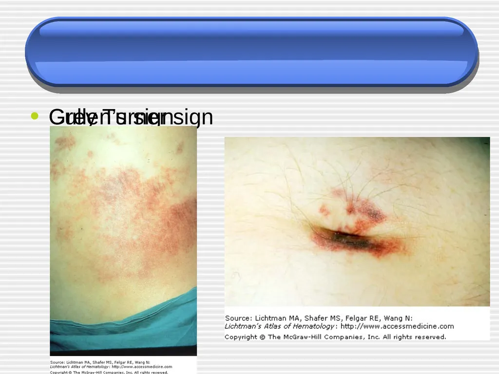
Mild icterus can be caused by biliary obstruction in gallstone pancreatitis, and an acute swinging pyrexia suggests cholangitis. Bleeding into the facial planes can produce bluish discoloration of the flanks (Grey Turner’s sign) or umbilicus (Cullen’s sign)
Subcutaneous fat necrosis may produce small, red, tender nodules on the skin of the legs.
Abdominal examination may reveal distension due to ileus or, more rarely, ascites with shifting dullness. A mass can develop in the epigastrium due to inflammation. There is usually muscle guarding in the upper abdomen, although marked rigidity is unusual. A pleural effusion is present in 10–20 per cent of patients. Pulmonary oedema and pneumonitis are also described and may give rise to the differential diagnosis of pneumonia or myocardial infarction. The patient may be confused and exhibit
the signs of metabolic derangement together with hypoxaemia.<br>
Subcutaneous fat necrosis may produce small, red, tender nodules on the skin of the legs.
Abdominal examination may reveal distension due to ileus or, more rarely, ascites with shifting dullness. A mass can develop in the epigastrium due to inflammation. There is usually muscle guarding in the upper abdomen, although marked rigidity is unusual. A pleural effusion is present in 10–20 per cent of patients. Pulmonary oedema and pneumonitis are also described and may give rise to the differential diagnosis of pneumonia or myocardial infarction. The patient may be confused and exhibit
the signs of metabolic derangement together with hypoxaemia.<br>
07
Grey Turner sign Cullen’s sign<br>
08
Differential Not all inclusive, but may include:
Biliary disease
Intestinal obstruction
Mesenteric Ischemia
MI (inferior)
AAA ( abdominal aortic aneurism)
Distal aortic dissection
PUD<br>
Biliary disease
Intestinal obstruction
Mesenteric Ischemia
MI (inferior)
AAA ( abdominal aortic aneurism)
Distal aortic dissection
PUD<br>
09
Evaluation amylase…Nonspecific !!!
Amylase levels > 3x normal very suggestive of pancreatitis
May be normal in chronic pancreatitis!!!
Enzyme level severity
False (-): acute on chronic alcoholism ; HyperTG
False (+): renal failure, other abdominal or salivary gland process, acidemia
lipase
More sensitive & specific than amylase<br>
Amylase levels > 3x normal very suggestive of pancreatitis
May be normal in chronic pancreatitis!!!
Enzyme level severity
False (-): acute on chronic alcoholism ; HyperTG
False (+): renal failure, other abdominal or salivary gland process, acidemia
lipase
More sensitive & specific than amylase<br>
10
Evaluation Other inflammatory markers will be elevated
CRP, IL-6, IL-8 (studies hoping to use these markers to aid in detecting severity of disease)
ALT > 3x normal gallstone pancreatitis
(96% specific, but only 48% sensitive)
Depending on severity may see:
Ca
WBC
BUN
Hct
glucose<br>
CRP, IL-6, IL-8 (studies hoping to use these markers to aid in detecting severity of disease)
ALT > 3x normal gallstone pancreatitis
(96% specific, but only 48% sensitive)
Depending on severity may see:
Ca
WBC
BUN
Hct
glucose<br>
11
Radiographic Evaluation AXR - “sentinel loop” or small bowel ileus
US or CT may show enlarged pancreas with stranding, abscess, fluid collections, hemorrhage, necrosis or pseudocyst
MRI/MRCP Better visualization of fluid collections
MRCP allows visualization of bile ducts for stones
Does not allow stone extraction or stent insertion<br>
US or CT may show enlarged pancreas with stranding, abscess, fluid collections, hemorrhage, necrosis or pseudocyst
MRI/MRCP Better visualization of fluid collections
MRCP allows visualization of bile ducts for stones
Does not allow stone extraction or stent insertion<br>
12
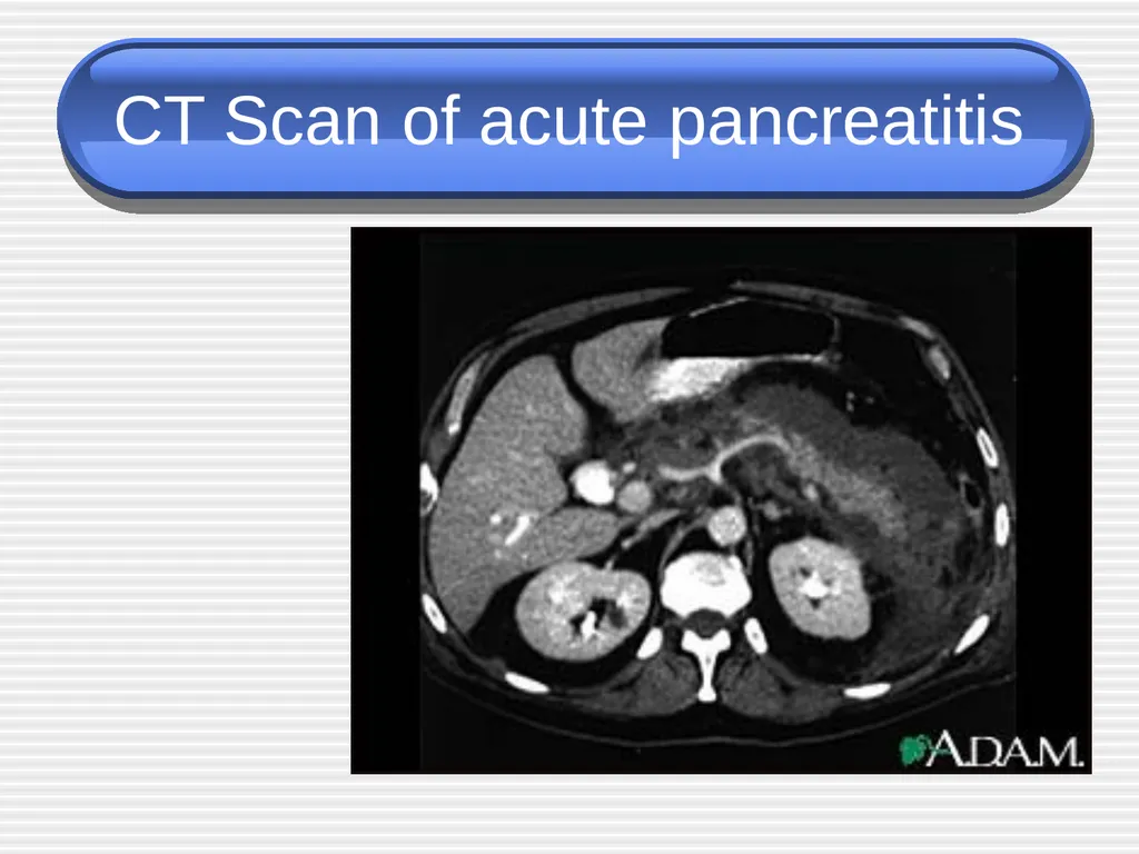
CT Scan of acute pancreatitis CT shows
significant
swelling
and
inflammation
of the
pancreas<br>
significant
swelling
and
inflammation
of the
pancreas<br>
13
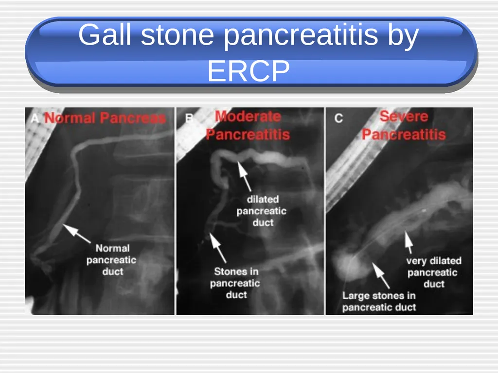
Gall stone pancreatitis by ERCP<br>
14
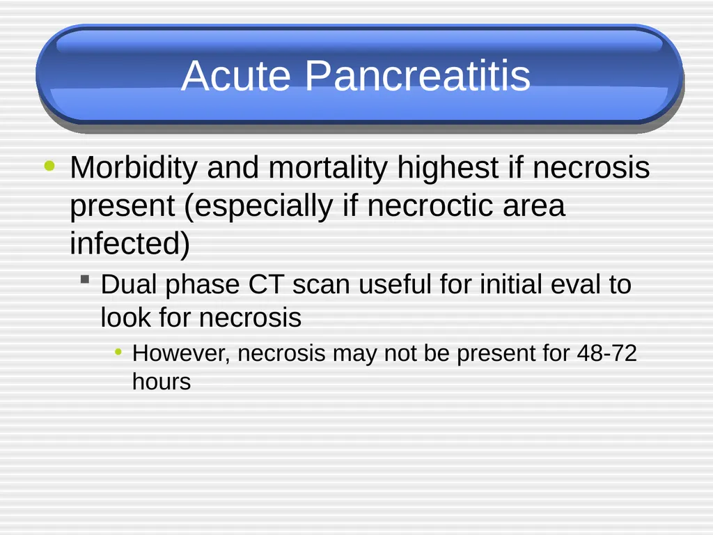
Acute Pancreatitis Morbidity and mortality highest if necrosis present (especially if necroctic area infected)
Dual phase CT scan useful for initial eval to look for necrosis
However, necrosis may not be present for 48-72 hours<br>
Dual phase CT scan useful for initial eval to look for necrosis
However, necrosis may not be present for 48-72 hours<br>
15
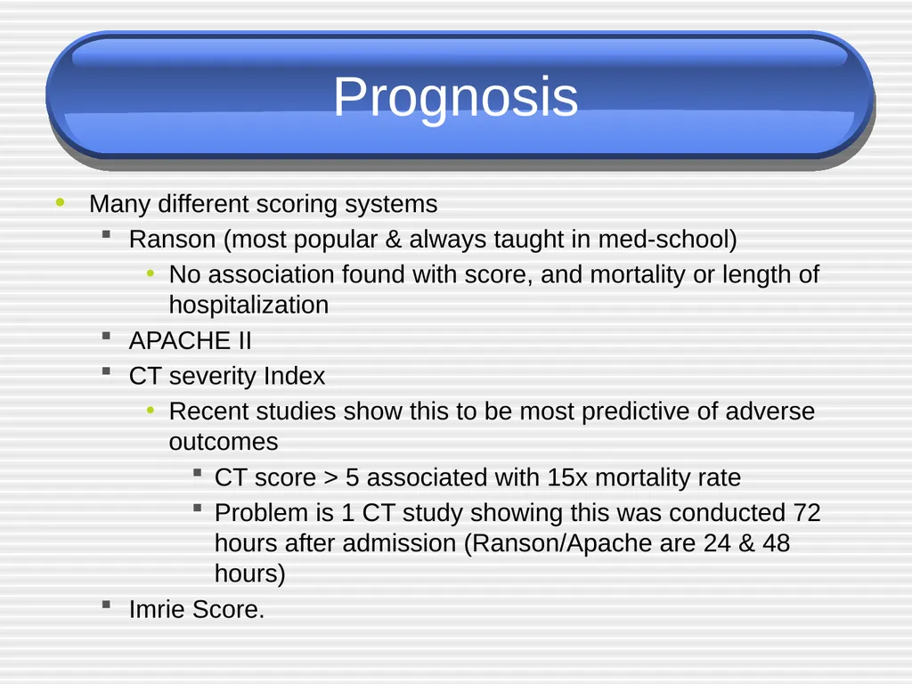
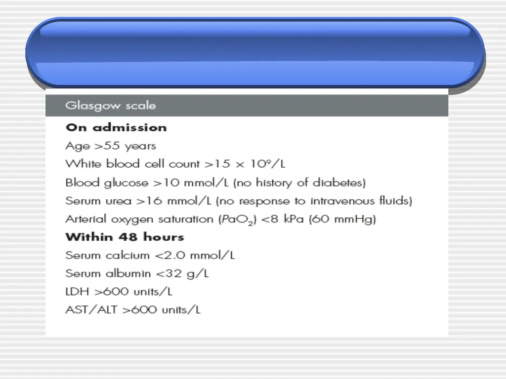
Prognosis Many different scoring systems
Ranson (most popular & always taught in med-school)
No association found with score, and mortality or length of hospitalization
APACHE II
CT severity Index
Recent studies show this to be most predictive of adverse outcomes
CT score > 5 associated with 15x mortality rate
Problem is 1 CT study showing this was conducted 72 hours after admission (Ranson/Apache are 24 & 48 hours)
Imrie Score.<br>
Ranson (most popular & always taught in med-school)
No association found with score, and mortality or length of hospitalization
APACHE II
CT severity Index
Recent studies show this to be most predictive of adverse outcomes
CT score > 5 associated with 15x mortality rate
Problem is 1 CT study showing this was conducted 72 hours after admission (Ranson/Apache are 24 & 48 hours)
Imrie Score.<br>
16
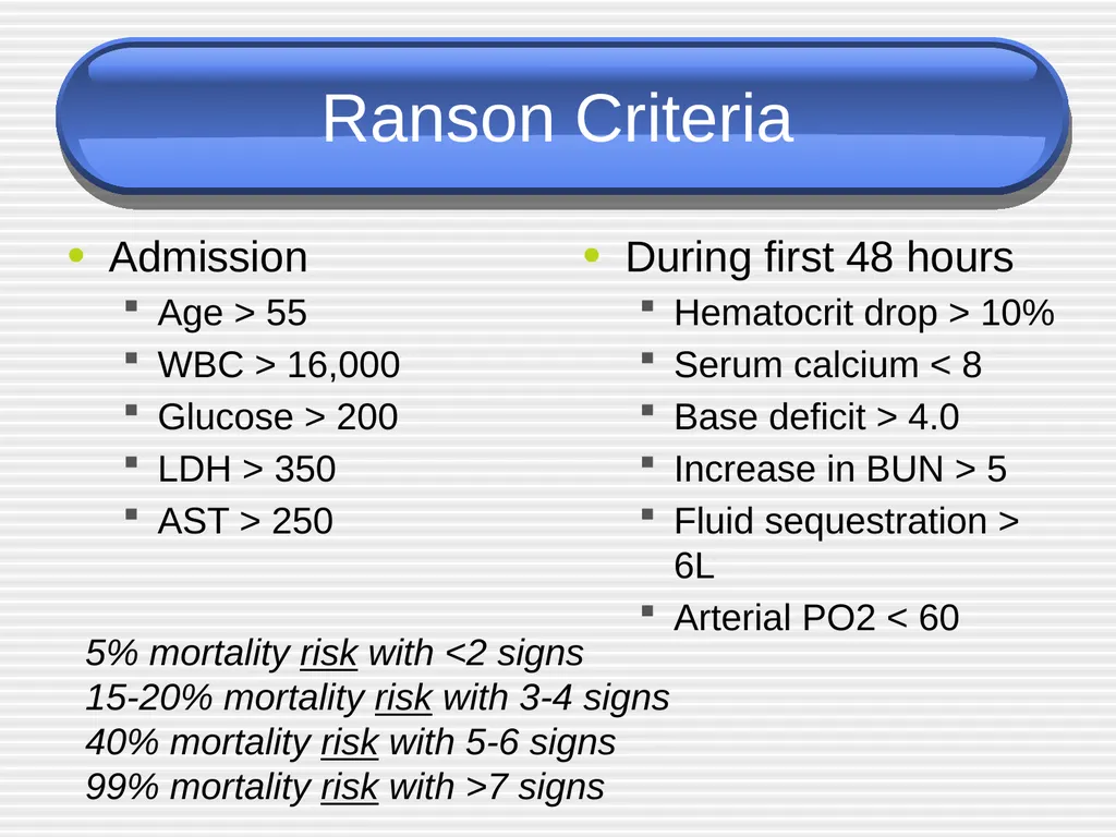
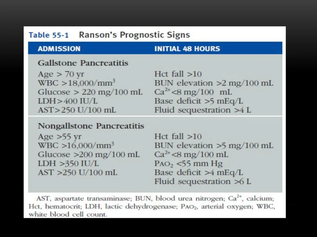
Ranson Criteria Admission
Age > 55
WBC > 16,000
Glucose > 200
LDH > 350
AST > 250 During first 48 hours
Hematocrit drop > 10%
Serum calcium < 8
Base deficit > 4.0
Increase in BUN > 5
Fluid sequestration > 6L
Arterial PO2 < 60 5% mortality risk with <2 signs
15-20% mortality risk with 3-4 signs
40% mortality risk with 5-6 signs
99% mortality risk with >7 signs<br>
Age > 55
WBC > 16,000
Glucose > 200
LDH > 350
AST > 250 During first 48 hours
Hematocrit drop > 10%
Serum calcium < 8
Base deficit > 4.0
Increase in BUN > 5
Fluid sequestration > 6L
Arterial PO2 < 60 5% mortality risk with <2 signs
15-20% mortality risk with 3-4 signs
40% mortality risk with 5-6 signs
99% mortality risk with >7 signs<br>
17
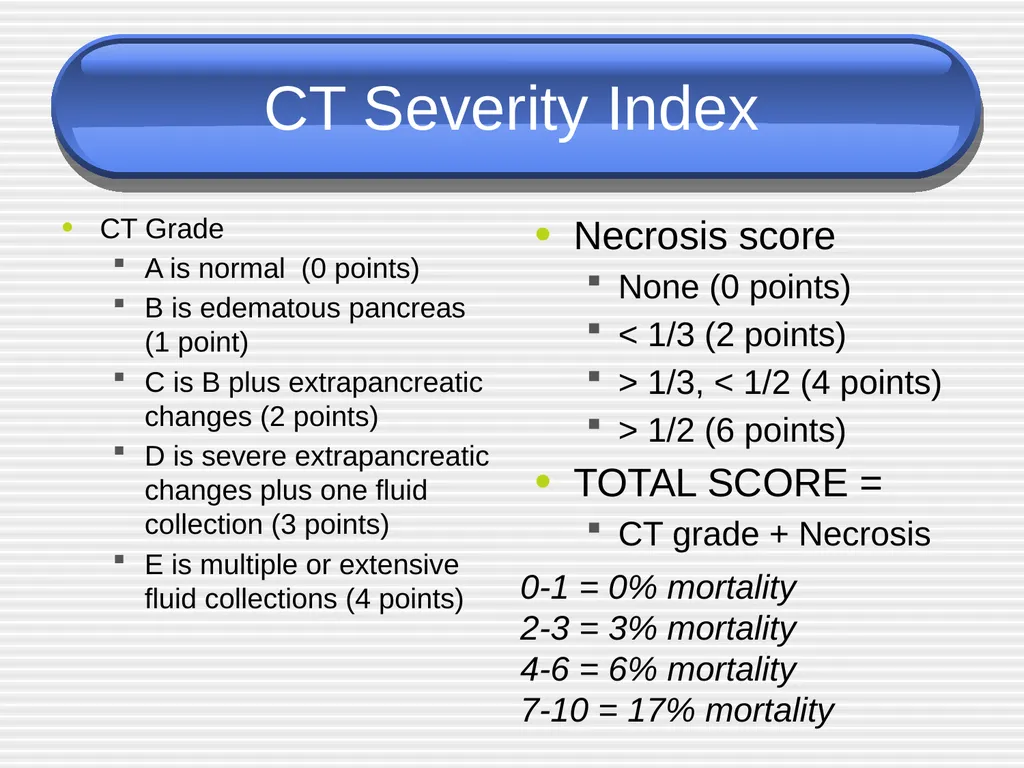
CT Severity Index CT Grade
A is normal (0 points)
B is edematous pancreas (1 point)
C is B plus extrapancreatic changes (2 points)
D is severe extrapancreatic changes plus one fluid collection (3 points)
E is multiple or extensive fluid collections (4 points) Necrosis score
None (0 points)
< 1/3 (2 points)
> 1/3, < 1/2 (4 points)
> 1/2 (6 points)
TOTAL SCORE =
CT grade + Necrosis 0-1 = 0% mortality
2-3 = 3% mortality
4-6 = 6% mortality
7-10 = 17% mortality<br>
A is normal (0 points)
B is edematous pancreas (1 point)
C is B plus extrapancreatic changes (2 points)
D is severe extrapancreatic changes plus one fluid collection (3 points)
E is multiple or extensive fluid collections (4 points) Necrosis score
None (0 points)
< 1/3 (2 points)
> 1/3, < 1/2 (4 points)
> 1/2 (6 points)
TOTAL SCORE =
CT grade + Necrosis 0-1 = 0% mortality
2-3 = 3% mortality
4-6 = 6% mortality
7-10 = 17% mortality<br>
18
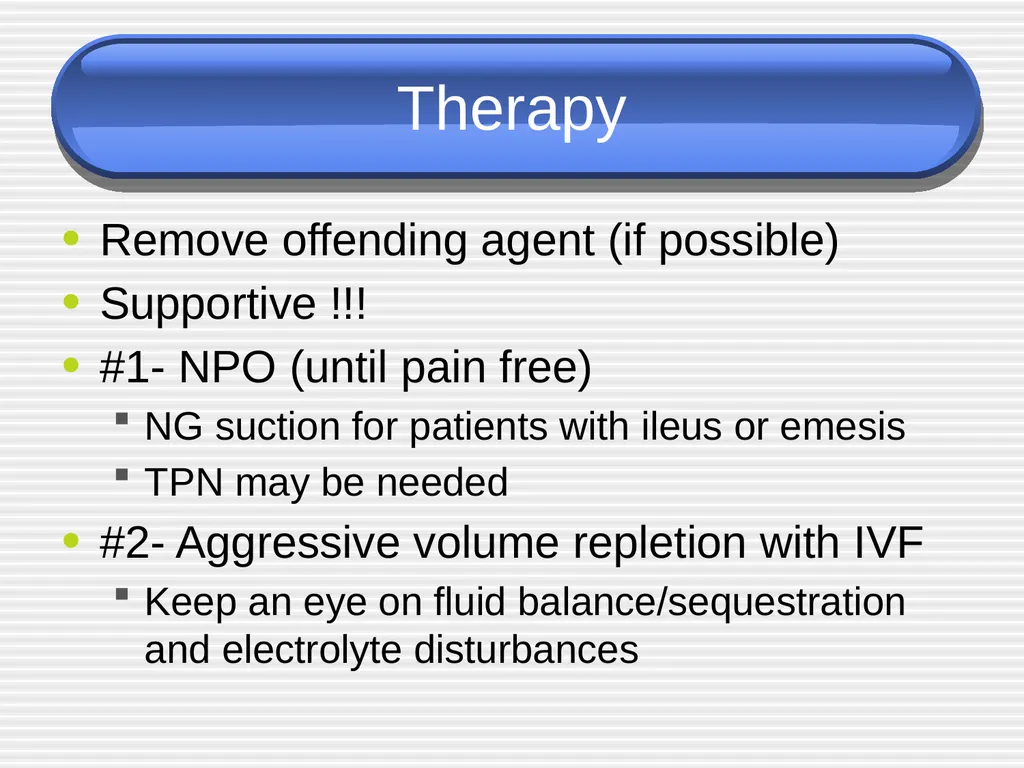
Therapy Remove offending agent (if possible)
Supportive !!!
#1- NPO (until pain free)
NG suction for patients with ileus or emesis
TPN may be needed
#2- Aggressive volume repletion with IVF
Keep an eye on fluid balance/sequestration and electrolyte disturbances<br>
Supportive !!!
#1- NPO (until pain free)
NG suction for patients with ileus or emesis
TPN may be needed
#2- Aggressive volume repletion with IVF
Keep an eye on fluid balance/sequestration and electrolyte disturbances<br>
19
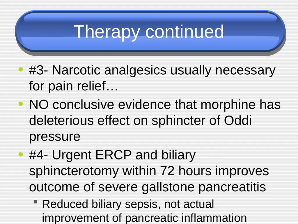
Therapy continued #3- Narcotic analgesics usually necessary for pain relief…
NO conclusive evidence that morphine has deleterious effect on sphincter of Oddi pressure
#4- Urgent ERCP and biliary sphincterotomy within 72 hours improves outcome of severe gallstone pancreatitis
Reduced biliary sepsis, not actual improvement of pancreatic inflammation
#5- Don’t forget PPI to prevent stress ulcer<br>
NO conclusive evidence that morphine has deleterious effect on sphincter of Oddi pressure
#4- Urgent ERCP and biliary sphincterotomy within 72 hours improves outcome of severe gallstone pancreatitis
Reduced biliary sepsis, not actual improvement of pancreatic inflammation
#5- Don’t forget PPI to prevent stress ulcer<br>
20
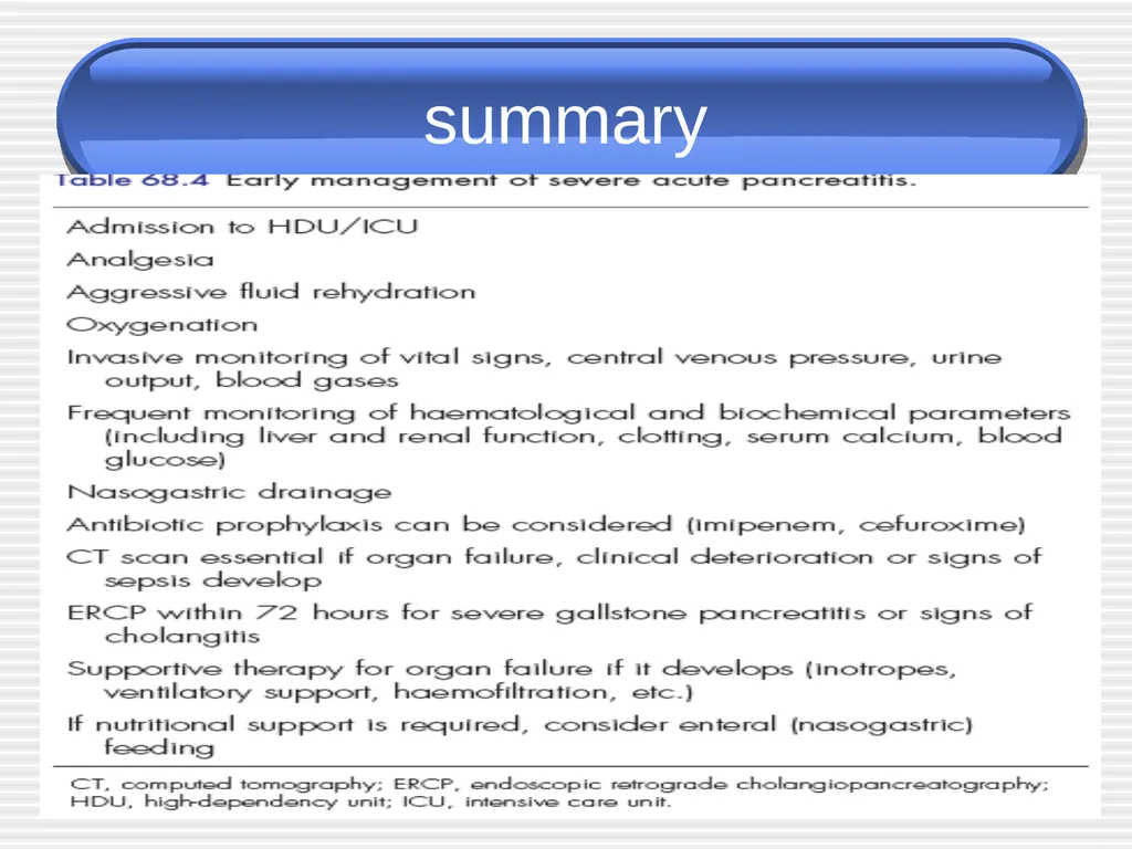
summary<br>
21
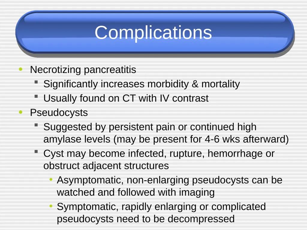
Complications Necrotizing pancreatitis
Significantly increases morbidity & mortality
Usually found on CT with IV contrast
Pseudocysts
Suggested by persistent pain or continued high amylase levels (may be present for 4-6 wks afterward)
Cyst may become infected, rupture, hemorrhage or obstruct adjacent structures
Asymptomatic, non-enlarging pseudocysts can be watched and followed with imaging
Symptomatic, rapidly enlarging or complicated pseudocysts need to be decompressed<br>
Significantly increases morbidity & mortality
Usually found on CT with IV contrast
Pseudocysts
Suggested by persistent pain or continued high amylase levels (may be present for 4-6 wks afterward)
Cyst may become infected, rupture, hemorrhage or obstruct adjacent structures
Asymptomatic, non-enlarging pseudocysts can be watched and followed with imaging
Symptomatic, rapidly enlarging or complicated pseudocysts need to be decompressed<br>
22
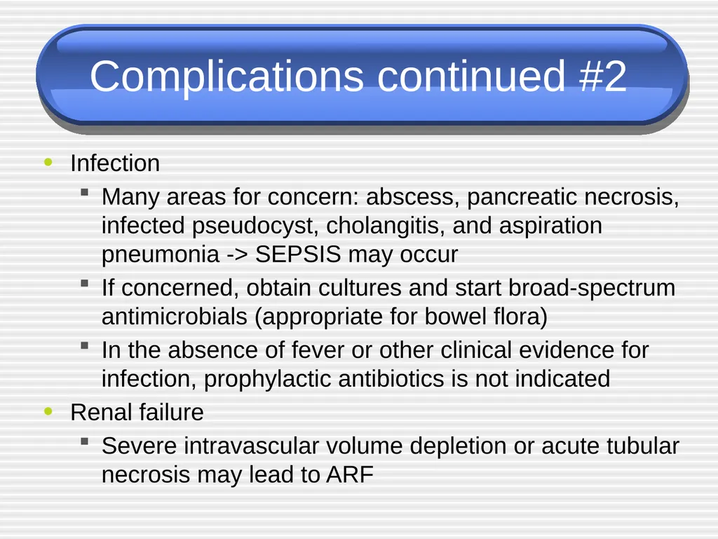
Complications continued #2 Infection
Many areas for concern: abscess, pancreatic necrosis, infected pseudocyst, cholangitis, and aspiration pneumonia -> SEPSIS may occur
If concerned, obtain cultures and start broad-spectrum antimicrobials (appropriate for bowel flora)
In the absence of fever or other clinical evidence for infection, prophylactic antibiotics is not indicated
Renal failure
Severe intravascular volume depletion or acute tubular necrosis may lead to ARF<br>
Many areas for concern: abscess, pancreatic necrosis, infected pseudocyst, cholangitis, and aspiration pneumonia -> SEPSIS may occur
If concerned, obtain cultures and start broad-spectrum antimicrobials (appropriate for bowel flora)
In the absence of fever or other clinical evidence for infection, prophylactic antibiotics is not indicated
Renal failure
Severe intravascular volume depletion or acute tubular necrosis may lead to ARF<br>
23
Complications continued #3 Pulmonary
Atelectasis, pleural effusion, pneumonia and ARDS can develop in severe cases
Other
Metabolic disturbances
hypocalcemia, hypomagnesemia, hyperglycemia
GI bleeds
Stress gastritis
Fistula formation<br>
Atelectasis, pleural effusion, pneumonia and ARDS can develop in severe cases
Other
Metabolic disturbances
hypocalcemia, hypomagnesemia, hyperglycemia
GI bleeds
Stress gastritis
Fistula formation<br>
24
Prognosis 85-90% mild, self-limited
Usually resolves in 3-7 days
10-15% severe requiring ICU admission
Mortality may approach 50% in severe cases<br>
Usually resolves in 3-7 days
10-15% severe requiring ICU admission
Mortality may approach 50% in severe cases<br>
25
Chronic pancreatitis Pathophys - irreversible parenchymal destruction leading to pancreatic dysfunction andloss of exocrine acinar cells and a marked increase in interstitial fibrous connective tissue. The islets of Langerhans are preserved and constitute a relatively greater proportion of the pancreatic tissue. Hyperplasia of islet cells is also seen. The sizes and number of nerves are increased, but the protective perineural sheath is damaged, and nerves are found in proximity to inflammatory foci. There appear to be selective increases in certain peptidergic nerves. These histologic observations may be related to the cause of pain in chronic pancreatitis<br>
26
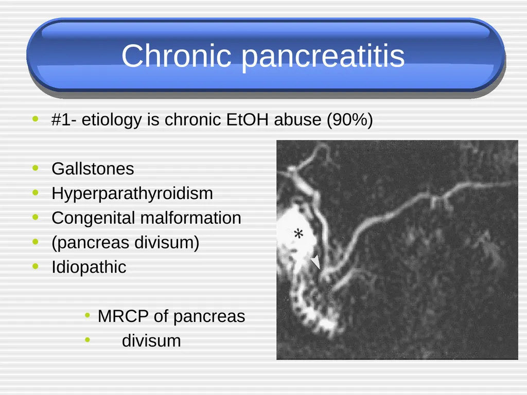
Chronic pancreatitis #1- etiology is chronic EtOH abuse (90%)
Gallstones
Hyperparathyroidism
Congenital malformation
(pancreas divisum)
Idiopathic
MRCP of pancreas
divisum<br>
Gallstones
Hyperparathyroidism
Congenital malformation
(pancreas divisum)
Idiopathic
MRCP of pancreas
divisum<br>
27
Presentations Pain .Why? Persistent, recurrent episodes of severe pain
Anorexia, nausea
Constipation, flatulence
Steatorrhea
Diabetes<br>
Anorexia, nausea
Constipation, flatulence
Steatorrhea
Diabetes<br>
28
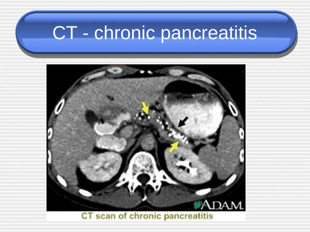
Evaluation or normal amylase and lipase
Plain AXR / CT may show calcified pancreas<br>
Plain AXR / CT may show calcified pancreas<br>
29
CT - chronic pancreatitis<br>
30
Complications Exocrine insufficiency typically manifests as weight loss and steatorrhea
If steatorrhea present, a trypsinogen level < 10 is diagnostic for chronic pancreatitis
Manage with low-fat diet and pancreatic enzyme supplements (Pancrease, Creon)
Endocrine insufficiency may result from islet cell destruction which leads to diabetes<br>
If steatorrhea present, a trypsinogen level < 10 is diagnostic for chronic pancreatitis
Manage with low-fat diet and pancreatic enzyme supplements (Pancrease, Creon)
Endocrine insufficiency may result from islet cell destruction which leads to diabetes<br>
31
management No cure . Symptomatic treatment.
Alcohol cessation may improve pain
Pain management critical but Narcotic dependency is common
Steatorrhea: Therapy involves limitation of fat intake and administration of adequate amounts of exogenous pancreatic enzyme preparations to provide at least 10% of normal lipolytic activity in the duodenum at the time that the food substrate is present<br>
Alcohol cessation may improve pain
Pain management critical but Narcotic dependency is common
Steatorrhea: Therapy involves limitation of fat intake and administration of adequate amounts of exogenous pancreatic enzyme preparations to provide at least 10% of normal lipolytic activity in the duodenum at the time that the food substrate is present<br>
32
Management. surgery Pain is the primary indication for surgery. Selection of the best operation for a particular patient must include consideration of the anatomy of the gland, preexisting endocrine or exocrine dysfunction, compliance and the rehabilitative capacity of the patient, postoperative endocrine or exocrine deficiency, and the likelihood of postoperative pain relief.
Patients with a dilated duct (>6 mm) are candidates for ductal drainage, with lateral pancreaticojejunostomy being the best choice of these procedures. It is important to achieve adequate decompression of the enlarged pancreatic head and uncinate
process during drainage procedures<br>
Patients with a dilated duct (>6 mm) are candidates for ductal drainage, with lateral pancreaticojejunostomy being the best choice of these procedures. It is important to achieve adequate decompression of the enlarged pancreatic head and uncinate
process during drainage procedures<br>
33
Conclusion Pancreatitis is common
YOU WILL SEE IT!!!
10-15% are severe = ICU admission
Mortality may approach 50% in severe cases
These are the cases where knowing future complications would be great (ie finding a marker that correlates with severity…and that’s what the clinical researchers are attempting to do)<br>
YOU WILL SEE IT!!!
10-15% are severe = ICU admission
Mortality may approach 50% in severe cases
These are the cases where knowing future complications would be great (ie finding a marker that correlates with severity…and that’s what the clinical researchers are attempting to do)<br>
34
Suggested questions???? The aims in investigating patients with suspected acute pancreatitis.
Enumerates three therapeutic differences between the management of mild and severe forms of acute pancreatitis .
Causes of renal failure in acute pancreatitis.<br>
Enumerates three therapeutic differences between the management of mild and severe forms of acute pancreatitis .
Causes of renal failure in acute pancreatitis.<br>