
Autoimmune Encephalitis Update William Kilgo, MD
MT
Published · 27 slides · 0 views

1 / 1


Description
Autoimmune Encephalitis Update William Kilgo, MD University of South Alabama Key Largo, Florida January 13-16, 2024 Disclosures Contracted Research (Principal Investigators must provide information, even if received by the institution):
Related Topics
Share
Embed code
Download this presentation From Below
"Autoimmune Encephalitis Update William Kilgo, MD" is the property of its rightful owner. Permission is granted to download and print the materials on this website for personal, non-commercial use only, and to display it on your personal computer provided you do not modify the materials and that you retain all copyright notices contained in the materials. By downloading content from our website, you accept the terms of this agreement.
Presentation Transcript
01
Autoimmune Encephalitis Update
William Kilgo, MD
University of South Alabama Key Largo, Florida January 13-16, 2024<br>
William Kilgo, MD
University of South Alabama Key Largo, Florida January 13-16, 2024<br>
02
Disclosures Contracted Research (Principal Investigators must provide information, even if received by the institution):
Genentech, EMD Serono, Alexion, Bristol Meyers Squibb
Honoraria: Octave Biosciences, TG Therapeutics
Speakers' Bureau: Genentech, Biogen, TG Therapeutics<br>
Honoraria: Octave Biosciences, TG Therapeutics
Speakers' Bureau: Genentech, Biogen, TG Therapeutics<br>
03
Objectives Review the most common subtypes of autoimmune encephalitis
Describe the clinical features that should raise concern for autoimmune encephalitis, with or without known antibody status
Review the diagnostic approach for autoimmune encephalitis
Review treatment options for autoimmune encephalitis<br>
Describe the clinical features that should raise concern for autoimmune encephalitis, with or without known antibody status
Review the diagnostic approach for autoimmune encephalitis
Review treatment options for autoimmune encephalitis<br>
04
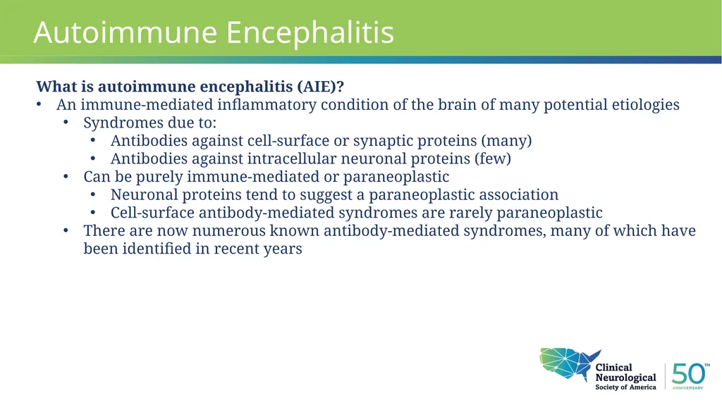
Autoimmune Encephalitis What is autoimmune encephalitis (AIE)?
An immune-mediated inflammatory condition of the brain of many potential etiologies
Syndromes due to:
Antibodies against cell-surface or synaptic proteins (many)
Antibodies against intracellular neuronal proteins (few)
Can be purely immune-mediated or paraneoplastic
Neuronal proteins tend to suggest a paraneoplastic association
Cell-surface antibody-mediated syndromes are rarely paraneoplastic
There are now numerous known antibody-mediated syndromes, many of which have been identified in recent years<br>
An immune-mediated inflammatory condition of the brain of many potential etiologies
Syndromes due to:
Antibodies against cell-surface or synaptic proteins (many)
Antibodies against intracellular neuronal proteins (few)
Can be purely immune-mediated or paraneoplastic
Neuronal proteins tend to suggest a paraneoplastic association
Cell-surface antibody-mediated syndromes are rarely paraneoplastic
There are now numerous known antibody-mediated syndromes, many of which have been identified in recent years<br>
05
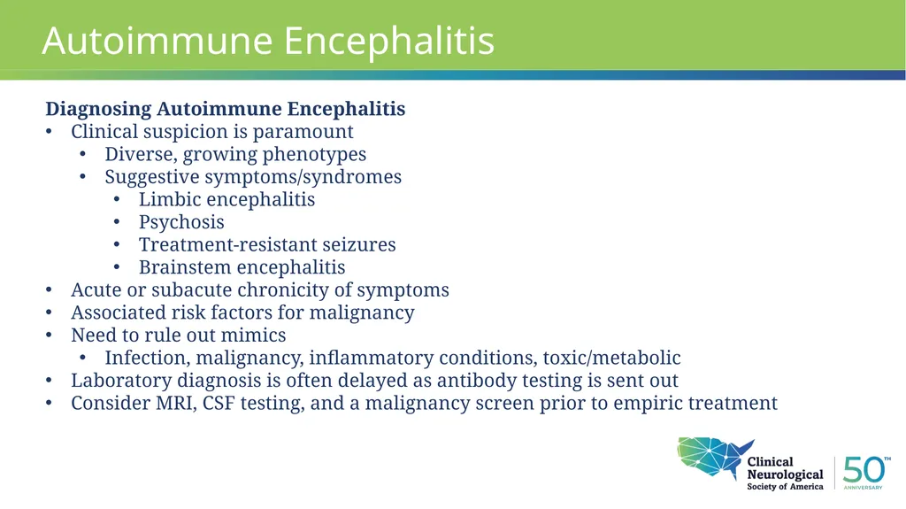
Autoimmune Encephalitis Diagnosing Autoimmune Encephalitis
Clinical suspicion is paramount
Diverse, growing phenotypes
Suggestive symptoms/syndromes
Limbic encephalitis
Psychosis
Treatment-resistant seizures
Brainstem encephalitis
Acute or subacute chronicity of symptoms
Associated risk factors for malignancy
Need to rule out mimics
Infection, malignancy, inflammatory conditions, toxic/metabolic
Laboratory diagnosis is often delayed as antibody testing is sent out
Consider MRI, CSF testing, and a malignancy screen prior to empiric treatment<br>
Clinical suspicion is paramount
Diverse, growing phenotypes
Suggestive symptoms/syndromes
Limbic encephalitis
Psychosis
Treatment-resistant seizures
Brainstem encephalitis
Acute or subacute chronicity of symptoms
Associated risk factors for malignancy
Need to rule out mimics
Infection, malignancy, inflammatory conditions, toxic/metabolic
Laboratory diagnosis is often delayed as antibody testing is sent out
Consider MRI, CSF testing, and a malignancy screen prior to empiric treatment<br>
06
Intracellular Targets
ANNA-1, ANNA-2, ANNA-3
CRMP-5
Ma1 and Ma2
PCA1 (Yo), PCA2, anti-DNER Autoimmune Encephalitis Synaptic Proteins and Cell-Surface antigens
GAD65
NMDAR
GABA-A & GABA-B
AMPA-R
VGKC complex associated proteins
LGI-1
CASPR2
Anti-IGLON5
Anti-DPPX encephalitis
Anti-GlyR encephalopathy
Anti-GFAP
Anti-mGluR1 and mGluR5<br>
ANNA-1, ANNA-2, ANNA-3
CRMP-5
Ma1 and Ma2
PCA1 (Yo), PCA2, anti-DNER Autoimmune Encephalitis Synaptic Proteins and Cell-Surface antigens
GAD65
NMDAR
GABA-A & GABA-B
AMPA-R
VGKC complex associated proteins
LGI-1
CASPR2
Anti-IGLON5
Anti-DPPX encephalitis
Anti-GlyR encephalopathy
Anti-GFAP
Anti-mGluR1 and mGluR5<br>
07
Autoimmune Encephalitis Diagnosing Autoimmune Encephalitis
Concurrent CSF and Serum testing is recommended when ordering broad antibody panels
Consider potential for false positive results
Serum positive result alone with negative CSF should prompt careful consideration of if the clinical syndrome is consistent with the result
Diagnostic Criteria for Definite Autoimmune Limbic Encephalitis (expert consensus)
Subacute onset memory deficits, seizure, or psychiatric symptoms localizing to limbic system
Bilateral medial temporal lobe hyperintensities on T2/FLAIR MRI sequences
CSF Pleocytosis (>5 cells) OR abnormal EEG suggesting temporal lobe involvement
Exclusion of alternative causes<br>
Concurrent CSF and Serum testing is recommended when ordering broad antibody panels
Consider potential for false positive results
Serum positive result alone with negative CSF should prompt careful consideration of if the clinical syndrome is consistent with the result
Diagnostic Criteria for Definite Autoimmune Limbic Encephalitis (expert consensus)
Subacute onset memory deficits, seizure, or psychiatric symptoms localizing to limbic system
Bilateral medial temporal lobe hyperintensities on T2/FLAIR MRI sequences
CSF Pleocytosis (>5 cells) OR abnormal EEG suggesting temporal lobe involvement
Exclusion of alternative causes<br>
08
Autoimmune Encephalitis Anti-NMDA receptor encephalitis
Most common etiology of AIE
Mostly impacts young adults and children (female predominance in ages < 45)
Often presents initially with psychiatric manifestations
Bizarre behavior, psychosis
Many patients will present first to a psychiatrist or psychiatric facility
Memory deficits
Movement disorders
Dyskinesias (orofacial, choreoathetosis, dystonia, rigidity)
Autonomic instability
Treatment resistant seizures
High association with ovarian teratoma in young females<br>
Most common etiology of AIE
Mostly impacts young adults and children (female predominance in ages < 45)
Often presents initially with psychiatric manifestations
Bizarre behavior, psychosis
Many patients will present first to a psychiatrist or psychiatric facility
Memory deficits
Movement disorders
Dyskinesias (orofacial, choreoathetosis, dystonia, rigidity)
Autonomic instability
Treatment resistant seizures
High association with ovarian teratoma in young females<br>
09
Associated with:
Normal Imaging (90%)
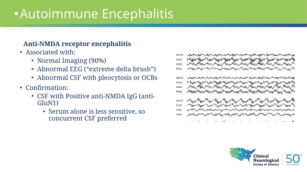
Abnormal EEG (“extreme delta brush”)
Abnormal CSF with pleocytosis or OCBs
Confirmation:
CSF with Positive anti-NMDA IgG (anti-GluN1)
Serum alone is less sensitive, so concurrent CSF preferred Autoimmune Encephalitis Anti-NMDA receptor encephalitis<br>
Normal Imaging (90%)
Abnormal EEG (“extreme delta brush”)
Abnormal CSF with pleocytosis or OCBs
Confirmation:
CSF with Positive anti-NMDA IgG (anti-GluN1)
Serum alone is less sensitive, so concurrent CSF preferred Autoimmune Encephalitis Anti-NMDA receptor encephalitis<br>
10
Case 1 58 year old male
Presents to hospital as a transfer from outside facility with several weeks of anterograde amnesia. He had been becoming more forgetful per his wife, who had been away on business. He was making mistakes at work, prompting their concern to contact her. He is aware of the memory issue and distressed by it.
Local hospital admitted and discharged him after initial work-up. Patient re-presented to hospital at behest of his PCP and was transferred to our facility.
He describes premonitory symptoms of an internal “rush” toward his face at times followed by involuntary movements of his face and left arm that occur at random, several times daily<br>
Presents to hospital as a transfer from outside facility with several weeks of anterograde amnesia. He had been becoming more forgetful per his wife, who had been away on business. He was making mistakes at work, prompting their concern to contact her. He is aware of the memory issue and distressed by it.
Local hospital admitted and discharged him after initial work-up. Patient re-presented to hospital at behest of his PCP and was transferred to our facility.
He describes premonitory symptoms of an internal “rush” toward his face at times followed by involuntary movements of his face and left arm that occur at random, several times daily<br>
11
Case 1 58 year old male
Work-up:
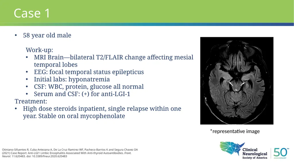
MRI Brain—bilateral T2/FLAIR change affecting mesial temporal lobes
EEG: focal temporal status epilepticus
Initial labs: hyponatremia
CSF: WBC, protein, glucose all normal
Serum and CSF: (+) for anti-LGI-1
Treatment:
High dose steroids inpatient, single relapse within one year. Stable on oral mycophenolate Otiniano-Sifuentes R, Cuba Antezana A, De La Cruz Ramirez WF, Pacheco-Barrios K and Segura Chavez DA (2021) Case Report: Anti-LGI1 Limbic Encephalitis Associated With Anti-thyroid Autoantibodies. Front. Neurol. 11:620483. doi: 10.3389/fneur.2020.620483 *representative image<br>
Work-up:
MRI Brain—bilateral T2/FLAIR change affecting mesial temporal lobes
EEG: focal temporal status epilepticus
Initial labs: hyponatremia
CSF: WBC, protein, glucose all normal
Serum and CSF: (+) for anti-LGI-1
Treatment:
High dose steroids inpatient, single relapse within one year. Stable on oral mycophenolate Otiniano-Sifuentes R, Cuba Antezana A, De La Cruz Ramirez WF, Pacheco-Barrios K and Segura Chavez DA (2021) Case Report: Anti-LGI1 Limbic Encephalitis Associated With Anti-thyroid Autoantibodies. Front. Neurol. 11:620483. doi: 10.3389/fneur.2020.620483 *representative image<br>
12
Autoimmune Encephalitis Anti-LGI-1 encephalitis
Most common form of AIE in patients over 50
Patients present with memory disturbances, confusion, and seizures
Cognitive symptoms may be preceded by characteristic movement disorder/seizure
Faciobrachial dystonic seizures
Patients may present with rapidly progressive dementia without associated symptoms, MRI, or CSF abnormalities<br>
Most common form of AIE in patients over 50
Patients present with memory disturbances, confusion, and seizures
Cognitive symptoms may be preceded by characteristic movement disorder/seizure
Faciobrachial dystonic seizures
Patients may present with rapidly progressive dementia without associated symptoms, MRI, or CSF abnormalities<br>
13
Autoimmune Encephalitis Anti-LGI-1 encephalitis
Imaging often normal
If abnormal, shows characteristic findings of limbic encephalitis
CSF: +/- oligoclonal bands and pleocytosis. Often normal
Patients are responsive to early immunotherapy
Long-term cognitive effects may persist
Relapse may occur in 1/3 of patients
Diagnosis: anti-LGI-1 antibodies in blood/CSF (blood more common)
Tumor association: Thymoma is rare, other types are usually incidental.<br>
Imaging often normal
If abnormal, shows characteristic findings of limbic encephalitis
CSF: +/- oligoclonal bands and pleocytosis. Often normal
Patients are responsive to early immunotherapy
Long-term cognitive effects may persist
Relapse may occur in 1/3 of patients
Diagnosis: anti-LGI-1 antibodies in blood/CSF (blood more common)
Tumor association: Thymoma is rare, other types are usually incidental.<br>
14
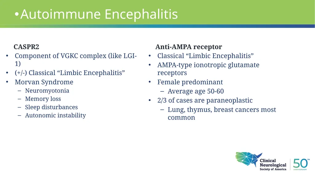
CASPR2 Component of VGKC complex (like LGI-1)
(+/-) Classical “Limbic Encephalitis”
Morvan Syndrome
Neuromyotonia
Memory loss
Sleep disturbances
Autonomic instability Autoimmune Encephalitis Classical “Limbic Encephalitis”
AMPA-type ionotropic glutamate receptors
Female predominant
Average age 50-60
2/3 of cases are paraneoplastic
Lung, thymus, breast cancers most common Anti-AMPA receptor<br>
(+/-) Classical “Limbic Encephalitis”
Morvan Syndrome
Neuromyotonia
Memory loss
Sleep disturbances
Autonomic instability Autoimmune Encephalitis Classical “Limbic Encephalitis”
AMPA-type ionotropic glutamate receptors
Female predominant
Average age 50-60
2/3 of cases are paraneoplastic
Lung, thymus, breast cancers most common Anti-AMPA receptor<br>
15
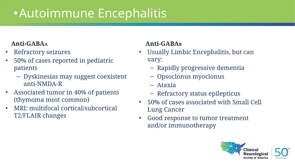
Anti-GABAA Refractory seizures
50% of cases reported in pediatric patients
Dyskinesias may suggest coexistent anti-NMDA-R
Associated tumor in 40% of patients (thymoma most common)
MRI: multifocal cortical/subcortical T2/FLAIR changes Autoimmune Encephalitis Usually Limbic Encephalitis, but can vary:
Rapidly progressive dementia
Opsoclonus myoclonus
Ataxia
Refractory status epilepticus
50% of cases associated with Small Cell Lung Cancer
Good response to tumor treatment and/or immunotherapy Anti-GABAB<br>
50% of cases reported in pediatric patients
Dyskinesias may suggest coexistent anti-NMDA-R
Associated tumor in 40% of patients (thymoma most common)
MRI: multifocal cortical/subcortical T2/FLAIR changes Autoimmune Encephalitis Usually Limbic Encephalitis, but can vary:
Rapidly progressive dementia
Opsoclonus myoclonus
Ataxia
Refractory status epilepticus
50% of cases associated with Small Cell Lung Cancer
Good response to tumor treatment and/or immunotherapy Anti-GABAB<br>
16
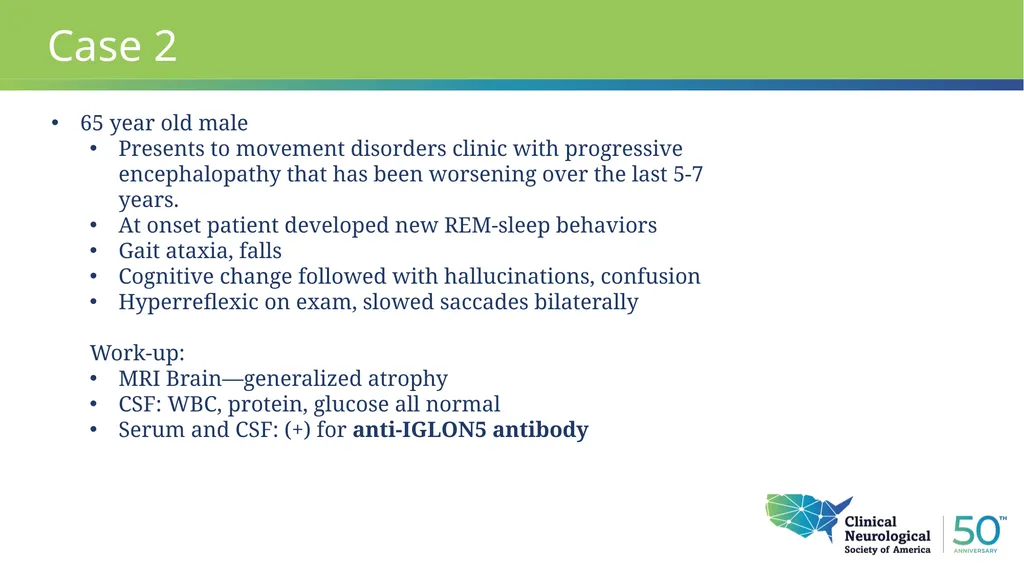
Case 2 65 year old male
Presents to movement disorders clinic with progressive encephalopathy that has been worsening over the last 5-7 years.
At onset patient developed new REM-sleep behaviors
Gait ataxia, falls
Cognitive change followed with hallucinations, confusion
Hyperreflexic on exam, slowed saccades bilaterally
Work-up:
MRI Brain—generalized atrophy
CSF: WBC, protein, glucose all normal
Serum and CSF: (+) for anti-IGLON5 antibody<br>
Presents to movement disorders clinic with progressive encephalopathy that has been worsening over the last 5-7 years.
At onset patient developed new REM-sleep behaviors
Gait ataxia, falls
Cognitive change followed with hallucinations, confusion
Hyperreflexic on exam, slowed saccades bilaterally
Work-up:
MRI Brain—generalized atrophy
CSF: WBC, protein, glucose all normal
Serum and CSF: (+) for anti-IGLON5 antibody<br>
17
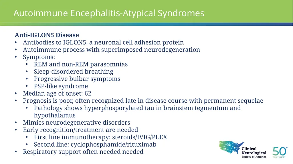
Autoimmune Encephalitis-Atypical Syndromes Anti-IGLON5 Disease
Antibodies to IGLON5, a neuronal cell adhesion protein
Autoimmune process with superimposed neurodegeneration
Symptoms:
REM and non-REM parasomnias
Sleep-disordered breathing
Progressive bulbar symptoms
PSP-like syndrome
Median age of onset: 62
Prognosis is poor, often recognized late in disease course with permanent sequelae
Pathology shows hyperphosporylated tau in brainstem tegmentum and hypothalamus
Mimics neurodegenerative disorders
Early recognition/treatment are needed
First line immunotherapy: steroids/IVIG/PLEX
Second line: cyclophosphamide/rituximab
Respiratory support often needed needed<br>
Antibodies to IGLON5, a neuronal cell adhesion protein
Autoimmune process with superimposed neurodegeneration
Symptoms:
REM and non-REM parasomnias
Sleep-disordered breathing
Progressive bulbar symptoms
PSP-like syndrome
Median age of onset: 62
Prognosis is poor, often recognized late in disease course with permanent sequelae
Pathology shows hyperphosporylated tau in brainstem tegmentum and hypothalamus
Mimics neurodegenerative disorders
Early recognition/treatment are needed
First line immunotherapy: steroids/IVIG/PLEX
Second line: cyclophosphamide/rituximab
Respiratory support often needed needed<br>
18
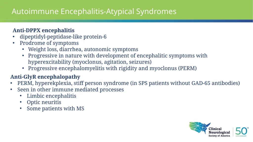
Autoimmune Encephalitis-Atypical Syndromes Anti-DPPX encephalitis
dipeptidyl-peptidase-like protein-6
Prodrome of symptoms
Weight loss, diarrhea, autonomic symptoms
Progressive in nature with development of encephalitic symptoms with hyperexcitability (myoclonus, agitation, seizures)
Progressive encephalomyelitis with rigidity and myoclonus (PERM) Anti-GlyR encephalopathy
PERM, hyperekplexia, stiff person syndrome (in SPS patients without GAD-65 antibodies)
Seen in other immune mediated processes
Limbic encephalitis
Optic neuritis
Some patients with MS<br>
dipeptidyl-peptidase-like protein-6
Prodrome of symptoms
Weight loss, diarrhea, autonomic symptoms
Progressive in nature with development of encephalitic symptoms with hyperexcitability (myoclonus, agitation, seizures)
Progressive encephalomyelitis with rigidity and myoclonus (PERM) Anti-GlyR encephalopathy
PERM, hyperekplexia, stiff person syndrome (in SPS patients without GAD-65 antibodies)
Seen in other immune mediated processes
Limbic encephalitis
Optic neuritis
Some patients with MS<br>
19
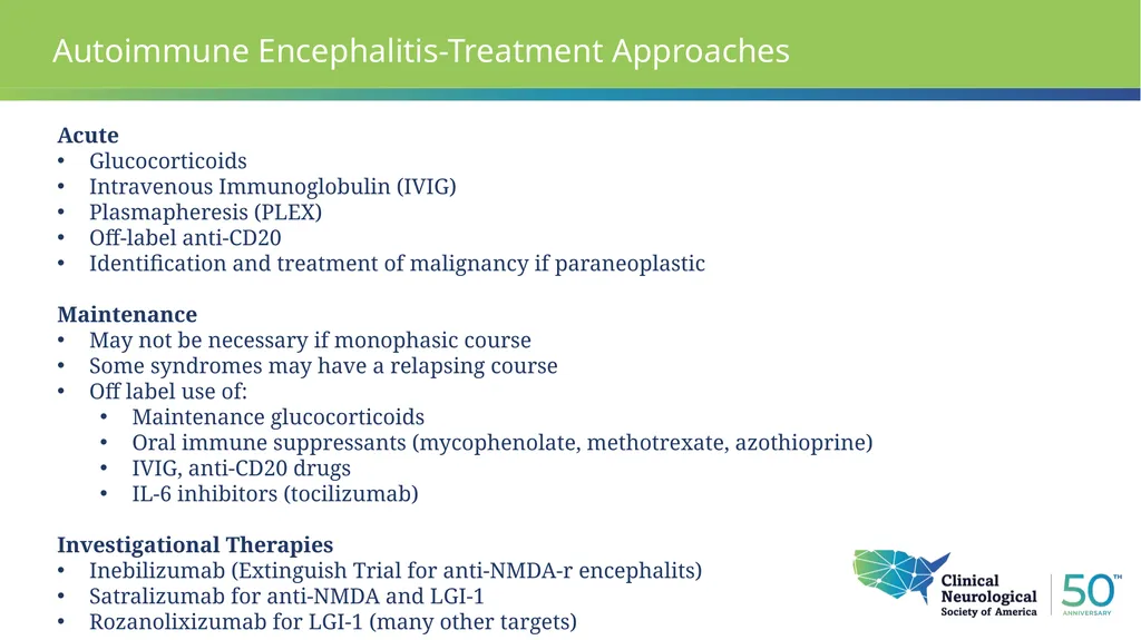
Autoimmune Encephalitis-Treatment Approaches Acute
Glucocorticoids
Intravenous Immunoglobulin (IVIG)
Plasmapheresis (PLEX)
Off-label anti-CD20
Identification and treatment of malignancy if paraneoplastic
Maintenance
May not be necessary if monophasic course
Some syndromes may have a relapsing course
Off label use of:
Maintenance glucocorticoids
Oral immune suppressants (mycophenolate, methotrexate, azothioprine)
IVIG, anti-CD20 drugs
IL-6 inhibitors (tocilizumab)
Investigational Therapies
Inebilizumab (Extinguish Trial for anti-NMDA-r encephalits)
Satralizumab for anti-NMDA and LGI-1
Rozanolixizumab for LGI-1 (many other targets)<br>
Glucocorticoids
Intravenous Immunoglobulin (IVIG)
Plasmapheresis (PLEX)
Off-label anti-CD20
Identification and treatment of malignancy if paraneoplastic
Maintenance
May not be necessary if monophasic course
Some syndromes may have a relapsing course
Off label use of:
Maintenance glucocorticoids
Oral immune suppressants (mycophenolate, methotrexate, azothioprine)
IVIG, anti-CD20 drugs
IL-6 inhibitors (tocilizumab)
Investigational Therapies
Inebilizumab (Extinguish Trial for anti-NMDA-r encephalits)
Satralizumab for anti-NMDA and LGI-1
Rozanolixizumab for LGI-1 (many other targets)<br>
20
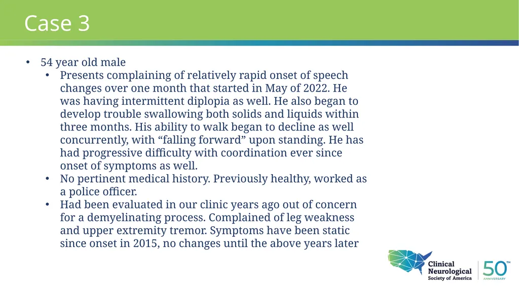
Case 3 54 year old male
Presents complaining of relatively rapid onset of speech changes over one month that started in May of 2022. He was having intermittent diplopia as well. He also began to develop trouble swallowing both solids and liquids within three months. His ability to walk began to decline as well concurrently, with “falling forward” upon standing. He has had progressive difficulty with coordination ever since onset of symptoms as well.
No pertinent medical history. Previously healthy, worked as a police officer.
Had been evaluated in our clinic years ago out of concern for a demyelinating process. Complained of leg weakness and upper extremity tremor. Symptoms have been static since onset in 2015, no changes until the above years later<br>
Presents complaining of relatively rapid onset of speech changes over one month that started in May of 2022. He was having intermittent diplopia as well. He also began to develop trouble swallowing both solids and liquids within three months. His ability to walk began to decline as well concurrently, with “falling forward” upon standing. He has had progressive difficulty with coordination ever since onset of symptoms as well.
No pertinent medical history. Previously healthy, worked as a police officer.
Had been evaluated in our clinic years ago out of concern for a demyelinating process. Complained of leg weakness and upper extremity tremor. Symptoms have been static since onset in 2015, no changes until the above years later<br>
21
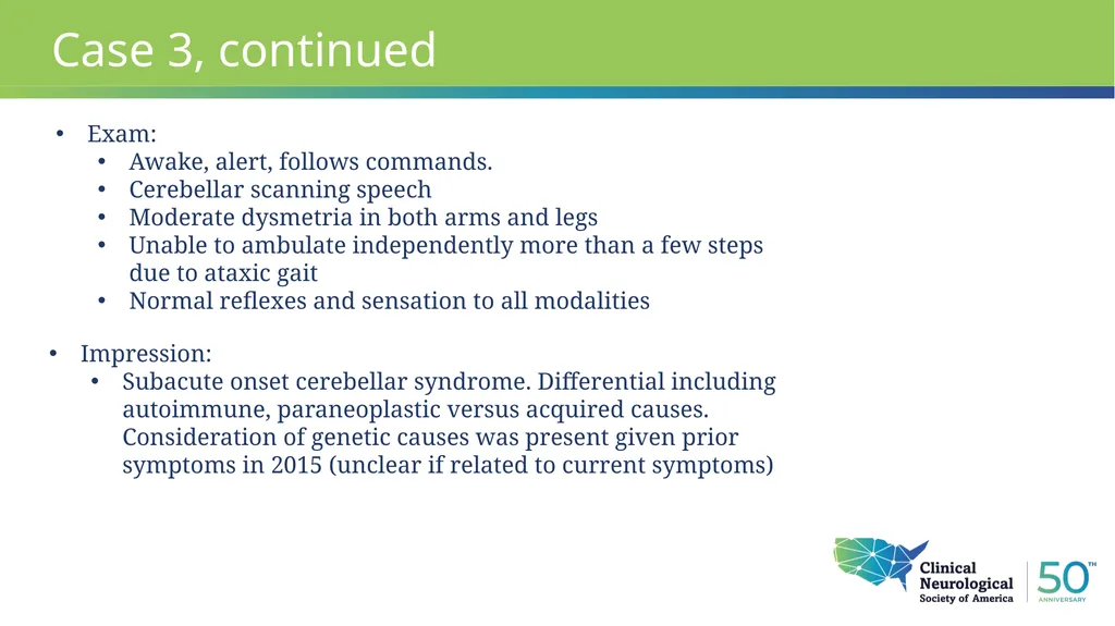
Case 3, continued Exam:
Awake, alert, follows commands.
Cerebellar scanning speech
Moderate dysmetria in both arms and legs
Unable to ambulate independently more than a few steps due to ataxic gait
Normal reflexes and sensation to all modalities Impression:
Subacute onset cerebellar syndrome. Differential including autoimmune, paraneoplastic versus acquired causes. Consideration of genetic causes was present given prior symptoms in 2015 (unclear if related to current symptoms)<br>
Awake, alert, follows commands.
Cerebellar scanning speech
Moderate dysmetria in both arms and legs
Unable to ambulate independently more than a few steps due to ataxic gait
Normal reflexes and sensation to all modalities Impression:
Subacute onset cerebellar syndrome. Differential including autoimmune, paraneoplastic versus acquired causes. Consideration of genetic causes was present given prior symptoms in 2015 (unclear if related to current symptoms)<br>
22
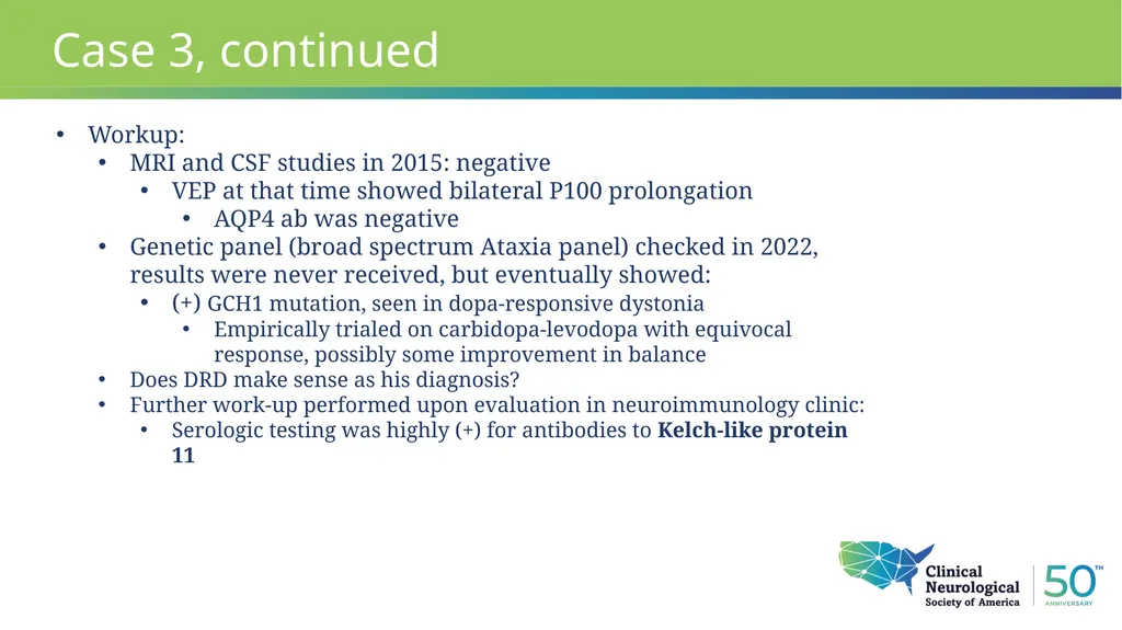
Case 3, continued Workup:
MRI and CSF studies in 2015: negative
VEP at that time showed bilateral P100 prolongation
AQP4 ab was negative
Genetic panel (broad spectrum Ataxia panel) checked in 2022, results were never received, but eventually showed:
(+) GCH1 mutation, seen in dopa-responsive dystonia
Empirically trialed on carbidopa-levodopa with equivocal response, possibly some improvement in balance
Does DRD make sense as his diagnosis?
Further work-up performed upon evaluation in neuroimmunology clinic:
Serologic testing was highly (+) for antibodies to Kelch-like protein 11<br>
MRI and CSF studies in 2015: negative
VEP at that time showed bilateral P100 prolongation
AQP4 ab was negative
Genetic panel (broad spectrum Ataxia panel) checked in 2022, results were never received, but eventually showed:
(+) GCH1 mutation, seen in dopa-responsive dystonia
Empirically trialed on carbidopa-levodopa with equivocal response, possibly some improvement in balance
Does DRD make sense as his diagnosis?
Further work-up performed upon evaluation in neuroimmunology clinic:
Serologic testing was highly (+) for antibodies to Kelch-like protein 11<br>
23
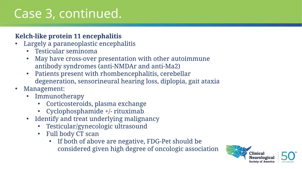
Case 3, continued. Kelch-like protein 11 encephalitis
Largely a paraneoplastic encephalitis
Testicular seminoma
May have cross-over presentation with other autoimmune antibody syndromes (anti-NMDAr and anti-Ma2)
Patients present with rhombencephalitis, cerebellar degeneration, sensorineural hearing loss, diplopia, gait ataxia
Management:
Immunotherapy
Corticosteroids, plasma exchange
Cyclophosphamide +/- rituximab
Identify and treat underlying malignancy
Testicular/gynecologic ultrasound
Full body CT scan
If both of above are negative, FDG-Pet should be considered given high degree of oncologic association<br>
Largely a paraneoplastic encephalitis
Testicular seminoma
May have cross-over presentation with other autoimmune antibody syndromes (anti-NMDAr and anti-Ma2)
Patients present with rhombencephalitis, cerebellar degeneration, sensorineural hearing loss, diplopia, gait ataxia
Management:
Immunotherapy
Corticosteroids, plasma exchange
Cyclophosphamide +/- rituximab
Identify and treat underlying malignancy
Testicular/gynecologic ultrasound
Full body CT scan
If both of above are negative, FDG-Pet should be considered given high degree of oncologic association<br>
24
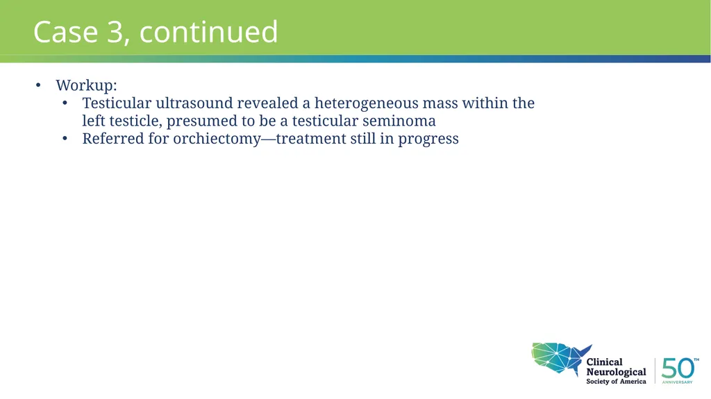
Case 3, continued Workup:
Testicular ultrasound revealed a heterogeneous mass within the left testicle, presumed to be a testicular seminoma
Referred for orchiectomy—treatment still in progress<br>
Testicular ultrasound revealed a heterogeneous mass within the left testicle, presumed to be a testicular seminoma
Referred for orchiectomy—treatment still in progress<br>
25
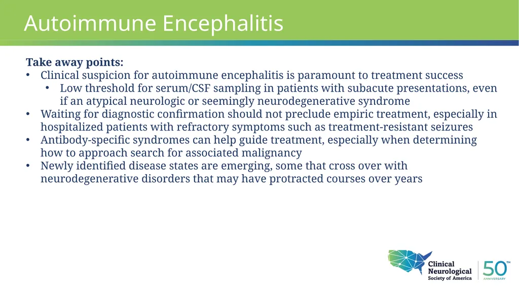
Autoimmune Encephalitis Take away points:
Clinical suspicion for autoimmune encephalitis is paramount to treatment success
Low threshold for serum/CSF sampling in patients with subacute presentations, even if an atypical neurologic or seemingly neurodegenerative syndrome
Waiting for diagnostic confirmation should not preclude empiric treatment, especially in hospitalized patients with refractory symptoms such as treatment-resistant seizures
Antibody-specific syndromes can help guide treatment, especially when determining how to approach search for associated malignancy
Newly identified disease states are emerging, some that cross over with neurodegenerative disorders that may have protracted courses over years<br>
Clinical suspicion for autoimmune encephalitis is paramount to treatment success
Low threshold for serum/CSF sampling in patients with subacute presentations, even if an atypical neurologic or seemingly neurodegenerative syndrome
Waiting for diagnostic confirmation should not preclude empiric treatment, especially in hospitalized patients with refractory symptoms such as treatment-resistant seizures
Antibody-specific syndromes can help guide treatment, especially when determining how to approach search for associated malignancy
Newly identified disease states are emerging, some that cross over with neurodegenerative disorders that may have protracted courses over years<br>
26
Subhead
Text
• Bullet Dubey D, Wilson MR, Clarkson B, et al. Expanded Clinical Phenotype, Oncological Associations, and Immunopathologic Insights of Paraneoplastic Kelch-like Protein-11 Encephalitis. JAMA Neurol. 2020;77(11):1420–1429. doi:10.1001/jamaneurol.2020.2231
https://blogs.neurology.org/epearls/paraneoplastic-kelch-like-protein-11-encephalitis/
Lancet Neurol. 2016 Apr;15(4):391-404. Epub 2016 Feb 20.
Neurol Neuroimmunol Neuroinflamm. 2021;8(4) Epub 2021 May 18.
Graus F, et al. A clinical approach to diagnosis of autoimmune encephalitis. Lancet Neurol. 2016 Apr;15(4):391-404. doi: 10.1016/S1474-4422(15)00401-9. Epub 2016 Feb 20. PMID: 26906964; PMCID: PMC5066574. References<br>
Text
• Bullet Dubey D, Wilson MR, Clarkson B, et al. Expanded Clinical Phenotype, Oncological Associations, and Immunopathologic Insights of Paraneoplastic Kelch-like Protein-11 Encephalitis. JAMA Neurol. 2020;77(11):1420–1429. doi:10.1001/jamaneurol.2020.2231
https://blogs.neurology.org/epearls/paraneoplastic-kelch-like-protein-11-encephalitis/
Lancet Neurol. 2016 Apr;15(4):391-404. Epub 2016 Feb 20.
Neurol Neuroimmunol Neuroinflamm. 2021;8(4) Epub 2021 May 18.
Graus F, et al. A clinical approach to diagnosis of autoimmune encephalitis. Lancet Neurol. 2016 Apr;15(4):391-404. doi: 10.1016/S1474-4422(15)00401-9. Epub 2016 Feb 20. PMID: 26906964; PMCID: PMC5066574. References<br>
27
Questions<br>