Bimanual pelvic examination Prepared by: Shaimaa Shahab Definition Is the stage of palpation during the clinical examination of the pelvis, in the female genital sphere. It is a examination done per vagina to assess the status of Vagina,
"Bimanual pelvic examination Prepared by: Shaimaa" is the property of its rightful owner. Permission is granted to
download and print the materials on this website for personal, non-commercial use only, and to display it
on your personal computer provided you do not modify the materials and that you retain all copyright
notices contained in the materials. By downloading content from our website, you accept the terms of this
agreement.
Definition Is the stage of palpation during the clinical examination of the pelvis, in the female genital sphere. It is a examination done per vagina to assess the status of Vagina, Cervix and progress of descents of fetus through birth canal.<br>
03
Purpose To cheak for changes or abnormalities in the reproductive organs.
To find possible signs of ovarian cyst.
To assess the progress and delay of labour.<br>
04
Indication At the Onset of the labour
Pruritus
Vaginal discharge
Vaginal bleeding
Pain in the lower abdominal floor
Menstrual disorders
Assessment of local involvement in patients with cancer starting from the genital area<br>
05
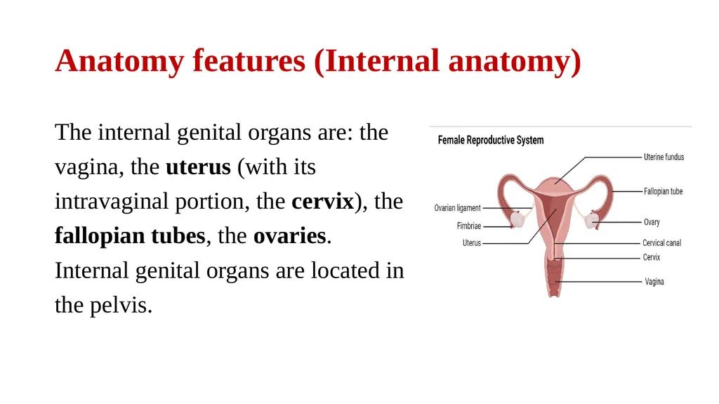
Anatomy features (Internal anatomy) The internal genital organs are: the vagina, the uterus (with its intravaginal portion, the cervix), the fallopian tubes, the ovaries. Internal genital organs are located in the pelvis.<br>
06
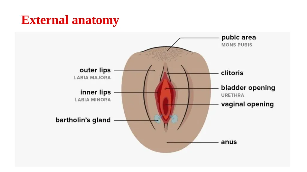
External anatomy<br>
07
Vulva : is the external Part of the female genitalia, it Protect a woman's sexual Organs, Urinary opening .
Bartholins gland: are to Pea sized Compound alveolar glands located slightly of the opening vagina, Secrete fluid that helps lubricate the vagina.
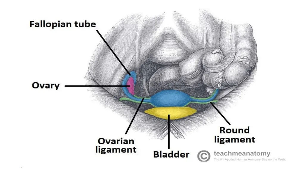
Adnexa: the parts of the adnexa are ( Fallopian tubes , Ovaries, and ligaments). they function to hold the uterus in the correct position during pregnancy.<br>
08
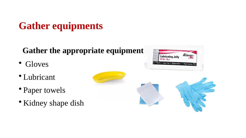
Gather equipments Gather the appropriate equipment:
Gloves
Lubricant
Paper towels
Kidney shape dish<br>
09
Inspect the vulva Inspect the vulva for abnormalities:
Ulcers typically associated with genital herpes.
Abnormal vaginal discharge
Scarring and Vaginal atrophy.
Masses: causes include Bartholin's cyst and vulval malignancy.
Inspect for evidence of vaginal prolapse (a bulge visible protruding from the vagina).<br>
10
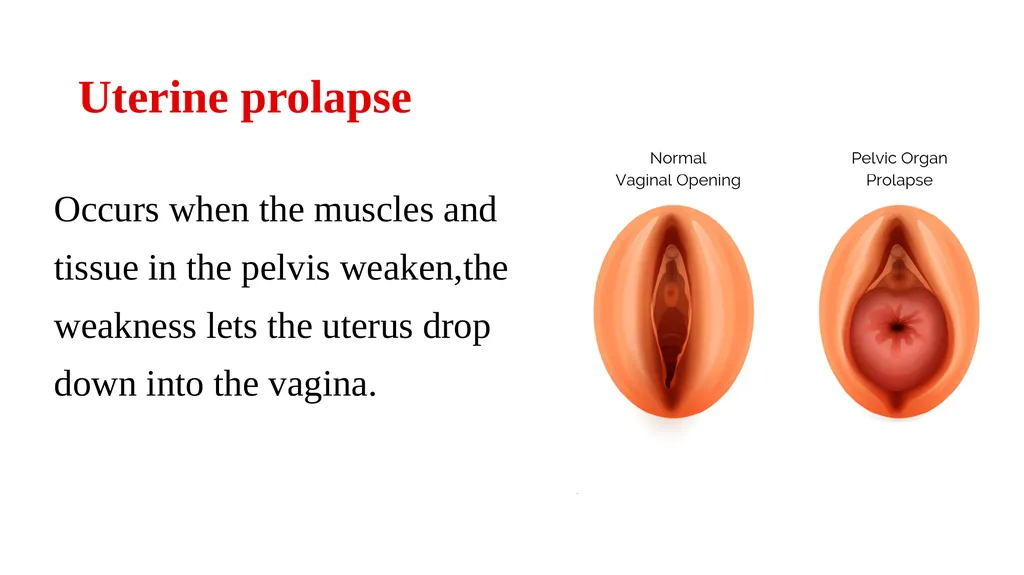
Uterine prolapse Occurs when the muscles and tissue in the pelvis weaken,the weakness lets the uterus drop down into the vagina.<br>
11
Palpation : Vaginal walls
Palpate the walls of the vagina for any irregularities or masses. Palpation by inserting the gloved index and middle finger of your dominant hand into the vagina.<br>
12
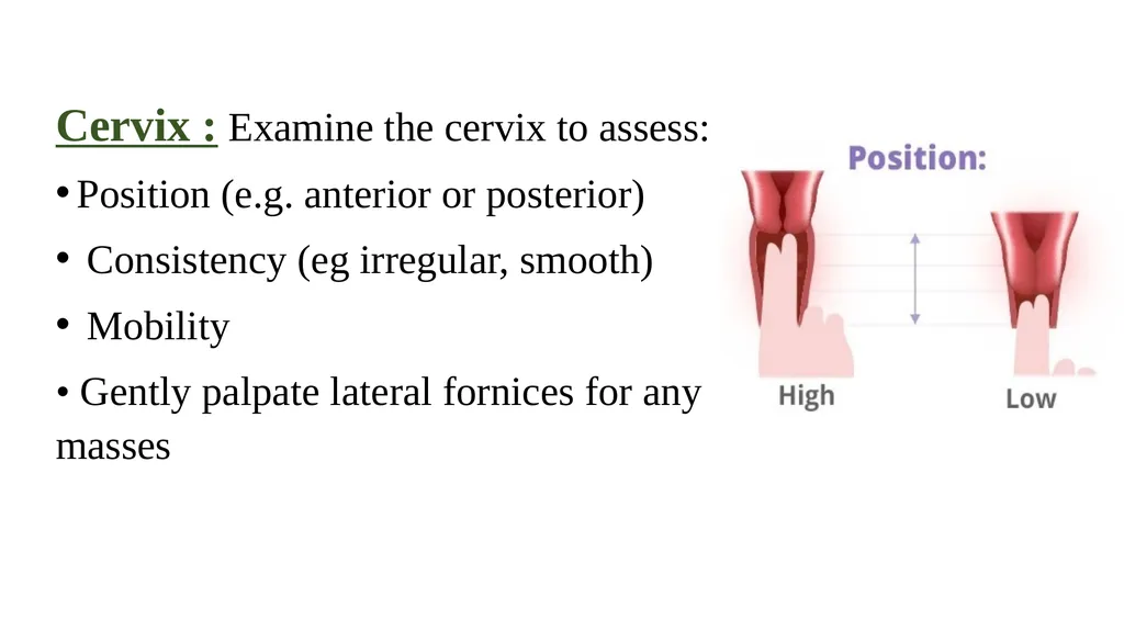
Cervix : Examine the cervix to assess:
Position (e.g. anterior or posterior)
Consistency (eg irregular, smooth)
Mobility
• Gently palpate lateral fornices for any masses<br>
13
Excitation test Is part of the bimanual examination, to assess for cervical motion tenderness CMT, Place fingers in the posterior fornix to lift the uterus whilst simultaneously pushing the fundus down by placing the left hand above the symphysis pubis. Gently move the cervix from side to side to check for cervical tenderness.<br>
14
NOTE! If the lady feels cervical tenderness, she has an infection and the Excitation test is positive.<br>
15
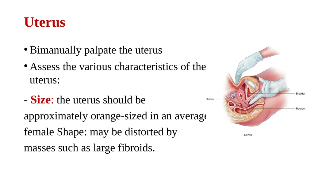
Uterus Bimanually palpate the uterus
Assess the various characteristics of the uterus:
- Size: the uterus should be approximately orange-sized in an average female Shape: may be distorted by masses such as large fibroids.<br>
16
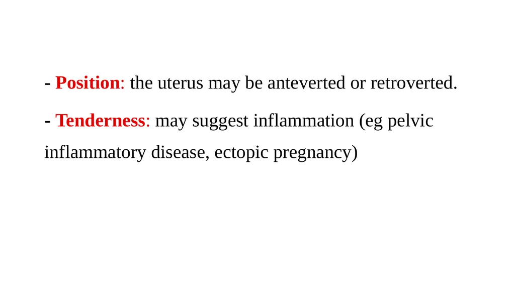
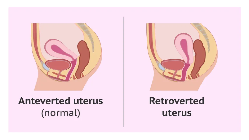
- Position: the uterus may be anteverted or retroverted.
- Tenderness: may suggest inflammation (eg pelvic inflammatory disease, ectopic pregnancy)<br>
17
Bimanually palpate right and left Adnexa:
feel for any palpable masses, noting their size and shape (e.g. ovarian cyst, ovarian tumour, fibroid).<br>
18
Vaginal Examination - Withdrawing Warn the patient that you are about to withdraw your fingers.
Withdraw your fingers and inspect the glove for blood or abnormal discharge.
Cover the patient with the sheet, explain that the examination is now complete and provide the patient with privacy so they can get dressed, provide paper towels for the patient to clean themselves.
Dispose of the used equipment into a clinical waste bin.<br>