
Bleeding CHAPTER 27: Focused Lecture National EMS







Description: Bleeding CHAPTER 27: Focused Lecture National EMS Education Standard Competencies (1 of 3) Trauma Applies fundamental knowledge to provide basic and selected advanced emergency care and transportation based on assessment findings for an
Related Topics
Download Presentation
"Bleeding CHAPTER 27: Focused Lecture National EMS" is the property of its rightful owner. Permission is granted to download and print the materials on this website for personal, non-commercial use only, and to display it on your personal computer provided you do not modify the materials and that you retain all copyright notices contained in the materials. By downloading content from our website, you accept the terms of this agreement.
Presentation Transcript
slide1. Bleeding CHAPTER 27: Focused Lecture<br>
slide2. National EMS Education Standard Competencies (1 of 3) Trauma
Applies fundamental knowledge to provide basic and selected advanced emergency care and transportation based on assessment findings for an acutely injured patient.<br>
slide3. National EMS Education Standard Competencies (2 of 3) Bleeding
Pathophysiology, assessment, and management of
Bleeding
Fluid resuscitation<br>
slide4. National EMS Education Standard Competencies (3 of 3) Pathophysiology
Applies comprehensive knowledge of the pathophysiology of respiration and perfusion to patient assessment and management.<br>
slide5. Introduction Bleeding can be one of the most time-sensitive conditions.
Can be external and obvious or internal and hidden
Can cause
Weakness
Shock
Death<br>
slide6. Physiology and Perfusion (1 of 4) Perfusion: Circulation of blood within an organ or tissue in adequate amounts
Hypoperfusion (shock): Cardiovascular system fails to provide sufficient circulation for body parts to function. © Jones & Bartlett Learning.<br>
slide7. Physiology and Perfusion (2 of 4) Emergency medical care is designed to support the:
Heart
Brain and spinal
Lungs
Kidneys<br>
slide8. Physiology and Perfusion (3 of 4) Cardiac cycle
Repetitive pumping process
Begins with onset of cardiac muscle contraction and ends with beginning of the next contraction
Preload: Amount of blood returned to the heart to be pumped out
Afterload: Pressure in the aorta or the peripheral vascular resistance (PVR), against which the left ventricle must pump blood<br>
slide9. Physiology and Perfusion (4 of 4) Stroke volume (SV): The amount of blood ejected per contraction
Cardiac cycle is connected to bleeding and shock through its relationship to blood pressure.
Delivery of oxygen to the tissues is dependent on an adequate heart rate, SV, hemoglobin levels, and arterial oxygen saturation.<br>
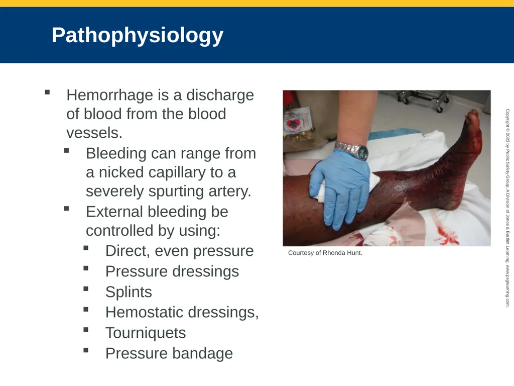
slide10. Pathophysiology Hemorrhage is a discharge of blood from the blood vessels.
Bleeding can range from a nicked capillary to a severely spurting artery.
External bleeding be controlled by using:
Direct, even pressure
Pressure dressings
Splints
Hemostatic dressings,
Tourniquets
Pressure bandage Courtesy of Rhonda Hunt.<br>
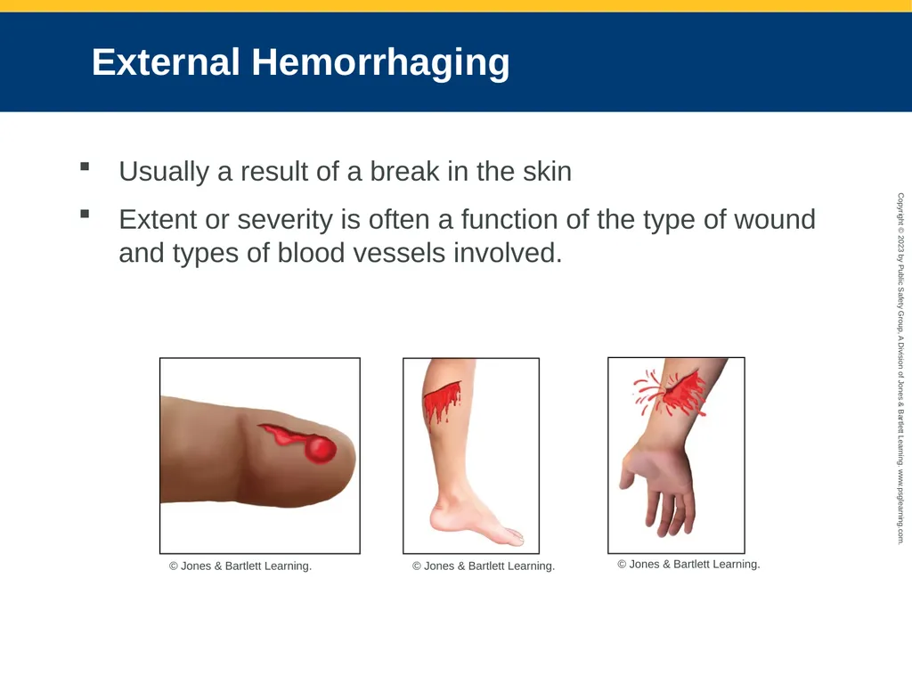
slide11. External Hemorrhaging Usually a result of a break in the skin
Extent or severity is often a function of the type of wound and types of blood vessels involved. © Jones & Bartlett Learning. © Jones & Bartlett Learning. © Jones & Bartlett Learning.<br>
slide12. Internal Hemorrhaging (1 of 3) May appear in any portion of the body
Nontraumatic internal hemorrhage often occurs in cases of GI bleeding, ruptured ectopic pregnancies, ruptured aneurysms, or other conditions.<br>
slide13. Internal Hemorrhaging (2 of 3) Mechanism of injury for internal bleeding
Falls
Blast injuries
Automobile or motorcycle crashes
Nature of illness for internal bleeding
Bleeding ulcers, bleeding from the colon, ruptured ectopic pregnancy, and aneurysms<br>
slide14. Internal Hemorrhaging (3 of 3) Signs and symptoms
Pain
Swelling in the area of bleeding
Distention
Bruising
Dyspnea, tachycardia, and hypotension
Hematoma
Other signs and symptoms: hematuria, hematemesis, hemoptysis, melena, hematochezia
First sign is generally a change in mental status<br>
slide15. The Importance of Bleeding Substantial changes in vital signs occur for approximately 2 pints (1 L) of blood loss.
Hemorrhagic shock results in inadequate perfusion and even death.
Considered serious with:
A significant MOI
Poor general appearance
Signs and symptoms of shock (hypoperfusion)
Rapid blood loss
Uncontrollable bleeding<br>
slide16. Physiologic Response to Hemorrhaging (1 of 2) Bleeding tends to stop quickly in response to internal clotting mechanisms.
Hemostasis: Platelets plug the hole and seal the injured portions of the vessel.
Medications can interfere with normal clotting.
A patient with hemophilia should be transported immediately.<br>
slide17. Physiologic Response to Hemorrhaging (2 of 2) Trauma triad of death
Combination of hypothermia, coagulopathy, and acidosis increases mortality.
Coagulopathy can result in further hemorrhage.
Identify the conditions of hypothermia, coagulopathy, and acidosis in the setting of trauma and intervene where possible.<br>
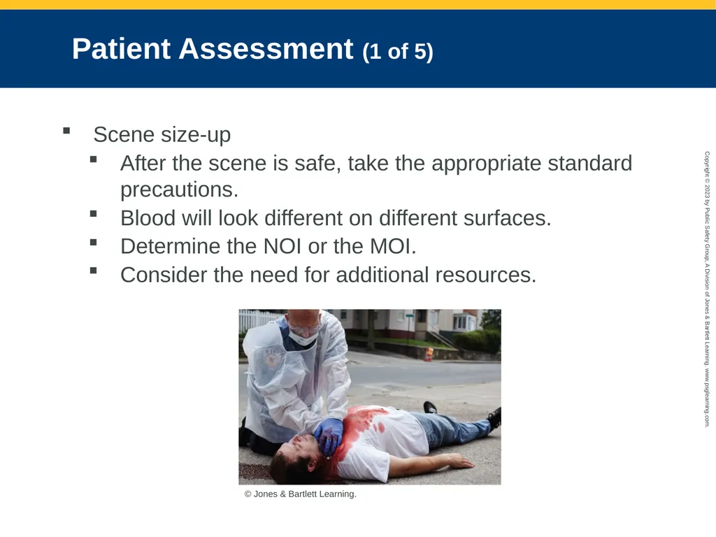
slide18. Patient Assessment (1 of 5) Scene size-up
After the scene is safe, take the appropriate standard precautions.
Blood will look different on different surfaces.
Determine the NOI or the MOI.
Consider the need for additional resources. © Jones & Bartlett Learning.<br>
slide19. Patient Assessment (2 of 5) Primary survey
Search for life-threatening bleeding.
Be aware of obvious signs of injury and distress.
Perform a rapid full-body scan of the patient.
Follow XABCDE mnemonic.
Determine level of consciousness.
Any internal bleeding must be treated promptly.
Determine how fast the transport decision should be made and where the patient should be taken.<br>
slide20. Patient Assessment (3 of 5) History taking
Investigate the chief complaint and be alert for signs or symptoms of other injuries due to the MOI and/or NOI.
Carefully assess the MOI in trauma patients.
Gather information on the patient’s chief complaint using the OPQRST-I mnemonic and obtain a SAMPLE history.<br>
slide21. Patient Assessment (4 of 5) Secondary assessment
Uncover injuries or illness that may have been missed.
Record vital signs, complete a focused assessment of pain, and attach appropriate monitoring devices.
Assess respiratory, cardiovascular, neurologic, and musculoskeletal system.
Obtain baseline vital signs.
Use monitoring devices.<br>
slide22. Patient Assessment (5 of 5) Reassessment
Assess the effectiveness of any interventions.
Obtain patient’s vital signs every 5 minutes en route to the ED.
Recognize, estimate, and report the amount of blood loss.
With internal bleeding, describe the MOI/NOI and the signs and symptoms.<br>
slide23. Emergency Medical Care (1 of 12) External hemorrhaging
Can be managed with direct pressure, combined with pressure dressings and/or splints
Use of a tourniquet is preferred for uncontrollable external bleeding in an extremity.
Provide high-flow oxygen and assist ventilation. © Jones & Bartlett Learning. Courtesy of MIEMSS.<br>
slide24. Emergency Medical Care (2 of 12) Direct pressure
Can control most instances of external bleeding
Apply pressure over the top of sterile dressing.
Create a pressure dressing.
Stretch the bandage tight.
If direct pressure fails, consider applying a tourniquet or wound packing with a hemostatic agent.<br>
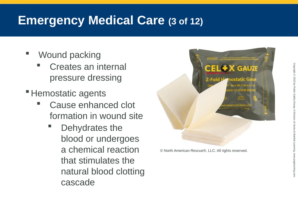
slide25. Emergency Medical Care (3 of 12) Wound packing
Creates an internal pressure dressing
Hemostatic agents
Cause enhanced clot formation in wound site
Dehydrates the blood or undergoes a chemical reaction that stimulates the natural blood clotting cascade © North American Rescue®, LLC. All rights reserved.<br>
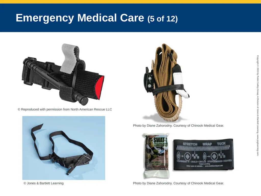
slide26. Emergency Medical Care (4 of 12) Tourniquets
Useful if a patient has severe hemorrhaging from an extremity injury below the axilla or groin
Commercially available tourniquets
Combat application tourniquet (CAT)
Special operations forces tactical tourniquet (SOFT-T)
Ratcheting medical tourniquet (RMT)
Stretch, wrap, and tuck tourniquet (SWAT-T)<br>
slide27. Emergency Medical Care (5 of 12) © Reproduced with permission from North American Rescue LLC © Jones & Bartlett Learning Photo by Diane Zahorodny. Courtesy of Chinook Medical Gear. Photo by Diane Zahorodny. Courtesy of Chinook Medical Gear.<br>
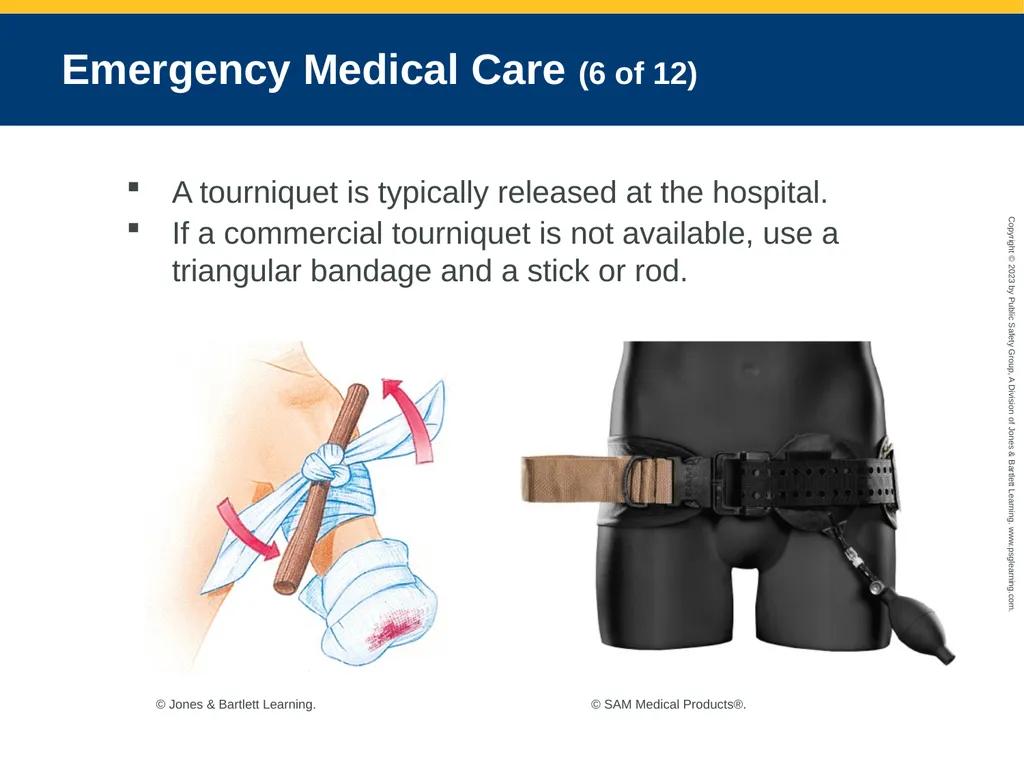
slide28. Emergency Medical Care (6 of 12) A tourniquet is typically released at the hospital.
If a commercial tourniquet is not available, use a triangular bandage and a stick or rod. © Jones & Bartlett Learning. © SAM Medical Products®.<br>
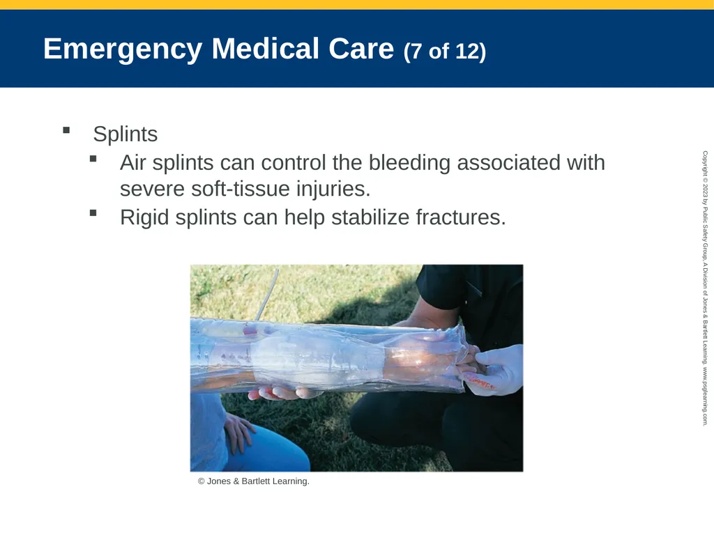
slide29. Emergency Medical Care (7 of 12) Splints
Air splints can control the bleeding associated with severe soft-tissue injuries.
Rigid splints can help stabilize fractures. © Jones & Bartlett Learning.<br>
slide30. Emergency Medical Care (8 of 12) Pneumatic antishock garment (PASG)
Inflatable device that covers the legs and abdomen
Rarely used, always follow local protocols<br>
slide31. Emergency Medical Care (9 of 12) Hemorrhaging from the nose, ears, and mouth
Can be caused by
Skull fracture
Facial injuries
Sinusitis, infections, nose drop use and abuse, dried or cracked nasal mucosa
High blood pressure
Coagulation disorders
Digital trauma © E.M. Singletary, M.D. Used with permission.<br>
slide32. Emergency Medical Care (10 of 12) Internal hemorrhaging
Definitive care includes rapid transport to the ED.
Treatment focuses on treatment of shock, minimizing movement of the injured part, and rapid transport
Ultrasonography may be used to locate bleeding.
Give nothing by mouth.
Keep the patient warm.
Monitor serial vital signs. Courtesy of Rhonda Hunt.<br>
slide33. Emergency Medical Care (11 of 12) Management of hemorrhagic shock
Focus on exsanguinating hemorrhage first.
Take standard precautions and ensure the scene is safe.
Identify and try to stop any major bleeding, and quickly evaluate the patient’s hemodynamic status.
Establish and maintain an open airway.
En route insert at least one, preferably two, 18-gauge peripheral IV line.<br>
slide34. Emergency Medical Care (12 of 12) Management of hemorrhagic shock (cont’d)
Obtain IV access at the scene only if transport is delayed.
Administer isotonic crystalloids of normal saline.
Monitor the level of consciousness, pulse, and blood pressure.<br>
slide2. National EMS Education Standard Competencies (1 of 3) Trauma
Applies fundamental knowledge to provide basic and selected advanced emergency care and transportation based on assessment findings for an acutely injured patient.<br>
slide3. National EMS Education Standard Competencies (2 of 3) Bleeding
Pathophysiology, assessment, and management of
Bleeding
Fluid resuscitation<br>
slide4. National EMS Education Standard Competencies (3 of 3) Pathophysiology
Applies comprehensive knowledge of the pathophysiology of respiration and perfusion to patient assessment and management.<br>
slide5. Introduction Bleeding can be one of the most time-sensitive conditions.
Can be external and obvious or internal and hidden
Can cause
Weakness
Shock
Death<br>
slide6. Physiology and Perfusion (1 of 4) Perfusion: Circulation of blood within an organ or tissue in adequate amounts
Hypoperfusion (shock): Cardiovascular system fails to provide sufficient circulation for body parts to function. © Jones & Bartlett Learning.<br>
slide7. Physiology and Perfusion (2 of 4) Emergency medical care is designed to support the:
Heart
Brain and spinal
Lungs
Kidneys<br>
slide8. Physiology and Perfusion (3 of 4) Cardiac cycle
Repetitive pumping process
Begins with onset of cardiac muscle contraction and ends with beginning of the next contraction
Preload: Amount of blood returned to the heart to be pumped out
Afterload: Pressure in the aorta or the peripheral vascular resistance (PVR), against which the left ventricle must pump blood<br>
slide9. Physiology and Perfusion (4 of 4) Stroke volume (SV): The amount of blood ejected per contraction
Cardiac cycle is connected to bleeding and shock through its relationship to blood pressure.
Delivery of oxygen to the tissues is dependent on an adequate heart rate, SV, hemoglobin levels, and arterial oxygen saturation.<br>
slide10. Pathophysiology Hemorrhage is a discharge of blood from the blood vessels.
Bleeding can range from a nicked capillary to a severely spurting artery.
External bleeding be controlled by using:
Direct, even pressure
Pressure dressings
Splints
Hemostatic dressings,
Tourniquets
Pressure bandage Courtesy of Rhonda Hunt.<br>
slide11. External Hemorrhaging Usually a result of a break in the skin
Extent or severity is often a function of the type of wound and types of blood vessels involved. © Jones & Bartlett Learning. © Jones & Bartlett Learning. © Jones & Bartlett Learning.<br>
slide12. Internal Hemorrhaging (1 of 3) May appear in any portion of the body
Nontraumatic internal hemorrhage often occurs in cases of GI bleeding, ruptured ectopic pregnancies, ruptured aneurysms, or other conditions.<br>
slide13. Internal Hemorrhaging (2 of 3) Mechanism of injury for internal bleeding
Falls
Blast injuries
Automobile or motorcycle crashes
Nature of illness for internal bleeding
Bleeding ulcers, bleeding from the colon, ruptured ectopic pregnancy, and aneurysms<br>
slide14. Internal Hemorrhaging (3 of 3) Signs and symptoms
Pain
Swelling in the area of bleeding
Distention
Bruising
Dyspnea, tachycardia, and hypotension
Hematoma
Other signs and symptoms: hematuria, hematemesis, hemoptysis, melena, hematochezia
First sign is generally a change in mental status<br>
slide15. The Importance of Bleeding Substantial changes in vital signs occur for approximately 2 pints (1 L) of blood loss.
Hemorrhagic shock results in inadequate perfusion and even death.
Considered serious with:
A significant MOI
Poor general appearance
Signs and symptoms of shock (hypoperfusion)
Rapid blood loss
Uncontrollable bleeding<br>
slide16. Physiologic Response to Hemorrhaging (1 of 2) Bleeding tends to stop quickly in response to internal clotting mechanisms.
Hemostasis: Platelets plug the hole and seal the injured portions of the vessel.
Medications can interfere with normal clotting.
A patient with hemophilia should be transported immediately.<br>
slide17. Physiologic Response to Hemorrhaging (2 of 2) Trauma triad of death
Combination of hypothermia, coagulopathy, and acidosis increases mortality.
Coagulopathy can result in further hemorrhage.
Identify the conditions of hypothermia, coagulopathy, and acidosis in the setting of trauma and intervene where possible.<br>
slide18. Patient Assessment (1 of 5) Scene size-up
After the scene is safe, take the appropriate standard precautions.
Blood will look different on different surfaces.
Determine the NOI or the MOI.
Consider the need for additional resources. © Jones & Bartlett Learning.<br>
slide19. Patient Assessment (2 of 5) Primary survey
Search for life-threatening bleeding.
Be aware of obvious signs of injury and distress.
Perform a rapid full-body scan of the patient.
Follow XABCDE mnemonic.
Determine level of consciousness.
Any internal bleeding must be treated promptly.
Determine how fast the transport decision should be made and where the patient should be taken.<br>
slide20. Patient Assessment (3 of 5) History taking
Investigate the chief complaint and be alert for signs or symptoms of other injuries due to the MOI and/or NOI.
Carefully assess the MOI in trauma patients.
Gather information on the patient’s chief complaint using the OPQRST-I mnemonic and obtain a SAMPLE history.<br>
slide21. Patient Assessment (4 of 5) Secondary assessment
Uncover injuries or illness that may have been missed.
Record vital signs, complete a focused assessment of pain, and attach appropriate monitoring devices.
Assess respiratory, cardiovascular, neurologic, and musculoskeletal system.
Obtain baseline vital signs.
Use monitoring devices.<br>
slide22. Patient Assessment (5 of 5) Reassessment
Assess the effectiveness of any interventions.
Obtain patient’s vital signs every 5 minutes en route to the ED.
Recognize, estimate, and report the amount of blood loss.
With internal bleeding, describe the MOI/NOI and the signs and symptoms.<br>
slide23. Emergency Medical Care (1 of 12) External hemorrhaging
Can be managed with direct pressure, combined with pressure dressings and/or splints
Use of a tourniquet is preferred for uncontrollable external bleeding in an extremity.
Provide high-flow oxygen and assist ventilation. © Jones & Bartlett Learning. Courtesy of MIEMSS.<br>
slide24. Emergency Medical Care (2 of 12) Direct pressure
Can control most instances of external bleeding
Apply pressure over the top of sterile dressing.
Create a pressure dressing.
Stretch the bandage tight.
If direct pressure fails, consider applying a tourniquet or wound packing with a hemostatic agent.<br>
slide25. Emergency Medical Care (3 of 12) Wound packing
Creates an internal pressure dressing
Hemostatic agents
Cause enhanced clot formation in wound site
Dehydrates the blood or undergoes a chemical reaction that stimulates the natural blood clotting cascade © North American Rescue®, LLC. All rights reserved.<br>
slide26. Emergency Medical Care (4 of 12) Tourniquets
Useful if a patient has severe hemorrhaging from an extremity injury below the axilla or groin
Commercially available tourniquets
Combat application tourniquet (CAT)
Special operations forces tactical tourniquet (SOFT-T)
Ratcheting medical tourniquet (RMT)
Stretch, wrap, and tuck tourniquet (SWAT-T)<br>
slide27. Emergency Medical Care (5 of 12) © Reproduced with permission from North American Rescue LLC © Jones & Bartlett Learning Photo by Diane Zahorodny. Courtesy of Chinook Medical Gear. Photo by Diane Zahorodny. Courtesy of Chinook Medical Gear.<br>
slide28. Emergency Medical Care (6 of 12) A tourniquet is typically released at the hospital.
If a commercial tourniquet is not available, use a triangular bandage and a stick or rod. © Jones & Bartlett Learning. © SAM Medical Products®.<br>
slide29. Emergency Medical Care (7 of 12) Splints
Air splints can control the bleeding associated with severe soft-tissue injuries.
Rigid splints can help stabilize fractures. © Jones & Bartlett Learning.<br>
slide30. Emergency Medical Care (8 of 12) Pneumatic antishock garment (PASG)
Inflatable device that covers the legs and abdomen
Rarely used, always follow local protocols<br>
slide31. Emergency Medical Care (9 of 12) Hemorrhaging from the nose, ears, and mouth
Can be caused by
Skull fracture
Facial injuries
Sinusitis, infections, nose drop use and abuse, dried or cracked nasal mucosa
High blood pressure
Coagulation disorders
Digital trauma © E.M. Singletary, M.D. Used with permission.<br>
slide32. Emergency Medical Care (10 of 12) Internal hemorrhaging
Definitive care includes rapid transport to the ED.
Treatment focuses on treatment of shock, minimizing movement of the injured part, and rapid transport
Ultrasonography may be used to locate bleeding.
Give nothing by mouth.
Keep the patient warm.
Monitor serial vital signs. Courtesy of Rhonda Hunt.<br>
slide33. Emergency Medical Care (11 of 12) Management of hemorrhagic shock
Focus on exsanguinating hemorrhage first.
Take standard precautions and ensure the scene is safe.
Identify and try to stop any major bleeding, and quickly evaluate the patient’s hemodynamic status.
Establish and maintain an open airway.
En route insert at least one, preferably two, 18-gauge peripheral IV line.<br>
slide34. Emergency Medical Care (12 of 12) Management of hemorrhagic shock (cont’d)
Obtain IV access at the scene only if transport is delayed.
Administer isotonic crystalloids of normal saline.
Monitor the level of consciousness, pulse, and blood pressure.<br>