
Books For References: Prokaryotic and Eukaryotic
SN
Published · 66 slides · 0 views

1 / 1






Description
Books For References: Prokaryotic and Eukaryotic cells Organization Different types What is a cell? A basic unit of organization or Structure of all living matter. A.G.Coewy and P. Sickevits (1963) Unit of Biological activity, delimited by
Related Topics
Share
Embed code
Download this presentation From Below
"Books For References: Prokaryotic and Eukaryotic" is the property of its rightful owner. Permission is granted to download and print the materials on this website for personal, non-commercial use only, and to display it on your personal computer provided you do not modify the materials and that you retain all copyright notices contained in the materials. By downloading content from our website, you accept the terms of this agreement.
Presentation Transcript
01
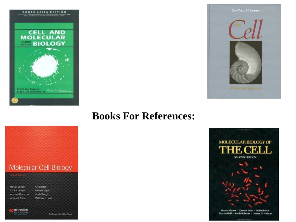
Books For References:<br>
02
Prokaryotic and Eukaryotic cells
Organization
Different types<br>
Organization
Different types<br>
03
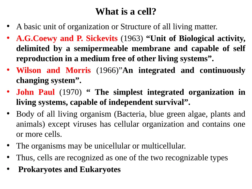
What is a cell? A basic unit of organization or Structure of all living matter.
A.G.Coewy and P. Sickevits (1963) “Unit of Biological activity, delimited by a semipermeable membrane and capable of self reproduction in a medium free of other living systems”.
Wilson and Morris (1966)”An integrated and continuously changing system”.
John Paul (1970) “ The simplest integrated organization in living systems, capable of independent survival”.
Body of all living organism (Bacteria, blue green algae, plants and animals) except viruses has cellular organization and contains one or more cells.
The organisms may be unicellular or multicellular.
Thus, cells are recognized as one of the two recognizable types
Prokaryotes and Eukaryotes<br>
A.G.Coewy and P. Sickevits (1963) “Unit of Biological activity, delimited by a semipermeable membrane and capable of self reproduction in a medium free of other living systems”.
Wilson and Morris (1966)”An integrated and continuously changing system”.
John Paul (1970) “ The simplest integrated organization in living systems, capable of independent survival”.
Body of all living organism (Bacteria, blue green algae, plants and animals) except viruses has cellular organization and contains one or more cells.
The organisms may be unicellular or multicellular.
Thus, cells are recognized as one of the two recognizable types
Prokaryotes and Eukaryotes<br>
04
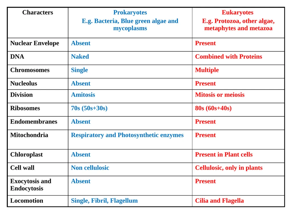
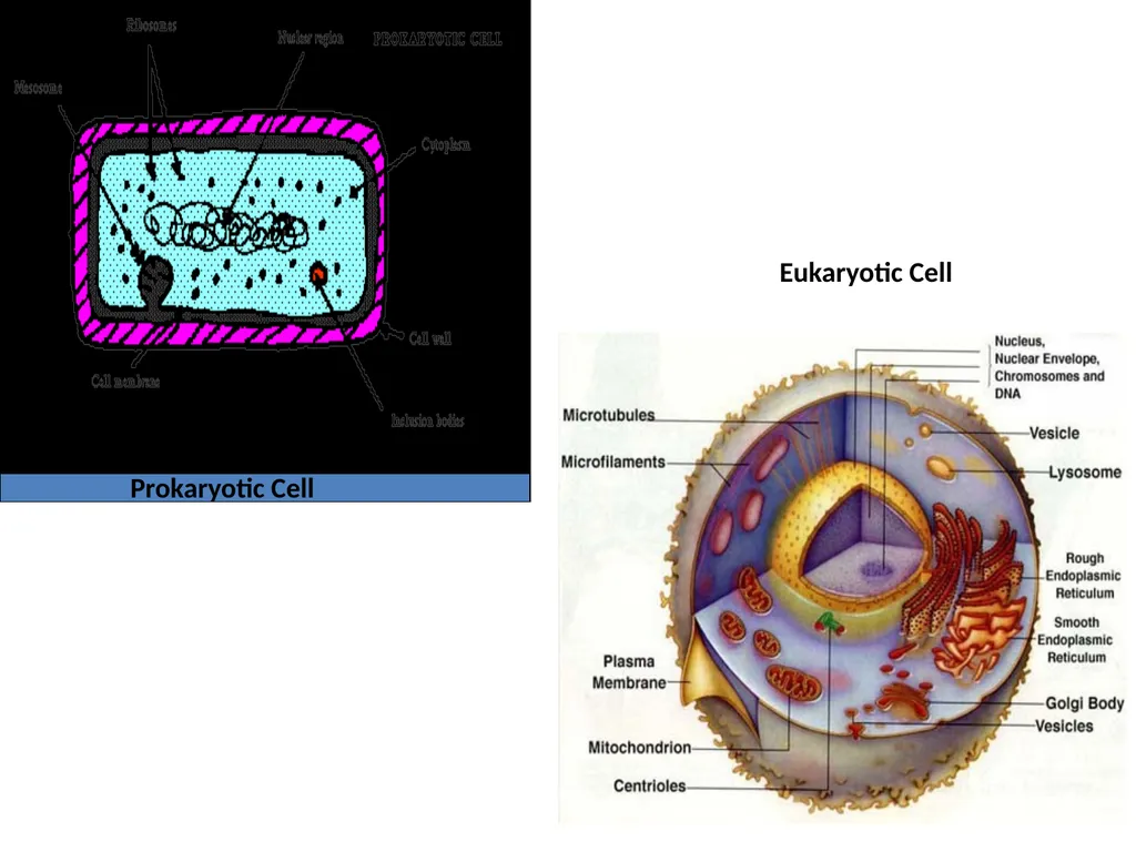
Prokaryotic Cell Eukaryotic Cell<br>
05
Evolutionary View – Prokaryotes are considered to be ancestors of eukaryotes.
Fossils 3 billion years old contained evidence of prokaryotes alone and eukaryotes probably appeared 1 million year ago.
In spite of the differences between prokaryotes and eukaryotes, there are considerable homologies in their molecular organization and function.
EG. All living organisms employ the same genetic code and a similar machinery for protein synthesis.
Prokaryote: eg. Mycoplasma, Bacteria, Cyanobacteria or Blue green algae<br>
Fossils 3 billion years old contained evidence of prokaryotes alone and eukaryotes probably appeared 1 million year ago.
In spite of the differences between prokaryotes and eukaryotes, there are considerable homologies in their molecular organization and function.
EG. All living organisms employ the same genetic code and a similar machinery for protein synthesis.
Prokaryote: eg. Mycoplasma, Bacteria, Cyanobacteria or Blue green algae<br>
06
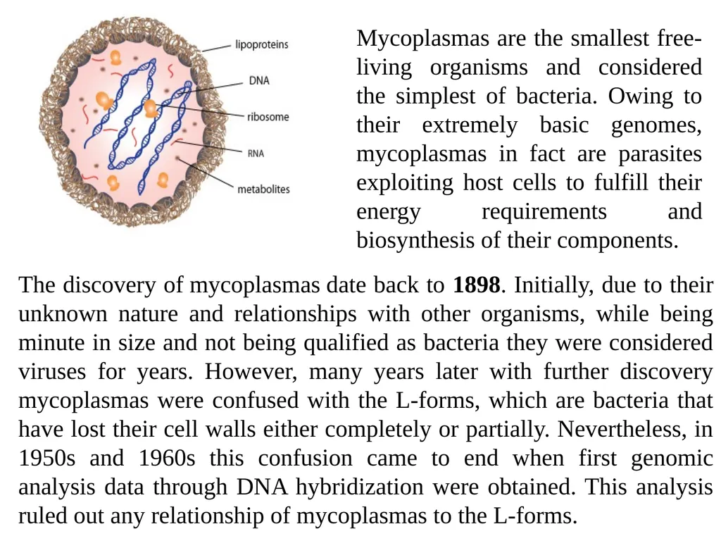
Mycoplasm Smallest mass, an older name for Mycoplasma was Pleuro pneumonia-Like Organisms (PPLO)
Infectious diseases in animals including humans
Size: 0.25 - 0.1μm in diameter
Unicellular, Free living, Saprophyte or Parasite
Shape – Spherical, bound by plasma membrane (75 Aº thick), Composed of protein and lipid. Easily cultured (Pleomorphic – many forms) Spheroid, thin branching filaments, stellate and asteroid
They differ from Bacteria in following respects:
Do not contain cell wall and mesosoms
(Like Viruses and animal cells)Resistance to antibiotics such as Penicillin
Filterable through the bacterial filters
Growth is inhibited by Tetracyclins and similar antibiotics that act on metabolic pathway.<br>
Infectious diseases in animals including humans
Size: 0.25 - 0.1μm in diameter
Unicellular, Free living, Saprophyte or Parasite
Shape – Spherical, bound by plasma membrane (75 Aº thick), Composed of protein and lipid. Easily cultured (Pleomorphic – many forms) Spheroid, thin branching filaments, stellate and asteroid
They differ from Bacteria in following respects:
Do not contain cell wall and mesosoms
(Like Viruses and animal cells)Resistance to antibiotics such as Penicillin
Filterable through the bacterial filters
Growth is inhibited by Tetracyclins and similar antibiotics that act on metabolic pathway.<br>
07
Discovered by French Scientist: E. Nocord and E. R. Roux (1898)
Pleural fluids of cattles suffering from plueropneumonia. Similar organisms were later isolated from other animals eg. Sheep, goats, dogs, rats, mice and human being and were named Pleuropneuminia like organism (PPLO)
According to Shellar and Bianchi 1987 – Between the Viruses and Bacteria
According to Albert et. al., 1989 - Simplest Bacteria
At one side of the cell occurs bleb (localized collection of fluid) of ill understood function.
PPLO contain many enzymes required for DNA replication, transcription, translation
Unlike Viruses they are free-living and do not require host cell for their replication., reproduce by binary fission & budding.<br>
Pleural fluids of cattles suffering from plueropneumonia. Similar organisms were later isolated from other animals eg. Sheep, goats, dogs, rats, mice and human being and were named Pleuropneuminia like organism (PPLO)
According to Shellar and Bianchi 1987 – Between the Viruses and Bacteria
According to Albert et. al., 1989 - Simplest Bacteria
At one side of the cell occurs bleb (localized collection of fluid) of ill understood function.
PPLO contain many enzymes required for DNA replication, transcription, translation
Unlike Viruses they are free-living and do not require host cell for their replication., reproduce by binary fission & budding.<br>
08
Currently, mycoplasmas are considered to have evolved from gram-positive, walled eubacteria by degenerative evolution, meaning their evolutionary history appears to include the loss of cell wall.
Further, in 1960s and 1970s knowledge about ultrastructure, cell membrane, genome and metabolic pathways of mycoplasmas resulted in conclusion that they are smallest and simplest self-replicating organisms.
The lack of cell wall convey some unique properties of mycoplasmas such as sensitivity to osmotic shock and detergents, resistance to penicillin, and formation of odd fried-egg shaped colonies.
Sections of mycoplasmas reveal that their cells are essentially built of three organelles, such as: the cell membrane, ribosomes, and circular double stranded DNA tightly packed molecule.
Their mode of replication is no different from that of prokaryotes dividing by binary fission.<br>
Further, in 1960s and 1970s knowledge about ultrastructure, cell membrane, genome and metabolic pathways of mycoplasmas resulted in conclusion that they are smallest and simplest self-replicating organisms.
The lack of cell wall convey some unique properties of mycoplasmas such as sensitivity to osmotic shock and detergents, resistance to penicillin, and formation of odd fried-egg shaped colonies.
Sections of mycoplasmas reveal that their cells are essentially built of three organelles, such as: the cell membrane, ribosomes, and circular double stranded DNA tightly packed molecule.
Their mode of replication is no different from that of prokaryotes dividing by binary fission.<br>
09
However for binary fission to happen, cytoplasmic division must fully synchronize with genome replication and in mycoplasmas cytoplasmic division lags behind genome replication, which ultimately results in the formation of multinucleated filaments.
For In vitro cultivation of mycoplasmas it has been discovered that they are difficult to cultivate.
The reasons for these difficulties for species such as Mycoplasma genitalium and Mycoplasma pneumoniae is that they lack all the genes involved in amino acid synthesis, making them dependent on exogenous supply of amino acids.
In order to get rid of these deficiencies mycoplasmas are grown on complex media, usually consisting of beef heart infusion, peptone, yeast extract, and serum with various supplements.<br>
For In vitro cultivation of mycoplasmas it has been discovered that they are difficult to cultivate.
The reasons for these difficulties for species such as Mycoplasma genitalium and Mycoplasma pneumoniae is that they lack all the genes involved in amino acid synthesis, making them dependent on exogenous supply of amino acids.
In order to get rid of these deficiencies mycoplasmas are grown on complex media, usually consisting of beef heart infusion, peptone, yeast extract, and serum with various supplements.<br>
10
Mycoplasmas are known to consist of just plasma membrane which makes them good models for membrane studies.
Due to this reason the availability of these membranes in pure state have enabled in their chemical, enzymatic and antigenic characterization.
The membrane mostly consists of 60% to 70% of proteins and rest 20% to 30% of lipid.
Dependence on exogenous supplies of fatty acids, and cholesterol serves as advantage to conduct further studies on these organisms.
Mycoplasma infections are commonly found in people with Lyme Disease.
Of the over 100 known species, more than a dozen are found in humans.<br>
Due to this reason the availability of these membranes in pure state have enabled in their chemical, enzymatic and antigenic characterization.
The membrane mostly consists of 60% to 70% of proteins and rest 20% to 30% of lipid.
Dependence on exogenous supplies of fatty acids, and cholesterol serves as advantage to conduct further studies on these organisms.
Mycoplasma infections are commonly found in people with Lyme Disease.
Of the over 100 known species, more than a dozen are found in humans.<br>
11
Many of them cause disease.
Mycoplasmas don’t have a cell wall or cell nucleus, act like parasites within or outside host cells, and can take on different shapes.
This versatility allows them to hide from the immune system and affect it in many ways.
Because of these features, they are hard to diagnose and treat.
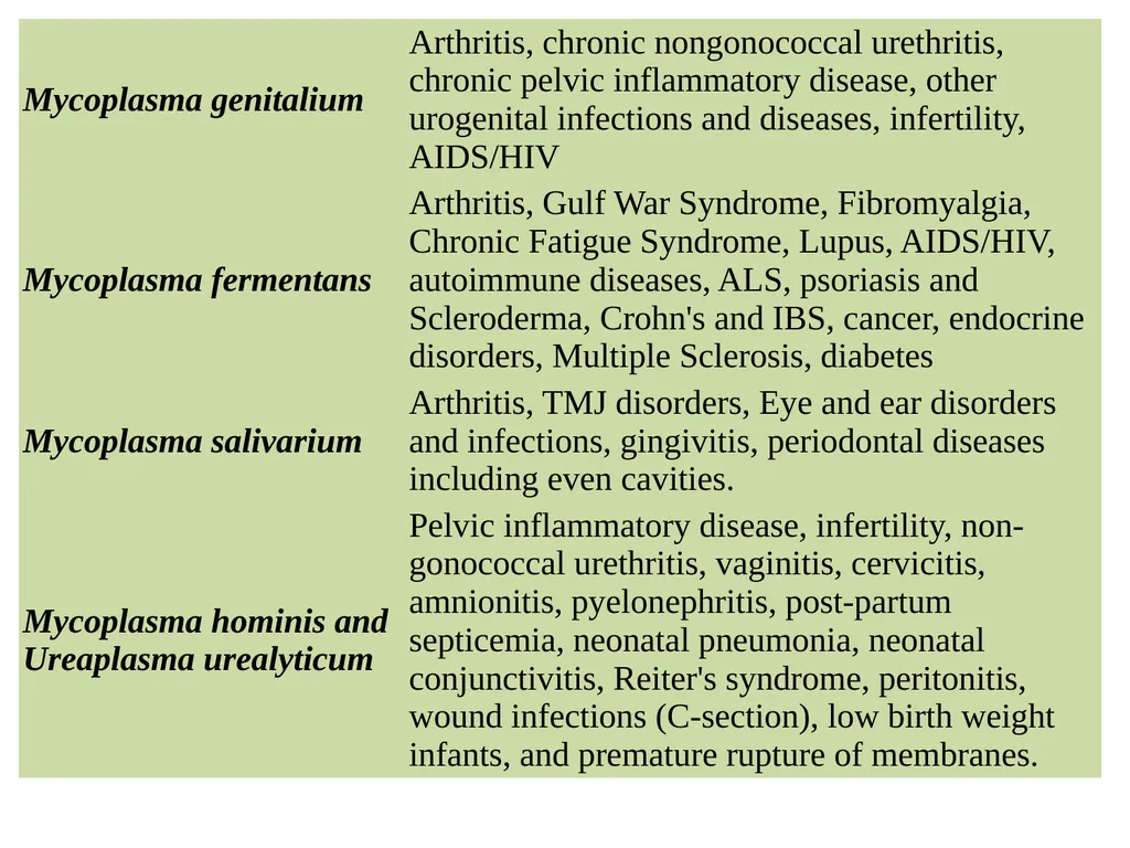
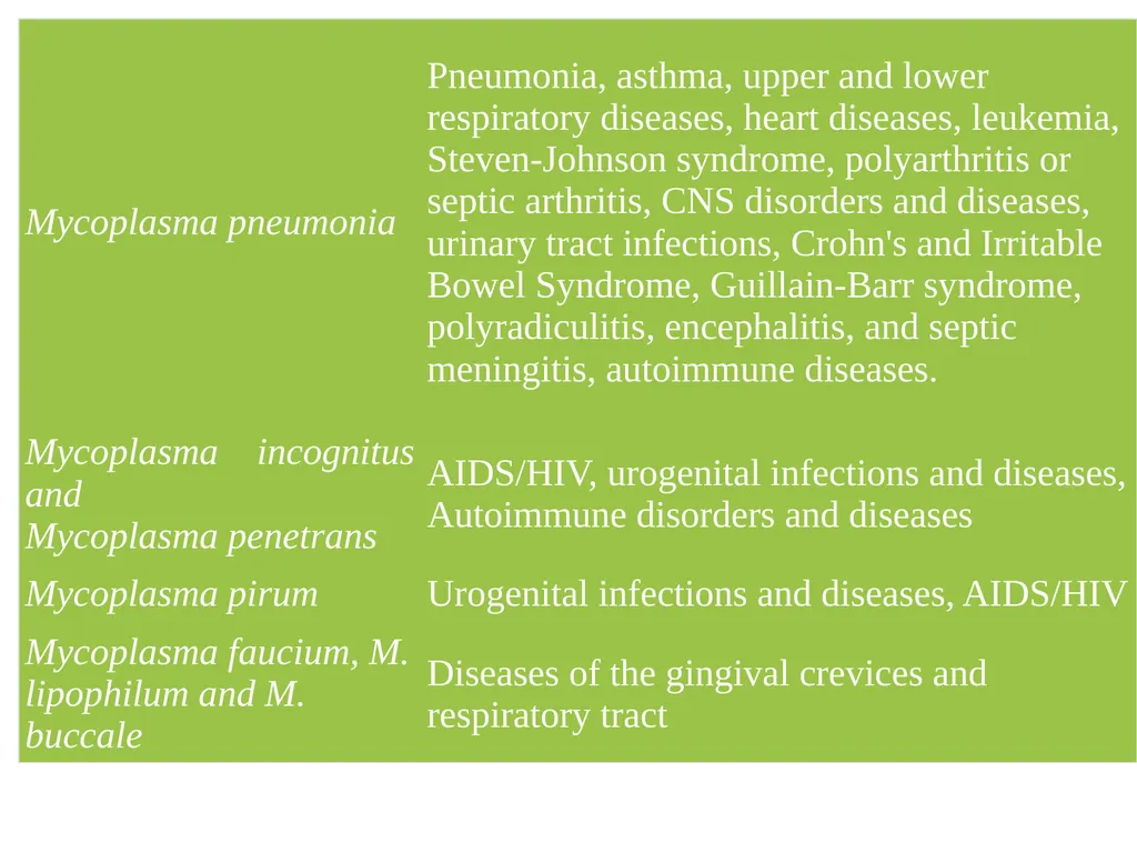
The most common species, Mycoplasma pneumoniae, typically causes respiratory infections like pneumonia, bronchitis, pharyngitis, and asthma.
But it’s a stealth pathogen that can also cause non-respiratory diseases affecting the nervous system, blood, joints, skin, heart, liver, and pancreas.
The other pathogenic species most typically found in humans are:
Mycloplasma fermentans, Mycoplasma genitalium, Mycoplasma hominis, Mycoplasma pirum, Mycoplasma salivarium, Ureaplasma urealyticum, Ureaplasma parvum<br>
Mycoplasmas don’t have a cell wall or cell nucleus, act like parasites within or outside host cells, and can take on different shapes.
This versatility allows them to hide from the immune system and affect it in many ways.
Because of these features, they are hard to diagnose and treat.
The most common species, Mycoplasma pneumoniae, typically causes respiratory infections like pneumonia, bronchitis, pharyngitis, and asthma.
But it’s a stealth pathogen that can also cause non-respiratory diseases affecting the nervous system, blood, joints, skin, heart, liver, and pancreas.
The other pathogenic species most typically found in humans are:
Mycloplasma fermentans, Mycoplasma genitalium, Mycoplasma hominis, Mycoplasma pirum, Mycoplasma salivarium, Ureaplasma urealyticum, Ureaplasma parvum<br>
12
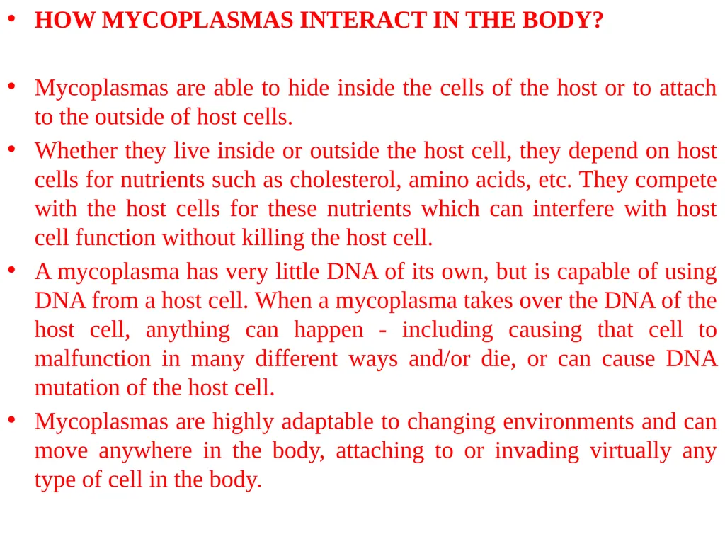
HOW MYCOPLASMAS INTERACT IN THE BODY?
Mycoplasmas are able to hide inside the cells of the host or to attach to the outside of host cells.
Whether they live inside or outside the host cell, they depend on host cells for nutrients such as cholesterol, amino acids, etc. They compete with the host cells for these nutrients which can interfere with host cell function without killing the host cell.
A mycoplasma has very little DNA of its own, but is capable of using DNA from a host cell. When a mycoplasma takes over the DNA of the host cell, anything can happen - including causing that cell to malfunction in many different ways and/or die, or can cause DNA mutation of the host cell.
Mycoplasmas are highly adaptable to changing environments and can move anywhere in the body, attaching to or invading virtually any type of cell in the body.<br>
Mycoplasmas are able to hide inside the cells of the host or to attach to the outside of host cells.
Whether they live inside or outside the host cell, they depend on host cells for nutrients such as cholesterol, amino acids, etc. They compete with the host cells for these nutrients which can interfere with host cell function without killing the host cell.
A mycoplasma has very little DNA of its own, but is capable of using DNA from a host cell. When a mycoplasma takes over the DNA of the host cell, anything can happen - including causing that cell to malfunction in many different ways and/or die, or can cause DNA mutation of the host cell.
Mycoplasmas are highly adaptable to changing environments and can move anywhere in the body, attaching to or invading virtually any type of cell in the body.<br>
13
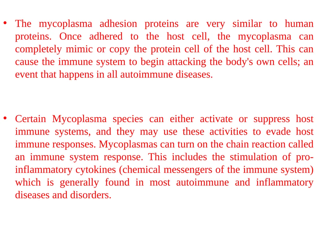
The mycoplasma adhesion proteins are very similar to human proteins. Once adhered to the host cell, the mycoplasma can completely mimic or copy the protein cell of the host cell. This can cause the immune system to begin attacking the body's own cells; an event that happens in all autoimmune diseases.
Certain Mycoplasma species can either activate or suppress host immune systems, and they may use these activities to evade host immune responses. Mycoplasmas can turn on the chain reaction called an immune system response. This includes the stimulation of pro-inflammatory cytokines (chemical messengers of the immune system) which is generally found in most autoimmune and inflammatory diseases and disorders.<br>
Certain Mycoplasma species can either activate or suppress host immune systems, and they may use these activities to evade host immune responses. Mycoplasmas can turn on the chain reaction called an immune system response. This includes the stimulation of pro-inflammatory cytokines (chemical messengers of the immune system) which is generally found in most autoimmune and inflammatory diseases and disorders.<br>
14
Mycoplasmas attach to host cells with a tiny arm coated in protein which attaches to the protein coating of host cells. For this reason, antibiotics like tetracycline, which are classified as "protein synthesis inhibitors" are often used against mycoplasma infections. While these antibiotics may block this protein attachment and very slowly starve it from the nutrients it needs from host cells to thrive and replicate, it still takes a healthy immune system to actually kill the mycoplasma for good.
Mycoplasma can also attach to or invade immune system cells, like the phagocytes (natural killer cells) that are supposed to kill them. Inside these phagocytes, they can be carried to new locations of inflammation or disease - hidden away like a spy who has infiltrated the defending army.
When a mycoplasma attaches to a host cell, it generates and releases hydrogen peroxide and superoxide radicals which cause oxidative stress and damage to the surrounding tissues.<br>
Mycoplasma can also attach to or invade immune system cells, like the phagocytes (natural killer cells) that are supposed to kill them. Inside these phagocytes, they can be carried to new locations of inflammation or disease - hidden away like a spy who has infiltrated the defending army.
When a mycoplasma attaches to a host cell, it generates and releases hydrogen peroxide and superoxide radicals which cause oxidative stress and damage to the surrounding tissues.<br>
15
Treatment Options For Mycoplasmal Infections
The negative impact of a mycoplasmal infection on the human immune system is undisputed. Due to it's ability to either activate or suppress the immune system, it is now being considered one of the culprits of many autoimmune diseases.
Yet, scientists still argue over the "chicken or egg first" type of sequence of events.
Do the mycoplasmas begin growing and replicating first and then weaken or deregulate the immune system? Or does a weakened immune system (caused by stress, poor diet or other illness) allow the mycoplasmas to take hold and begin their opportunistic growth resulting in chronic disease and to weaken and deregulate the immune system even further?
The answer is probably both, and it becomes one of the most critical treatment aspects of mycoplasmal infections. In immuno deficient patients it can be very difficult to treat these mycoplasma infections with appropriate broad spectrum antibiotics which are immunosuppressive themselves.<br>
The negative impact of a mycoplasmal infection on the human immune system is undisputed. Due to it's ability to either activate or suppress the immune system, it is now being considered one of the culprits of many autoimmune diseases.
Yet, scientists still argue over the "chicken or egg first" type of sequence of events.
Do the mycoplasmas begin growing and replicating first and then weaken or deregulate the immune system? Or does a weakened immune system (caused by stress, poor diet or other illness) allow the mycoplasmas to take hold and begin their opportunistic growth resulting in chronic disease and to weaken and deregulate the immune system even further?
The answer is probably both, and it becomes one of the most critical treatment aspects of mycoplasmal infections. In immuno deficient patients it can be very difficult to treat these mycoplasma infections with appropriate broad spectrum antibiotics which are immunosuppressive themselves.<br>
16
Although the tetracycline and erythromycin types of antibiotics are effective for some mycoplasmal infections, M. fermentans, M. hominis and M. pirum strains are usually resistant to erythromycin, and tetracycline-resistant strains of M. hominis and U. urealyticum have been reported.
However, these antibiotics have a very limited ability to directly kill these mycoplasmas, and their efficacy eventually depends on an intact host immune system to eliminate the mycoplasmas.
These types of protein inhibiting antibiotics will stop the protein adhesion of the mycoplasma to host cells but won't directly kill the mycoplasma itself.
With an already weaken immune system, many patients lack the ability to mount a strong antibody response against these deadly stealth pathogens to kill them effectively.
Regardless, many physicians and rheumatologists are treating their arthritis, CFISD, fibromyalgia and other mycoplasma infections with long term antibiotic therapy.<br>
However, these antibiotics have a very limited ability to directly kill these mycoplasmas, and their efficacy eventually depends on an intact host immune system to eliminate the mycoplasmas.
These types of protein inhibiting antibiotics will stop the protein adhesion of the mycoplasma to host cells but won't directly kill the mycoplasma itself.
With an already weaken immune system, many patients lack the ability to mount a strong antibody response against these deadly stealth pathogens to kill them effectively.
Regardless, many physicians and rheumatologists are treating their arthritis, CFISD, fibromyalgia and other mycoplasma infections with long term antibiotic therapy.<br>
17
One of the more popular conventional protocols involves rotating multiple 6 week cycles of Minocycline or Doxycycline (200-300 mg/day), Ciprofloxacin (1,500 mg/day), Azithromycin (250-500 mg/day, and/or Clarithromycin (750-1,000 mg/day) among others.
Sometimes the side effects of these strong antibiotics can be as bad as the symptoms of the diseases they are treating since a minimum of 6 months and up to two years of antibiotic therapy may be required.
Many doctors now believe that antibiotics should not be used solely or exclusively to treat mycoplasmal infections, without addressing rebuilding the immune system which is imperative for a complete recovery and eradication of infection.
Others are using more natural antibiotics found in plants which can be as effective or more effective with fewer side effects or negative impact on the body.
These include olive leaf extract products, urva ursi, and Neem leaf or seed extracts.<br>
Sometimes the side effects of these strong antibiotics can be as bad as the symptoms of the diseases they are treating since a minimum of 6 months and up to two years of antibiotic therapy may be required.
Many doctors now believe that antibiotics should not be used solely or exclusively to treat mycoplasmal infections, without addressing rebuilding the immune system which is imperative for a complete recovery and eradication of infection.
Others are using more natural antibiotics found in plants which can be as effective or more effective with fewer side effects or negative impact on the body.
These include olive leaf extract products, urva ursi, and Neem leaf or seed extracts.<br>
18
One of the main side effects of antibiotics, whether it is a natural plant antibiotic or a chemical antibiotic, is the loss of friendly bacteria that is needed in the gastrointestinal system for proper digestion and elimination.
No antibiotic can differentiate a friendly bacteria from a harmful one. Therefore, any time an antibiotic must be taken, especially long term, taking a probiotic formula to replace friendly bacteria is indicated and helpful in avoiding side effects like candida and fungi overgrowth which can cause digestive and elimination difficulties and other side effects.
Several probiotic products are widely available over-the-counter which combine these friendly bacteria - live cultures of Lactobacillus acidophilous, Lactobacillus bifidus and other bacteria with FOS (fructooligosaccharides) to promote growth in the gastrointestinal system.
It's important to take this type of supplement when taking antibiotics of any kind and best to be taken either 3-4 hours prior to, or after taking the antibiotic dosage. Full live-cultured yogurt contains acidophilous and is a good food source for these friendly bacteria.<br>
No antibiotic can differentiate a friendly bacteria from a harmful one. Therefore, any time an antibiotic must be taken, especially long term, taking a probiotic formula to replace friendly bacteria is indicated and helpful in avoiding side effects like candida and fungi overgrowth which can cause digestive and elimination difficulties and other side effects.
Several probiotic products are widely available over-the-counter which combine these friendly bacteria - live cultures of Lactobacillus acidophilous, Lactobacillus bifidus and other bacteria with FOS (fructooligosaccharides) to promote growth in the gastrointestinal system.
It's important to take this type of supplement when taking antibiotics of any kind and best to be taken either 3-4 hours prior to, or after taking the antibiotic dosage. Full live-cultured yogurt contains acidophilous and is a good food source for these friendly bacteria.<br>
19
Another common side effect when taking antibiotics is called a Herxheimer Reaction. This occurs from the organism die-off and generally is the first indication that the antibiotic therapy is working. Symptoms that are associated with a Herxheimer include: chills, fever, night sweats, muscle aches, joint pains, lymphatic pain, mental fog, and extreme fatigue. Depending on the severity of the infection and resulting die-off, these symptoms can last 1-2 weeks and sometimes longer and can vary in intensity.
Drinking at least two quarts of filtered or distilled water every day to flush the organisms from the body is helpful in reducing the length and severity of a Herxheimer reaction.
Another natural remedy to reduce Herxheimer reactions and thought to be helpful in helping the lymph glands to filter and remove dying organisms is a Whole Lemon-Olive Oil Drink. To prepare this natural remedy, place one whole unpeeled lemon (washed) in a blender with 1 cup of juice or water and 1 tablespoon of extra virgin olive oil. Blend in blender until smooth, then pour through a wire strainer. Discard pulp and drink liquid.<br>
Drinking at least two quarts of filtered or distilled water every day to flush the organisms from the body is helpful in reducing the length and severity of a Herxheimer reaction.
Another natural remedy to reduce Herxheimer reactions and thought to be helpful in helping the lymph glands to filter and remove dying organisms is a Whole Lemon-Olive Oil Drink. To prepare this natural remedy, place one whole unpeeled lemon (washed) in a blender with 1 cup of juice or water and 1 tablespoon of extra virgin olive oil. Blend in blender until smooth, then pour through a wire strainer. Discard pulp and drink liquid.<br>
20
Once the mycoplasmas are being controlled by some form of effective natural or chemical antibiotic, re-nourishing and replacing the nutrients drained from the infected host cells can help speed recovery and reduce symptoms. A general multi-vitamin supplement plus extra C, D, E, CoQ-10, beta-carotene, quercetin, folic acid, bioflavoids and biotin are necessary and helpful when recovering from a mycoplasmal infection.
Supplementing back the depleted amino acids has been reported to be helpful in some recovering from these infections. These include L-cysteine, L-tyrosine, L-glutamine, L-carnitine, and malic acid. Remember, however, that mycoplasmas thrive on arginine! Avoid L-arginine supplements and multi-amino acid formulas containing L-arginine, as well as foods rich in arginine to avoid feeding the mycoplasmas. The richest food sources of arginine (to avoid) are nuts and seeds, including the oils derived from seeds and nuts which should be eliminated or drastically reduced in the diet.<br>
Supplementing back the depleted amino acids has been reported to be helpful in some recovering from these infections. These include L-cysteine, L-tyrosine, L-glutamine, L-carnitine, and malic acid. Remember, however, that mycoplasmas thrive on arginine! Avoid L-arginine supplements and multi-amino acid formulas containing L-arginine, as well as foods rich in arginine to avoid feeding the mycoplasmas. The richest food sources of arginine (to avoid) are nuts and seeds, including the oils derived from seeds and nuts which should be eliminated or drastically reduced in the diet.<br>
21
Vitamins A, C and E, and other antioxidants found in natural plants, have also been reported to help speed recovery and to minimize the oxidative stress caused by mycoplasmas. One of the most popular antioxidants sold today are various extracts of grape seeds. Remember however, most seeds are rich in arginine, including grape seeds, and should generally be avoided.
Other helpful supplements to replenish drained nutrients from parasitic mycoplasmas are generally indicated based upon which specific cells the mycoplasma might be feeding on and which nutrients are being depleted. Specifically with fibromyalgia patients, leading research indicates that many of the hormones and enzymes produced in the neuroendocrine system and Hypothalamus-Pituitary-Adrenal Axis are depleted or malfunctioning which have the ability to cause many of the symptoms found in these patients.<br>
Other helpful supplements to replenish drained nutrients from parasitic mycoplasmas are generally indicated based upon which specific cells the mycoplasma might be feeding on and which nutrients are being depleted. Specifically with fibromyalgia patients, leading research indicates that many of the hormones and enzymes produced in the neuroendocrine system and Hypothalamus-Pituitary-Adrenal Axis are depleted or malfunctioning which have the ability to cause many of the symptoms found in these patients.<br>
22
Finally and most importantly is nutritionally supporting the immune system. There are various natural products sold today which can stimulate and support immune function. There are many natural products available in the market place today which nutritionally support immune function. Another important consideration is the elimination of drugs that might suppress immunity. Dr. Garth Nicolson, one of the world renown experts on mycoplasmas states: "We have recommended that patients be taken off antidepressants and other potentially immune-suppressing drugs. Some of these drugs are used to help alleviate certain signs and symptoms, but in our opinion they can interfere with therapy, and they should be gradually reduced or eliminated.” This of course would be indicated for many fibromyalgia and Chronic Fatigue patients who are routinely prescribed antidepressants.<br>
23
Bacteria Most primitive, simple, unicellular prokaryote
Occur almost everywhere (air, water, soil, inside other organisms)
Occur in vast number.
Either autotrophic or hetrotrophic.
High metabolic rate and Multiply at a rapid rate
Size - 1μm to 3 μm
Smallest bacteria – 0.15 to 0.3 μm (Dialister pneumosintes)
Largest bacteria – 13 to 15 μm (Spirillum voluntans)
Shape – varies – spherical or round, single or in pair (eg. Diplococci i.e. Diplococcus pneumonia)
In group of four either in cuboidal arrangement, in irregular clumps (eg. Staphylococci i.e. Staphylococcus aureus - boils)
Bead like chain (eg. Streptococci i.e. Streptococcus pyogenes – sore throat)<br>
Occur almost everywhere (air, water, soil, inside other organisms)
Occur in vast number.
Either autotrophic or hetrotrophic.
High metabolic rate and Multiply at a rapid rate
Size - 1μm to 3 μm
Smallest bacteria – 0.15 to 0.3 μm (Dialister pneumosintes)
Largest bacteria – 13 to 15 μm (Spirillum voluntans)
Shape – varies – spherical or round, single or in pair (eg. Diplococci i.e. Diplococcus pneumonia)
In group of four either in cuboidal arrangement, in irregular clumps (eg. Staphylococci i.e. Staphylococcus aureus - boils)
Bead like chain (eg. Streptococci i.e. Streptococcus pyogenes – sore throat)<br>
24
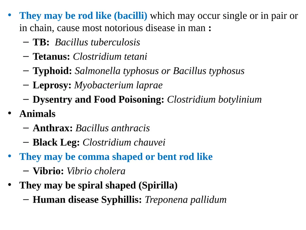
They may be rod like (bacilli) which may occur single or in pair or in chain, cause most notorious disease in man :
TB: Bacillus tuberculosis
Tetanus: Clostridium tetani
Typhoid: Salmonella typhosus or Bacillus typhosus
Leprosy: Myobacterium laprae
Dysentry and Food Poisoning: Clostridium botylinium
Animals
Anthrax: Bacillus anthracis
Black Leg: Clostridium chauvei
They may be comma shaped or bent rod like
Vibrio: Vibrio cholera
They may be spiral shaped (Spirilla)
Human disease Syphillis: Treponena pallidum<br>
TB: Bacillus tuberculosis
Tetanus: Clostridium tetani
Typhoid: Salmonella typhosus or Bacillus typhosus
Leprosy: Myobacterium laprae
Dysentry and Food Poisoning: Clostridium botylinium
Animals
Anthrax: Bacillus anthracis
Black Leg: Clostridium chauvei
They may be comma shaped or bent rod like
Vibrio: Vibrio cholera
They may be spiral shaped (Spirilla)
Human disease Syphillis: Treponena pallidum<br>
25
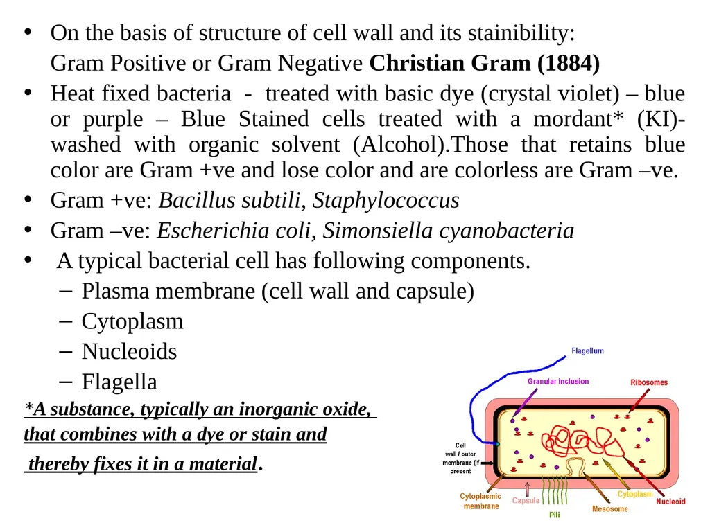
On the basis of structure of cell wall and its stainibility:
Gram Positive or Gram Negative Christian Gram (1884)
Heat fixed bacteria - treated with basic dye (crystal violet) – blue or purple – Blue Stained cells treated with a mordant* (KI)- washed with organic solvent (Alcohol).Those that retains blue color are Gram +ve and lose color and are colorless are Gram –ve.
Gram +ve: Bacillus subtili, Staphylococcus
Gram –ve: Escherichia coli, Simonsiella cyanobacteria
A typical bacterial cell has following components.
Plasma membrane (cell wall and capsule)
Cytoplasm
Nucleoids
Flagella
*A substance, typically an inorganic oxide,
that combines with a dye or stain and
thereby fixes it in a material.<br>
Gram Positive or Gram Negative Christian Gram (1884)
Heat fixed bacteria - treated with basic dye (crystal violet) – blue or purple – Blue Stained cells treated with a mordant* (KI)- washed with organic solvent (Alcohol).Those that retains blue color are Gram +ve and lose color and are colorless are Gram –ve.
Gram +ve: Bacillus subtili, Staphylococcus
Gram –ve: Escherichia coli, Simonsiella cyanobacteria
A typical bacterial cell has following components.
Plasma membrane (cell wall and capsule)
Cytoplasm
Nucleoids
Flagella
*A substance, typically an inorganic oxide,
that combines with a dye or stain and
thereby fixes it in a material.<br>
26
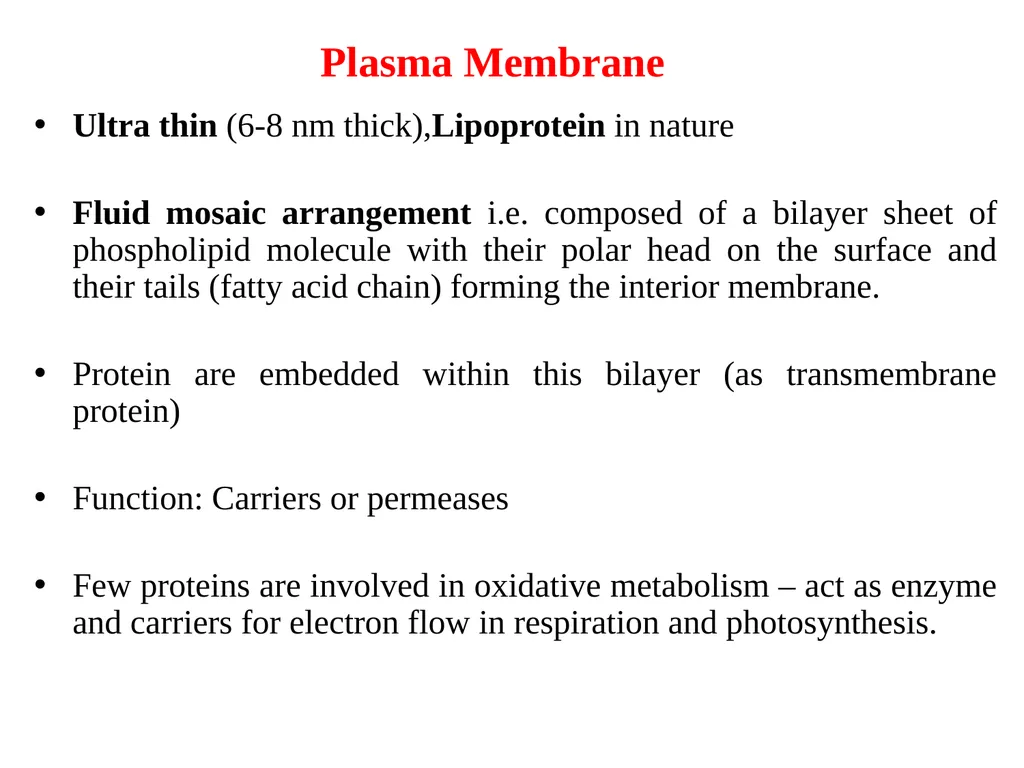
Plasma Membrane Ultra thin (6-8 nm thick),Lipoprotein in nature
Fluid mosaic arrangement i.e. composed of a bilayer sheet of phospholipid molecule with their polar head on the surface and their tails (fatty acid chain) forming the interior membrane.
Protein are embedded within this bilayer (as transmembrane protein)
Function: Carriers or permeases
Few proteins are involved in oxidative metabolism – act as enzyme and carriers for electron flow in respiration and photosynthesis.<br>
Fluid mosaic arrangement i.e. composed of a bilayer sheet of phospholipid molecule with their polar head on the surface and their tails (fatty acid chain) forming the interior membrane.
Protein are embedded within this bilayer (as transmembrane protein)
Function: Carriers or permeases
Few proteins are involved in oxidative metabolism – act as enzyme and carriers for electron flow in respiration and photosynthesis.<br>
27
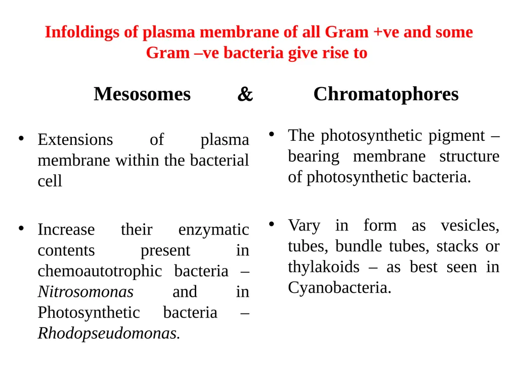
Infoldings of plasma membrane of all Gram +ve and some Gram –ve bacteria give rise to Extensions of plasma membrane within the bacterial cell
Increase their enzymatic contents present in chemoautotrophic bacteria – Nitrosomonas and in Photosynthetic bacteria – Rhodopseudomonas. The photosynthetic pigment – bearing membrane structure of photosynthetic bacteria.
Vary in form as vesicles, tubes, bundle tubes, stacks or thylakoids – as best seen in Cyanobacteria. Chromatophores Mesosomes <br>
Increase their enzymatic contents present in chemoautotrophic bacteria – Nitrosomonas and in Photosynthetic bacteria – Rhodopseudomonas. The photosynthetic pigment – bearing membrane structure of photosynthetic bacteria.
Vary in form as vesicles, tubes, bundle tubes, stacks or thylakoids – as best seen in Cyanobacteria. Chromatophores Mesosomes <br>
28
Cell Wall Plasma membrane is covered with a strong and rigid cell wall that renders mechanical protection and provides characteristic shape.
Absent in mycoplasm
Made up of: lipids, proteins, polysaccharides and chitin but rarely cellulose (Thus different from plant cell wall)
Cellwall of Gram –ve bacteria: Two layers, comprises of proteoglycan or peptidoglycan (muramic acid) containing periplasmic space around the plasma membrane
Cellwall of Gram +ve bacteria: Thicker, amorphous, homogenous and single layered and it contains techoic acid and techuronic acid.<br>
Absent in mycoplasm
Made up of: lipids, proteins, polysaccharides and chitin but rarely cellulose (Thus different from plant cell wall)
Cellwall of Gram –ve bacteria: Two layers, comprises of proteoglycan or peptidoglycan (muramic acid) containing periplasmic space around the plasma membrane
Cellwall of Gram +ve bacteria: Thicker, amorphous, homogenous and single layered and it contains techoic acid and techuronic acid.<br>
29
Capsule In some bacteria, cell wall is surrounded by a additional slime or gel layer called Capsule.
Thick, gummy, mucilaginous and secreted by plasma membrane.
It serves mainly as a protective layer against attack by phagocytes and viruses.
Helps in regulating concentration and uptake of essential ions and water.<br>
Thick, gummy, mucilaginous and secreted by plasma membrane.
It serves mainly as a protective layer against attack by phagocytes and viruses.
Helps in regulating concentration and uptake of essential ions and water.<br>
30
Cytoplasm Plasma membrane encloses a space consisting of hyaloplasm, matrix or cytosol.
Forms the ground substance and is the sheet of all metabolic activities.
Consists of water, protein, lipids, CHO, different types of RNA and various smaller molecules.
Differentiated into two distinct areas: Less electron dense and very dense area.
Reserve material of bacteria are stored in cytoplasm either as finely dispersed or distinct granules.
Storage granules:
Organic polymer (form of poly-β-hydroxybutyricacid)
Inorganic phosphate (form of granules of volutin)
Sulphur (form by oxidation from hydrogen sulphide)<br>
Forms the ground substance and is the sheet of all metabolic activities.
Consists of water, protein, lipids, CHO, different types of RNA and various smaller molecules.
Differentiated into two distinct areas: Less electron dense and very dense area.
Reserve material of bacteria are stored in cytoplasm either as finely dispersed or distinct granules.
Storage granules:
Organic polymer (form of poly-β-hydroxybutyricacid)
Inorganic phosphate (form of granules of volutin)
Sulphur (form by oxidation from hydrogen sulphide)<br>
31
Nucleoids Nuclear material – in form of single, circular, double stranded DNA molecule in a specific clear region of the cytoplasm called Nucleoid.
It has no ribosome and nucleolus.
Bacterial chromosomes do not contain histone protein however chromosomes of some species are found to contain small quantity of heat stable proteins (hsp) – analogous to eukaryotic histones.<br>
It has no ribosome and nucleolus.
Bacterial chromosomes do not contain histone protein however chromosomes of some species are found to contain small quantity of heat stable proteins (hsp) – analogous to eukaryotic histones.<br>
32
Many species of bacteria – have extra chromosomal genetic material in the form of small, circular and closed DNA molecule called Plasmids.
Functions:
Production of antibiotically active protiens or colcins which inhibit the growth of other strains of bacteria
Acts as sex or fertility factor (F factor) – bacterial conjugation
R Factor – genes for the resistance to drugs such as chloramphenicol, neomycin, penicilin, streptomycin, sulphonamides and tetracyclines.<br>
Functions:
Production of antibiotically active protiens or colcins which inhibit the growth of other strains of bacteria
Acts as sex or fertility factor (F factor) – bacterial conjugation
R Factor – genes for the resistance to drugs such as chloramphenicol, neomycin, penicilin, streptomycin, sulphonamides and tetracyclines.<br>
33
Flagella and other structures Cellular locomotion.
Simple in organisation (protein flagellin).
Gram -ve - non flagellar appendages – Fimbriae or Pilli, non-motile adhesive structure – mold, plants, animal cells.
Are known to be coded by the genes of the plasmid.
Gram +ve bacteria – tubular, pericellular and rigid appendages – protein spinin.
Helps the bacterial cells to tolerate environmental conditions (salinity, pH, temp., etc.)<br>
Simple in organisation (protein flagellin).
Gram -ve - non flagellar appendages – Fimbriae or Pilli, non-motile adhesive structure – mold, plants, animal cells.
Are known to be coded by the genes of the plasmid.
Gram +ve bacteria – tubular, pericellular and rigid appendages – protein spinin.
Helps the bacterial cells to tolerate environmental conditions (salinity, pH, temp., etc.)<br>
34
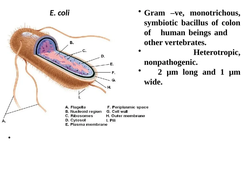
E. coli Gram –ve, monotrichous, symbiotic bacillus of colon of human beings and other vertebrates.
Heterotropic, nonpathogenic.
2 μm long and 1 μm wide.<br>
Heterotropic, nonpathogenic.
2 μm long and 1 μm wide.<br>
35
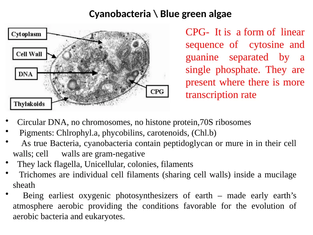
Circular DNA, no chromosomes, no histone protein,70S ribosomes
Pigments: Chlrophyl.a, phycobilins, carotenoids, (Chl.b)
As true Bacteria, cyanobacteria contain peptidoglycan or mure in in their cell walls; cell walls are gram-negative
They lack flagella, Unicellular, colonies, filaments
Trichomes are individual cell filaments (sharing cell walls) inside a mucilage sheath
Being earliest oxygenic photosynthesizers of earth – made early earth’s atmosphere aerobic providing the conditions favorable for the evolution of aerobic bacteria and eukaryotes. Cyanobacteria \ Blue green algae CPG- It is a form of linear sequence of cytosine and guanine separated by a single phosphate. They are present where there is more transcription rate<br>
Pigments: Chlrophyl.a, phycobilins, carotenoids, (Chl.b)
As true Bacteria, cyanobacteria contain peptidoglycan or mure in in their cell walls; cell walls are gram-negative
They lack flagella, Unicellular, colonies, filaments
Trichomes are individual cell filaments (sharing cell walls) inside a mucilage sheath
Being earliest oxygenic photosynthesizers of earth – made early earth’s atmosphere aerobic providing the conditions favorable for the evolution of aerobic bacteria and eukaryotes. Cyanobacteria \ Blue green algae CPG- It is a form of linear sequence of cytosine and guanine separated by a single phosphate. They are present where there is more transcription rate<br>
36
VIRUSES Occupy a unique space between the living and nonliving worlds – they are made of the same molecules as living cells; On the other hand they are incapable of independent existence, being completely dependent on a host cell to reproduce.
Almost all living organisms have viruses that infect them.
Human viruses include polio, Influenza, herpes, rabies, Ebola, smallpox, , and the AIDS (acquired immunodeficiency syndrome) virus HIV (human immunodeficiency virus).
Viruses are submicroscopic particles consisting of a core of genetic material enclosed within a protein coat called the capsid.
Viruses are metabolically inert until they enter a host cell, whereupon the viral genetic material directs the host cell machinery to produce viral protein and viral genetic material.<br>
Almost all living organisms have viruses that infect them.
Human viruses include polio, Influenza, herpes, rabies, Ebola, smallpox, , and the AIDS (acquired immunodeficiency syndrome) virus HIV (human immunodeficiency virus).
Viruses are submicroscopic particles consisting of a core of genetic material enclosed within a protein coat called the capsid.
Viruses are metabolically inert until they enter a host cell, whereupon the viral genetic material directs the host cell machinery to produce viral protein and viral genetic material.<br>
37
Viruses often insert their genome into that of the host, an ability that is widely made use of in molecular genetics .
Bacterial viruses, called bacteriophages are used by scientists to transfer genes between bacterial strains.
Human viruses are used as vehicles for gene therapy.
By exploiting the natural infection cycle of a virus such as adenovirus, it is possible to introduce a functional copy of a human gene into a patient suffering from a genetic disease such as cystic fibrosis.
The study of viruses is known as virology, a sub-speciality of microbiology.
Viruses rely on the host cell to make more virus.<br>
Bacterial viruses, called bacteriophages are used by scientists to transfer genes between bacterial strains.
Human viruses are used as vehicles for gene therapy.
By exploiting the natural infection cycle of a virus such as adenovirus, it is possible to introduce a functional copy of a human gene into a patient suffering from a genetic disease such as cystic fibrosis.
The study of viruses is known as virology, a sub-speciality of microbiology.
Viruses rely on the host cell to make more virus.<br>
38
Once viruses have entered cells, the cells’ machinery is used to copy the viral genome.
Depending on the virus type, the genome may be single- or double-stranded DNA, or even RNA.
A viral genome is packaged within a protective protein coat.
Viruses that infect bacteria are called bacteriophages.
One of these, called lambda, has a fixed-size DNA molecule of 4.5 × 10⁴ base pairs.
In contrast, the bacteriophage M13 can change its chromosome size, its protein coat expanding in parallel to accommodate the chromosome.
This makes M13 useful in genetic engineering.
Size: Range between 30 – 300 nm.<br>
Depending on the virus type, the genome may be single- or double-stranded DNA, or even RNA.
A viral genome is packaged within a protective protein coat.
Viruses that infect bacteria are called bacteriophages.
One of these, called lambda, has a fixed-size DNA molecule of 4.5 × 10⁴ base pairs.
In contrast, the bacteriophage M13 can change its chromosome size, its protein coat expanding in parallel to accommodate the chromosome.
This makes M13 useful in genetic engineering.
Size: Range between 30 – 300 nm.<br>
39
An infectious virus particle is known as Virion.
Virions are virus particles: they are the Inert carriers of the genome, and are Assembled inside cells, from virus-specified components: they do not grow, and do not form by division.
Basically, all virions have: genomic nucleic acid: this may be RNA or DNA, ss or ds, and may be all or part of the genome protein associated with the genome: this may be in the form of a helically-assembling or isometrically-assembling , Simple nucleocapsid only, or more complex structures with two or more layers of protein, with nucleoprotein .
If the virions are simple Nucleoproteins - that is, contain only nucleic acid and protein - then they are usually composed only of virus-specified components.
However, certain host components may be "trapped" within virions, such as polyamines: these are polycationic compounds which serve to neutralise charge on the viral nucleic acid as it is packed into the Capsid, or protein coat.<br>
Virions are virus particles: they are the Inert carriers of the genome, and are Assembled inside cells, from virus-specified components: they do not grow, and do not form by division.
Basically, all virions have: genomic nucleic acid: this may be RNA or DNA, ss or ds, and may be all or part of the genome protein associated with the genome: this may be in the form of a helically-assembling or isometrically-assembling , Simple nucleocapsid only, or more complex structures with two or more layers of protein, with nucleoprotein .
If the virions are simple Nucleoproteins - that is, contain only nucleic acid and protein - then they are usually composed only of virus-specified components.
However, certain host components may be "trapped" within virions, such as polyamines: these are polycationic compounds which serve to neutralise charge on the viral nucleic acid as it is packed into the Capsid, or protein coat.<br>
40
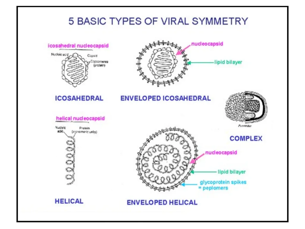
Structure of Virus Virus particles consist of two or three parts: the genetic material made from either DNA or RNA, long molecules that carry genetic information; a protein coat (capsid) that protects these genes; and in some cases an envelope of lipids that surrounds the protein coat when they are outside a cell.
The capsid is made from proteins encoded by the viral genome and its shape serves as the basis for morphological distinction.
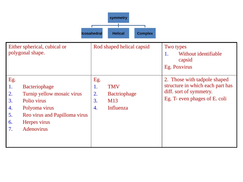
The shapes of viruses range from simple helical and icosahedral forms to more complex structures.
The elements of isosahedral symmetry involves 6- five fold rotation axes, 10- three fold rotation axes and 15- two fold rotion axes.
The capsid and entire virus structure can be mechanically (physically) probed through atomic force microscopy.<br>
The capsid is made from proteins encoded by the viral genome and its shape serves as the basis for morphological distinction.
The shapes of viruses range from simple helical and icosahedral forms to more complex structures.
The elements of isosahedral symmetry involves 6- five fold rotation axes, 10- three fold rotation axes and 15- two fold rotion axes.
The capsid and entire virus structure can be mechanically (physically) probed through atomic force microscopy.<br>
41
Atomic force microscopy (AFM) or scanning force microscopy (SFM) is a very high-resolution type of scanning probe microscopy, with demonstrates resolution on the order of fractions of a nanometer
Binnig, Quate and Gerber invented the first atomic force microscope (also abbreviated as AFM) in 1986.
The first commercially available atomic force microscope was introduced in 1989.
The AFM is one of the foremost tools for imaging, measuring, and manipulating matter at the nanoscale.<br>
Binnig, Quate and Gerber invented the first atomic force microscope (also abbreviated as AFM) in 1986.
The first commercially available atomic force microscope was introduced in 1989.
The AFM is one of the foremost tools for imaging, measuring, and manipulating matter at the nanoscale.<br>
42
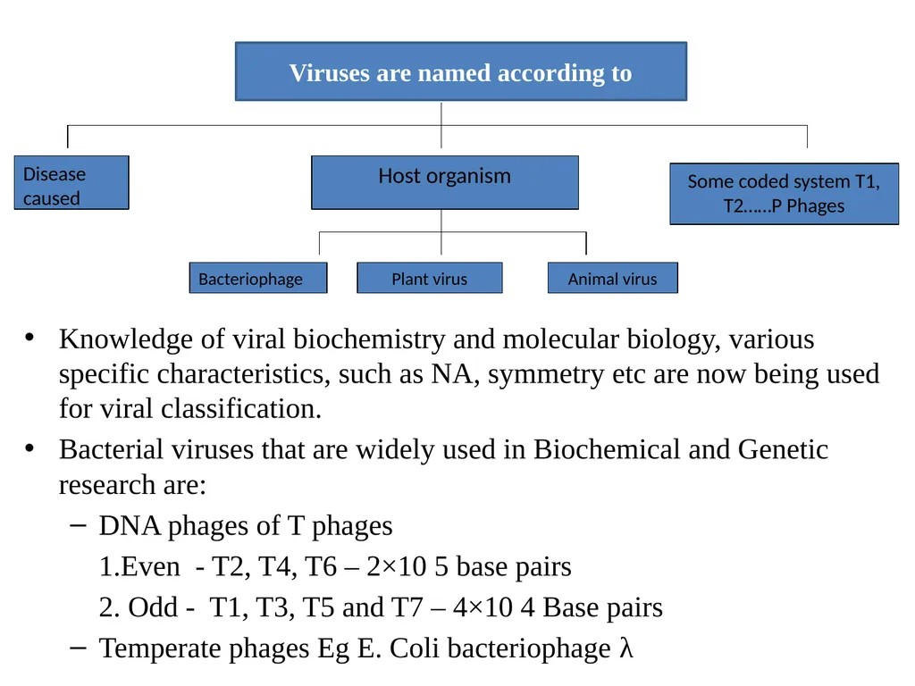
Knowledge of viral biochemistry and molecular biology, various specific characteristics, such as NA, symmetry etc are now being used for viral classification.
Bacterial viruses that are widely used in Biochemical and Genetic research are:
DNA phages of T phages
1.Even - T2, T4, T6 – 2×10 5 base pairs
2. Odd - T1, T3, T5 and T7 – 4×10 4 Base pairs
Temperate phages Eg E. Coli bacteriophage λ<br>
Bacterial viruses that are widely used in Biochemical and Genetic research are:
DNA phages of T phages
1.Even - T2, T4, T6 – 2×10 5 base pairs
2. Odd - T1, T3, T5 and T7 – 4×10 4 Base pairs
Temperate phages Eg E. Coli bacteriophage λ<br>
43
Small DNA phages Eg. Ø×174 & rod like M13
1st organism in which DNA sequence of the genome was determined permitting extensive understanding of viral life cycle.
Simple, do not encode most of the protein required for replication of their DNA but depend on cellular proteins for this purpose.
Useful in identifying and analyzing the cellular proteins involved in DNA replication.
RNA phages:
Contain RNA instead of DNA, Smallest known viruses
Easy to grow and in large number
Because their RNA genomes also serve as their m-RNA
1st long m-RNA molecule to be sequenced was the genome of an RNA phage.<br>
1st organism in which DNA sequence of the genome was determined permitting extensive understanding of viral life cycle.
Simple, do not encode most of the protein required for replication of their DNA but depend on cellular proteins for this purpose.
Useful in identifying and analyzing the cellular proteins involved in DNA replication.
RNA phages:
Contain RNA instead of DNA, Smallest known viruses
Easy to grow and in large number
Because their RNA genomes also serve as their m-RNA
1st long m-RNA molecule to be sequenced was the genome of an RNA phage.<br>
44
Smallest known virus, encodes 4 protein i.e
An RNA polymerase for replication of viral RNA
TWO capsid protein
An enzyme that dissolved the bacterial cell wall and allows the release of intracellular particles into the medium.<br>
An RNA polymerase for replication of viral RNA
TWO capsid protein
An enzyme that dissolved the bacterial cell wall and allows the release of intracellular particles into the medium.<br>
45
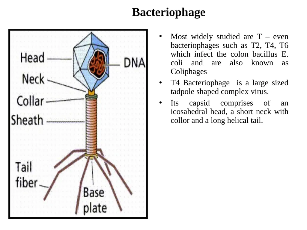
Bacteriophage Most widely studied are T – even bacteriophages such as T2, T4, T6 which infect the colon bacillus E. coli and are also known as Coliphages
T4 Bacteriophage is a large sized tadpole shaped complex virus.
Its capsid comprises of an icosahedral head, a short neck with collor and a long helical tail.<br>
T4 Bacteriophage is a large sized tadpole shaped complex virus.
Its capsid comprises of an icosahedral head, a short neck with collor and a long helical tail.<br>
46
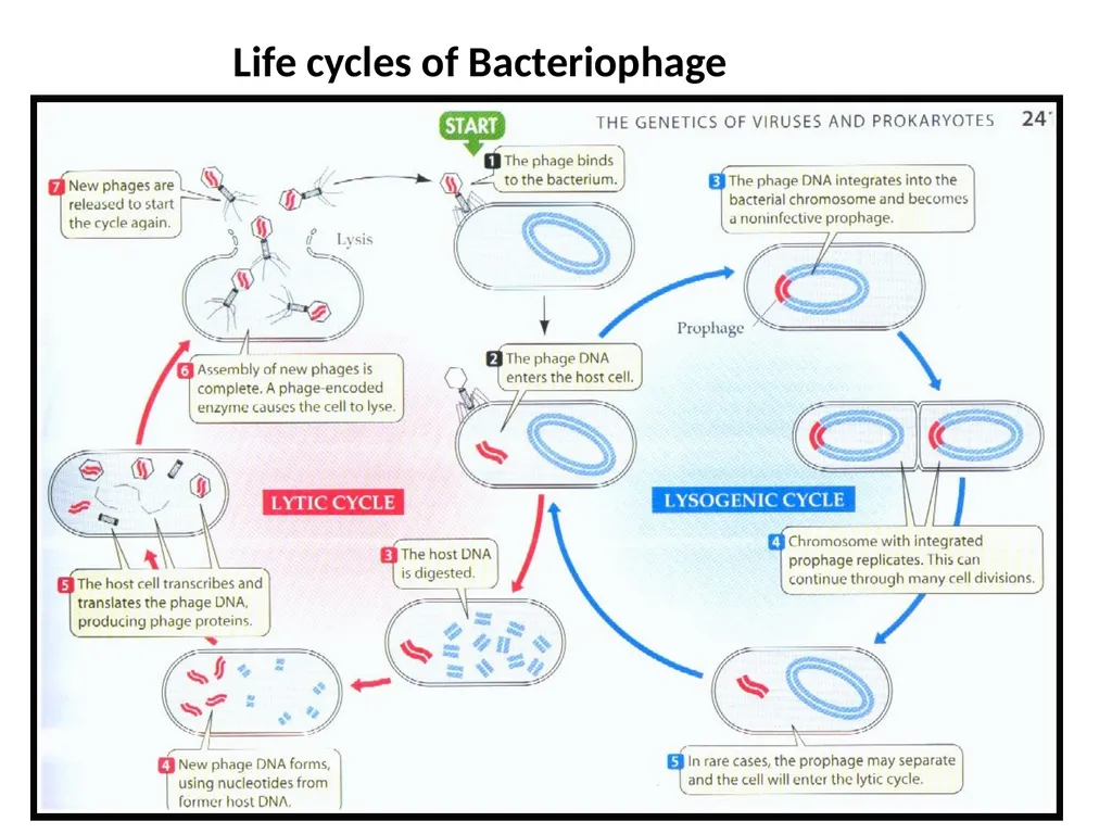
Life cycle is classified as:
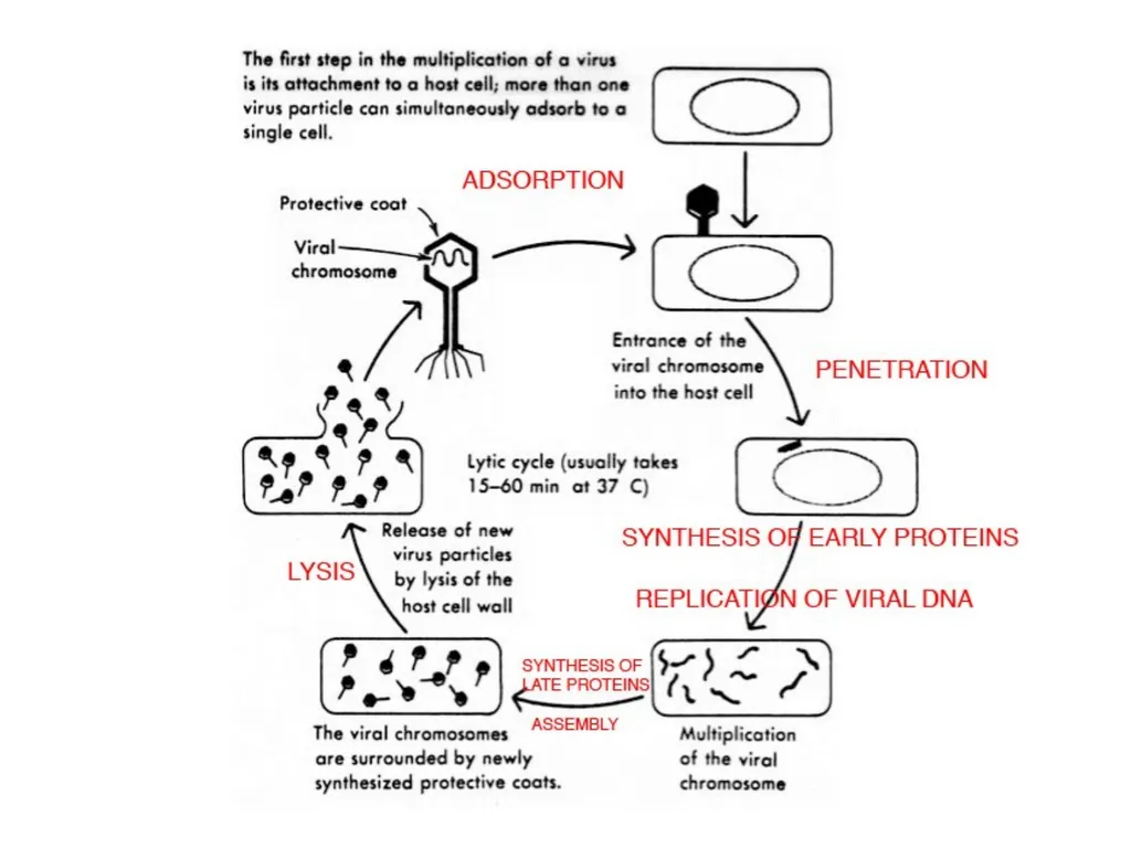
Lytic cycles: which involves four different events – Adsorption, Penetration, Replication, Release. Eg. T4 - Bacteriophage
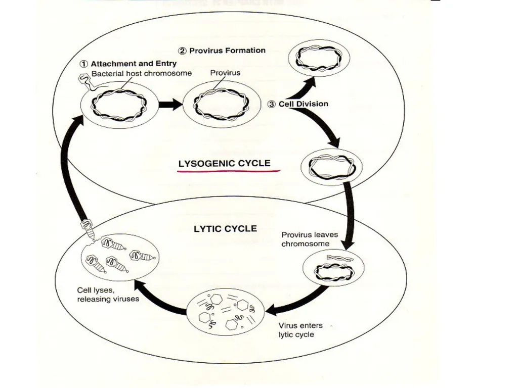
2. Lysogenic cycles: Characterized by delayed lysis after phage infection.Such viruses are called temperate viruses and infected host cell is said to be Lysogenic because dormant virus may at any time become active and begin directing the synthesis of new virus particles.<br>
Lytic cycles: which involves four different events – Adsorption, Penetration, Replication, Release. Eg. T4 - Bacteriophage
2. Lysogenic cycles: Characterized by delayed lysis after phage infection.Such viruses are called temperate viruses and infected host cell is said to be Lysogenic because dormant virus may at any time become active and begin directing the synthesis of new virus particles.<br>
47
Life cycles of Bacteriophage<br>
48
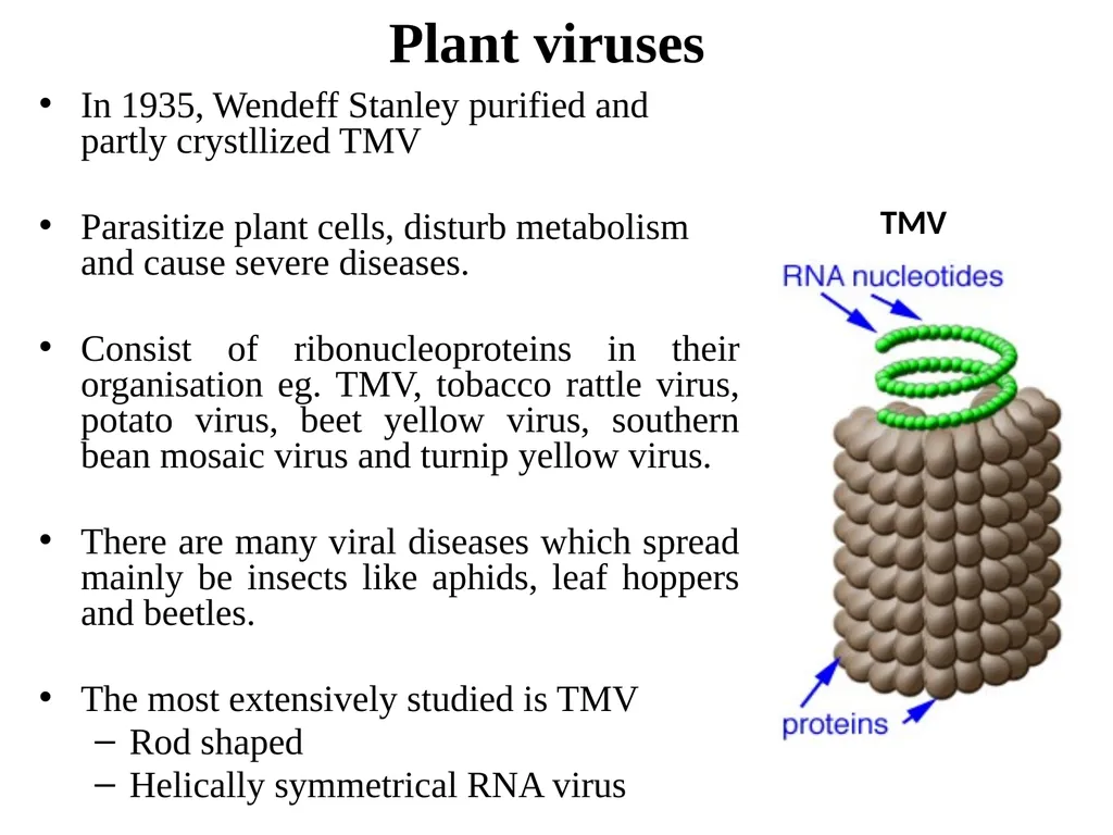
Plant viruses In 1935, Wendeff Stanley purified and partly crystllized TMV
Parasitize plant cells, disturb metabolism and cause severe diseases.
Consist of ribonucleoproteins in their organisation eg. TMV, tobacco rattle virus, potato virus, beet yellow virus, southern bean mosaic virus and turnip yellow virus.
There are many viral diseases which spread mainly be insects like aphids, leaf hoppers and beetles.
The most extensively studied is TMV
Rod shaped
Helically symmetrical RNA virus TMV<br>
Parasitize plant cells, disturb metabolism and cause severe diseases.
Consist of ribonucleoproteins in their organisation eg. TMV, tobacco rattle virus, potato virus, beet yellow virus, southern bean mosaic virus and turnip yellow virus.
There are many viral diseases which spread mainly be insects like aphids, leaf hoppers and beetles.
The most extensively studied is TMV
Rod shaped
Helically symmetrical RNA virus TMV<br>
49
Vast majority of plant viruses have a ss linear +RNA genome with a capsid having helical or icosahedral symmetry.
They have small genomes and 3-4 proteins.
1. Helicase – important in unwinding and seperation of the +ve and –ve RNA strands.
2. RNA replicase – An RNA dependent RNA polymerase is encoded in those viruses that are able to use host’s enzyme.
3. Cell to cell movement protein – Facilitates the spread of the viral RNA through plant tissue.
4. Capsomere – it is a protein subunit of the capsid.
Many plant virus depend on insect vectors to infect plant cells.
TMV is only dependent on mechanical damage to cell walls, which allows the virus to bind to the pl. memb. of the host cell.<br>
They have small genomes and 3-4 proteins.
1. Helicase – important in unwinding and seperation of the +ve and –ve RNA strands.
2. RNA replicase – An RNA dependent RNA polymerase is encoded in those viruses that are able to use host’s enzyme.
3. Cell to cell movement protein – Facilitates the spread of the viral RNA through plant tissue.
4. Capsomere – it is a protein subunit of the capsid.
Many plant virus depend on insect vectors to infect plant cells.
TMV is only dependent on mechanical damage to cell walls, which allows the virus to bind to the pl. memb. of the host cell.<br>
50
In 1956, A. Gierer & G. Schramm (German) and H.Fraenkel-Conrat and R.Williams (US) first demonstrated:
Pure RNA from TMV could direct the production of infectious TMV
Separate preparation of TMV protein and NA could reassemble infectious virions.
Protein from one TMV was mixed with RNA from another TMV strain, the reassembled hybrid virion also were infected.
Analysis of cells infected with such hybrid virions revealed that the source of the RNA determined which of the two viral strains was produced, further confirming RNA as the viral genetic material<br>
Pure RNA from TMV could direct the production of infectious TMV
Separate preparation of TMV protein and NA could reassemble infectious virions.
Protein from one TMV was mixed with RNA from another TMV strain, the reassembled hybrid virion also were infected.
Analysis of cells infected with such hybrid virions revealed that the source of the RNA determined which of the two viral strains was produced, further confirming RNA as the viral genetic material<br>
51
Animal Viruses Infects animal cells causing diseases in animals including man.
Shape – polyhedron or spherical
Genetic material – in the form of DNA or RNA.
RNA – Poliomyelits virus
DNA – Herpes virus
Have two types of life cycles of growth:
Permissive growth: Permits an animal virus to multiply lytically and kill the host cell.
Non-permissive growth: Permits an animal virus to enter the host cell but does not allow it to multiply lytically. The viral chromosome either become integrated into genome of the host cell, where it is replicated along with the host chromosome or it forms a Plasmid.
Plasmid – an extrachromosomal circular DNA molecule that replicates in a controlled fashion without killing.
Eg. Influenza virus
Semlike Forest virus
Retroviruses – (AIDS virus)<br>
Shape – polyhedron or spherical
Genetic material – in the form of DNA or RNA.
RNA – Poliomyelits virus
DNA – Herpes virus
Have two types of life cycles of growth:
Permissive growth: Permits an animal virus to multiply lytically and kill the host cell.
Non-permissive growth: Permits an animal virus to enter the host cell but does not allow it to multiply lytically. The viral chromosome either become integrated into genome of the host cell, where it is replicated along with the host chromosome or it forms a Plasmid.
Plasmid – an extrachromosomal circular DNA molecule that replicates in a controlled fashion without killing.
Eg. Influenza virus
Semlike Forest virus
Retroviruses – (AIDS virus)<br>
52
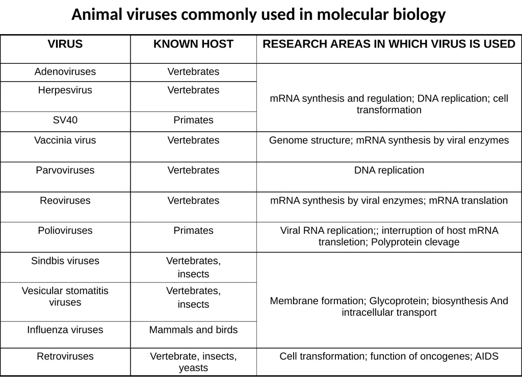
Animal viruses commonly used in molecular biology<br>
53
Viriods Term ‘Viriod’ was 1st coined by Diener (1971) who discovered the 1st viriod called Potato Spindle Tuber Viriod (PSTV)
They are small RNA circles,made up of only 300 – 400 nucleotides
Long and lacking AUG codon.,replicate autonomously despite they do not code for any proteins.
Naked RNA viruses – no protein coat or capsid.
Pass from plant to plant only when the surfaces of both recipient and donor are in contact.
They infect Eukaryotes but not Prokaryotes.
They form an infectious class of subviral pathogens which cause infections and diseases in many plants and animals.<br>
They are small RNA circles,made up of only 300 – 400 nucleotides
Long and lacking AUG codon.,replicate autonomously despite they do not code for any proteins.
Naked RNA viruses – no protein coat or capsid.
Pass from plant to plant only when the surfaces of both recipient and donor are in contact.
They infect Eukaryotes but not Prokaryotes.
They form an infectious class of subviral pathogens which cause infections and diseases in many plants and animals.<br>
54
Prions Proteinaceous infective particles.
Rod shaped
Cause number of diseases in animals
Scrapie disease of CNS in sheep and goats
Kuru in humans
Mad cow in cattle
Can survive heat, ionizing, UV radiations and chemical treatment.
Viruses, viriods and prions – Agents that invade cells, subverting normal cellular functions and often killing their unwilling hosts.<br>
Rod shaped
Cause number of diseases in animals
Scrapie disease of CNS in sheep and goats
Kuru in humans
Mad cow in cattle
Can survive heat, ionizing, UV radiations and chemical treatment.
Viruses, viriods and prions – Agents that invade cells, subverting normal cellular functions and often killing their unwilling hosts.<br>
55
Kuru is an incurable degenerative neurological disorder (brain disease) that is a type of transmissible spongiform encephalopathy found in humans.
Though the theory is not universally accepted, some researchers have posited that kuru was transmitted among members of the fore tribe of Papua New Guinea via cannibalism.
Simon Mead of University college London, and others, showed in their genetic and clinical assessment that people who survived the epidemic in Papua New Guinea were carriers of a prion resistant factor.
Mead's group has shown the source of immunity to be the inheritance of a genetic variant of prion protein G127V.
This work remains breaking news as of November 22, 2009.<br>
Though the theory is not universally accepted, some researchers have posited that kuru was transmitted among members of the fore tribe of Papua New Guinea via cannibalism.
Simon Mead of University college London, and others, showed in their genetic and clinical assessment that people who survived the epidemic in Papua New Guinea were carriers of a prion resistant factor.
Mead's group has shown the source of immunity to be the inheritance of a genetic variant of prion protein G127V.
This work remains breaking news as of November 22, 2009.<br>
56
Mad Cow Disease (MCD) is Bovine Spongiform Encephalopathy .
The disease is caused by prions.
Prions can cross between species (although not all species get diseases from them).
Cattle get the disease from eating infected food, such as feed that contains rendered parts of infected sheep.
Variant Creutzfeldt-Jakob disease (vCJD) is the "mad cow" disease that people contract when they are exposed to food contaminated with bovine spongiform encephalopathy (BSE).
Scrapie is a fatal, degenerative diseases that affects the nervous systems of sheep and goats.<br>
The disease is caused by prions.
Prions can cross between species (although not all species get diseases from them).
Cattle get the disease from eating infected food, such as feed that contains rendered parts of infected sheep.
Variant Creutzfeldt-Jakob disease (vCJD) is the "mad cow" disease that people contract when they are exposed to food contaminated with bovine spongiform encephalopathy (BSE).
Scrapie is a fatal, degenerative diseases that affects the nervous systems of sheep and goats.<br>
57
It is one of several transmissible spongiform encephalopathies(TSEs), which are related to bovine spongiform encephalopathy (BSE or "mad cow disease") and chronic wasting disease of deer.
Like other spongiform encephalopathies, scrapie is caused by a prion.
Scrapie has been known since the 18th century (1732) and does not appear to be transmissible to humans.
The name scrapie is derived from one of the clinical signs of the condition, wherein affected animals will compulsively scrape off their fleece against rocks, trees or fences.
The disease apparently causes an itching sensation in the animals.
Other clinical signs include excessive lip-smacking, altered gaits, and convulsive collapse.<br>
Like other spongiform encephalopathies, scrapie is caused by a prion.
Scrapie has been known since the 18th century (1732) and does not appear to be transmissible to humans.
The name scrapie is derived from one of the clinical signs of the condition, wherein affected animals will compulsively scrape off their fleece against rocks, trees or fences.
The disease apparently causes an itching sensation in the animals.
Other clinical signs include excessive lip-smacking, altered gaits, and convulsive collapse.<br>
58
Scrapie is infectious and transmissible among similar animals, and so one of the most common ways to contain scrapie (since it is incurable) is to quarantine and destroy those affected.
However, scrapie tends to persist in flocks and can also arise apparently spontaneously in flocks that have not previously had cases of the disease.
The mechanism of transmission between animals and other aspects of the biology of the disease are only poorly understood and these are active areas of research.
Recent studies suggest that prions may be spread through urine and persist in the environment for decades.
Viruses, viriods and prions – Agents that invade cells, subverting normal cellular functions and often killing their unwilling hosts<br>
However, scrapie tends to persist in flocks and can also arise apparently spontaneously in flocks that have not previously had cases of the disease.
The mechanism of transmission between animals and other aspects of the biology of the disease are only poorly understood and these are active areas of research.
Recent studies suggest that prions may be spread through urine and persist in the environment for decades.
Viruses, viriods and prions – Agents that invade cells, subverting normal cellular functions and often killing their unwilling hosts<br>