
BRADFORD VTS TEACHING AIDS Eye Emergencies in
LS
Published · 36 slides · 0 views

1 / 1

Description
BRADFORD VTS TEACHING AIDS Eye Emergencies in Primary Care A practical guide to recognising, treating and referring the acute eye for GP trainees, IMGs and primary care clinicians. June 2026 Adapted and updated from Ocular Emergencies
Related Topics
Share
Embed code
Download this presentation From Below
"BRADFORD VTS TEACHING AIDS Eye Emergencies in" is the property of its rightful owner. Permission is granted to download and print the materials on this website for personal, non-commercial use only, and to display it on your personal computer provided you do not modify the materials and that you retain all copyright notices contained in the materials. By downloading content from our website, you accept the terms of this agreement.
Presentation Transcript
01
BRADFORD VTS TEACHING AIDS Eye Emergencies in Primary Care A practical guide to recognising, treating and referring the acute eye — for GP trainees, IMGs and primary care clinicians. June 2026 Adapted and updated from “Ocular Emergencies” (Smith & Nephew) and “Common Eye Emergencies” (N. Kafil-Hussain)<br>
02
Important — please read This presentation is provided exclusively for education and training, as a teaching aid. It does not constitute formal clinical guidance.
Clinicians must independently verify all medical information, prescribing guidance, doses, procedures and legal requirements against current national guidance (NICE, BNF), local formularies and policies, and the relevant regulatory bodies before applying anything in practice.
Drug names, doses and pathways change. Where a dose is shown it is illustrative for teaching — always check the live BNF / NICE CKS and your local eye-unit referral pathway. Bradford VTS Teaching Aids · Educational use only — verify all guidance against NICE / BNF and your local policy before clinical use. 2<br>
03
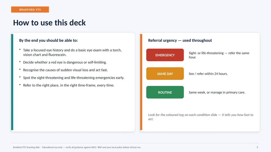
BRADFORD VTS How to use this deck By the end you should be able to: Take a focused eye history and do a basic eye exam with a torch, vision chart and fluorescein.
Decide whether a red eye is dangerous or self-limiting.
Recognise the causes of sudden visual loss and act fast.
Spot the sight-threatening and life-threatening emergencies early.
Refer to the right place, in the right time-frame, every time. Referral urgency — used throughout EMERGENCY Sight- or life-threatening — refer the same hour. SAME DAY See / refer within 24 hours. ROUTINE Same week, or manage in primary care. Look for the coloured tag on each condition slide — it tells you how fast to act. Bradford VTS Teaching Aids · Educational use only — verify all guidance against NICE / BNF and your local policy before clinical use. 3<br>
04
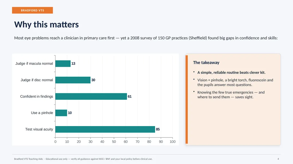
BRADFORD VTS Why this matters Most eye problems reach a clinician in primary care first — yet a 2008 survey of 150 GP practices (Sheffield) found big gaps in confidence and skills: The takeaway A simple, reliable routine beats clever kit.
Vision + pinhole, a bright torch, fluorescein and the pupils answer most questions.
Knowing the few true emergencies — and where to send them — saves sight. Bradford VTS Teaching Aids · Educational use only — verify all guidance against NICE / BNF and your local policy before clinical use. 4<br>
Vision + pinhole, a bright torch, fluorescein and the pupils answer most questions.
Knowing the few true emergencies — and where to send them — saves sight. Bradford VTS Teaching Aids · Educational use only — verify all guidance against NICE / BNF and your local policy before clinical use. 4<br>
05
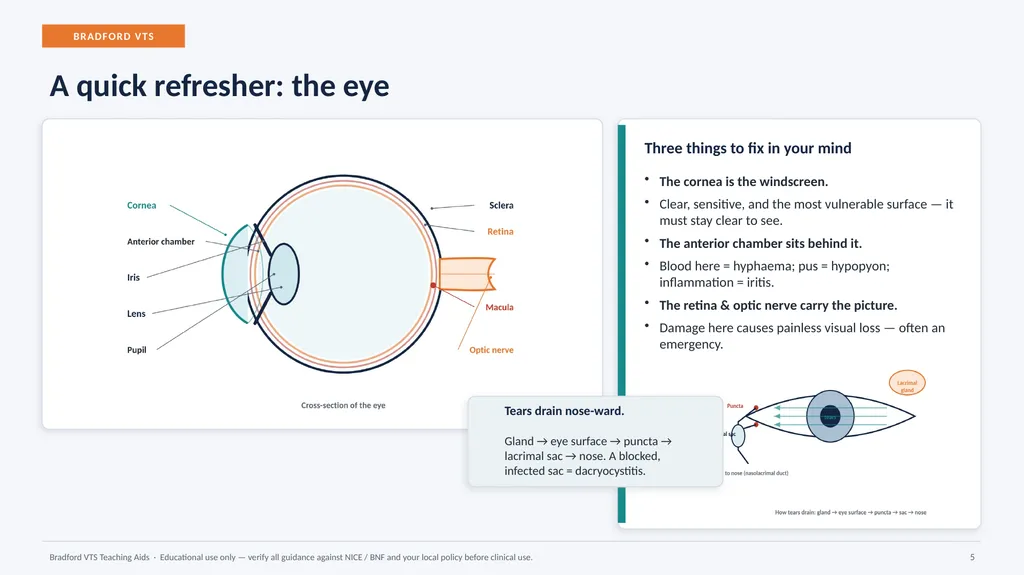
BRADFORD VTS A quick refresher: the eye Three things to fix in your mind The cornea is the windscreen.
Clear, sensitive, and the most vulnerable surface — it must stay clear to see.
The anterior chamber sits behind it.
Blood here = hyphaema; pus = hypopyon; inflammation = iritis.
The retina & optic nerve carry the picture.
Damage here causes painless visual loss — often an emergency. Bradford VTS Teaching Aids · Educational use only — verify all guidance against NICE / BNF and your local policy before clinical use. 5 Tears drain nose-ward.
Gland → eye surface → puncta → lacrimal sac → nose. A blocked, infected sac = dacryocystitis.<br>
06
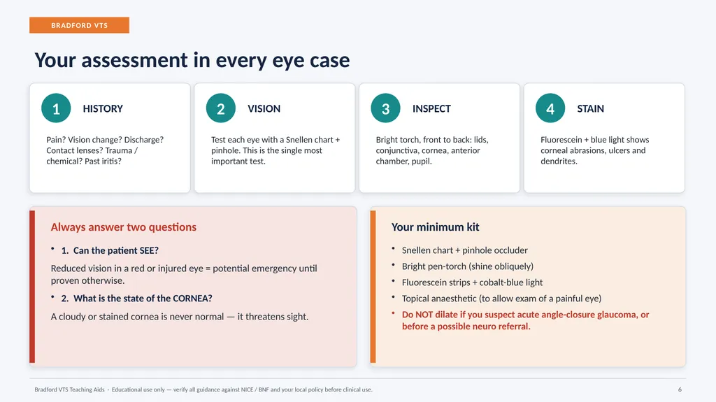
BRADFORD VTS Your assessment in every eye case 1 HISTORY Pain? Vision change? Discharge? Contact lenses? Trauma / chemical? Past iritis? 2 VISION Test each eye with a Snellen chart + pinhole. This is the single most important test. 3 INSPECT Bright torch, front to back: lids, conjunctiva, cornea, anterior chamber, pupil. 4 STAIN Fluorescein + blue light shows corneal abrasions, ulcers and dendrites. Always answer two questions 1. Can the patient SEE?
Reduced vision in a red or injured eye = potential emergency until proven otherwise.
2. What is the state of the CORNEA?
A cloudy or stained cornea is never normal — it threatens sight. Your minimum kit Snellen chart + pinhole occluder
Bright pen-torch (shine obliquely)
Fluorescein strips + cobalt-blue light
Topical anaesthetic (to allow exam of a painful eye)
Do NOT dilate if you suspect acute angle-closure glaucoma, or before a possible neuro referral. Bradford VTS Teaching Aids · Educational use only — verify all guidance against NICE / BNF and your local policy before clinical use. 6<br>
07
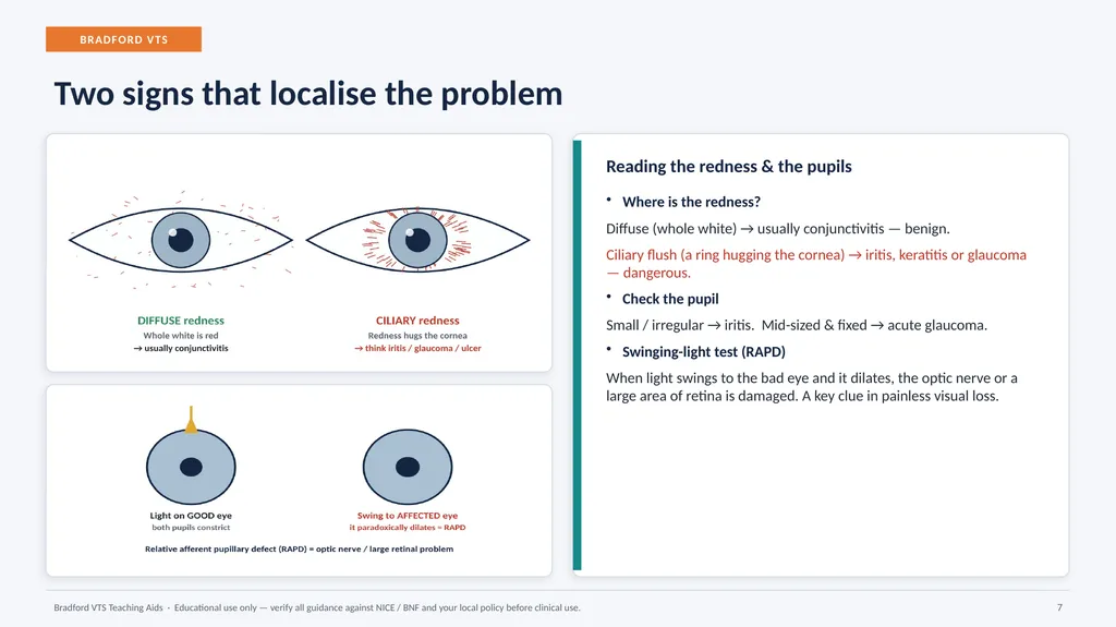
BRADFORD VTS Two signs that localise the problem Reading the redness & the pupils Where is the redness?
Diffuse (whole white) → usually conjunctivitis — benign.
Ciliary flush (a ring hugging the cornea) → iritis, keratitis or glaucoma — dangerous.
Check the pupil
Small / irregular → iritis. Mid-sized & fixed → acute glaucoma.
Swinging-light test (RAPD)
When light swings to the bad eye and it dilates, the optic nerve or a large area of retina is damaged. A key clue in painless visual loss. Bradford VTS Teaching Aids · Educational use only — verify all guidance against NICE / BNF and your local policy before clinical use. 7<br>
08
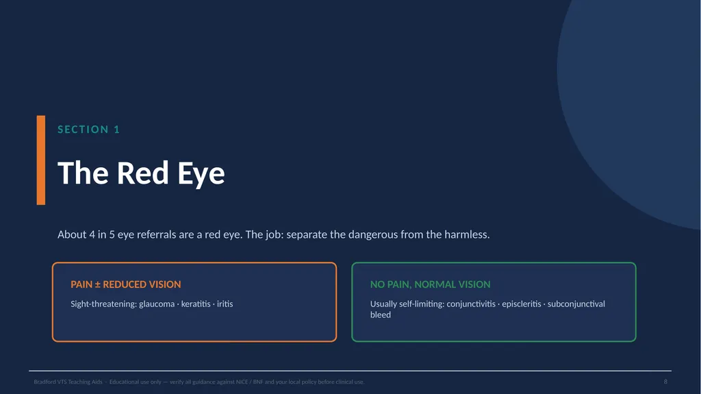
SECTION 1 The Red Eye About 4 in 5 eye referrals are a red eye. The job: separate the dangerous from the harmless. PAIN ± REDUCED VISION Sight-threatening: glaucoma · keratitis · iritis NO PAIN, NORMAL VISION Usually self-limiting: conjunctivitis · episcleritis · subconjunctival bleed Bradford VTS Teaching Aids · Educational use only — verify all guidance against NICE / BNF and your local policy before clinical use. 8<br>
09
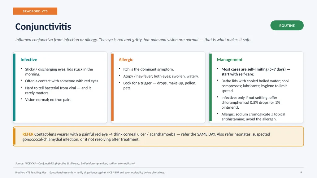
BRADFORD VTS Conjunctivitis ROUTINE Inflamed conjunctiva from infection or allergy. The eye is red and gritty, but pain and vision are normal — that is what makes it safe. Infective Sticky / discharging eyes; lids stuck in the morning.
Often a contact with someone with red eyes.
Hard to tell bacterial from viral — and it rarely matters.
Vision normal; no true pain. Allergic Itch is the dominant symptom.
Atopy / hay-fever; both eyes; swollen, watery.
Look for a trigger — drops, make-up, pollen, pets. Management Most cases are self-limiting (5–7 days) — start with self-care:
Bathe lids with cooled boiled water; cool compresses; lubricants; hygiene to limit spread.
Infective: only if not settling, offer chloramphenicol 0.5% drops (or 1% ointment).
Allergic: sodium cromoglicate ± topical antihistamine; avoid the allergen. REFER Contact-lens wearer with a painful red eye → think corneal ulcer / acanthamoeba — refer the SAME DAY. Also refer neonates, suspected gonococcal/chlamydial infection, or if not resolving after treatment. Source: NICE CKS – Conjunctivitis (infective & allergic); BNF (chloramphenicol, sodium cromoglicate). Bradford VTS Teaching Aids · Educational use only — verify all guidance against NICE / BNF and your local policy before clinical use. 9<br>
10
BRADFORD VTS Iritis (anterior uveitis) SAME DAY Inflammation inside the eye. Often young adults; may be linked to ankylosing spondylitis, sarcoidosis or IBD. Tends to recur. Conjunctivitis irritates — iritis truly hurts. Presentation Painful, aching red eye.
Photophobia with reduced vision.
May have been mislabelled “resistant conjunctivitis”. Signs Ciliary flush — redness hugging the cornea.
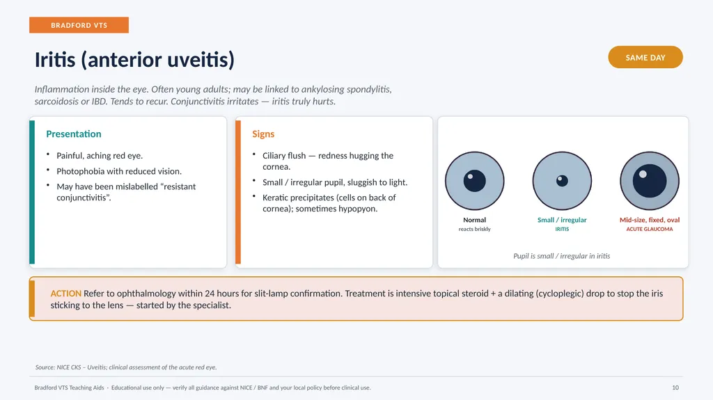
Small / irregular pupil, sluggish to light.
Keratic precipitates (cells on back of cornea); sometimes hypopyon. Pupil is small / irregular in iritis ACTION Refer to ophthalmology within 24 hours for slit-lamp confirmation. Treatment is intensive topical steroid + a dilating (cycloplegic) drop to stop the iris sticking to the lens — started by the specialist. Source: NICE CKS – Uveitis; clinical assessment of the acute red eye. Bradford VTS Teaching Aids · Educational use only — verify all guidance against NICE / BNF and your local policy before clinical use. 10<br>
11
BRADFORD VTS Episcleritis & scleritis ROUTINE → URGENT Episcleritis is common, mild and self-limiting. Scleritis is rare (≈0.1% of eye-clinic visits) but serious — and far more painful. Episcleritis Localised patch of redness; little or no discomfort.
Vision normal; no discharge.
Self-limiting — settles in ~2 weeks; often recurs.
If sore, refer same week; mild topical steroid may help. Scleritis Severe, boring, deep ache that wakes the patient.
Often linked to rheumatoid / autoimmune disease.
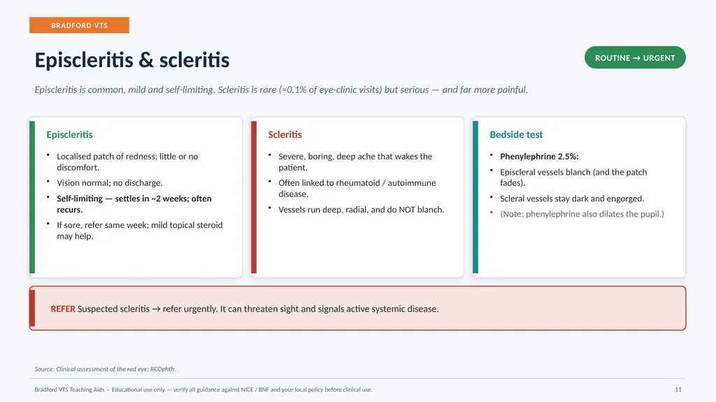
Vessels run deep, radial, and do NOT blanch. Bedside test Phenylephrine 2.5%:
Episcleral vessels blanch (and the patch fades).
Scleral vessels stay dark and engorged.
(Note: phenylephrine also dilates the pupil.) REFER Suspected scleritis → refer urgently. It can threaten sight and signals active systemic disease. Source: Clinical assessment of the red eye; RCOphth. Bradford VTS Teaching Aids · Educational use only — verify all guidance against NICE / BNF and your local policy before clinical use. 11<br>
12
BRADFORD VTS Acute angle-closure glaucoma EMERGENCY Drainage angle blocks → pressure shoots up. Uncommon but a true emergency: it can blind within hours. Typically over-50s, long-sighted, often in dim light. Presentation Severe pain, red eye; haloes around lights.
Headache, nausea & vomiting (can mimic a GI bug).
Rapidly reduced vision. Signs Hazy (steamy) cornea; iris hard to see.
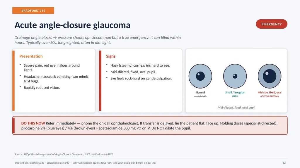
Mid-dilated, fixed, oval pupil.
Eye feels rock-hard on gentle palpation. Mid-dilated, fixed, oval pupil DO THIS NOW Refer immediately — phone the on-call ophthalmologist. If transfer is delayed: lie the patient flat, face up. Holding doses (specialist-directed): pilocarpine 2% (blue eyes) / 4% (brown eyes) + acetazolamide 500 mg PO or IV. Do NOT dilate the pupil. Source: RCOphth – Management of Angle-Closure Glaucoma; NICE; verify doses in BNF. Bradford VTS Teaching Aids · Educational use only — verify all guidance against NICE / BNF and your local policy before clinical use. 12<br>
13
BRADFORD VTS Corneal infection (keratitis) SAME DAY A potentially sight-threatening ulcer of the cornea. Two patterns matter most in primary care: bacterial (often contact-lens related) and herpes simplex (dendritic). Presentation Painful red eye, photophobia, watering.
Reduced vision.
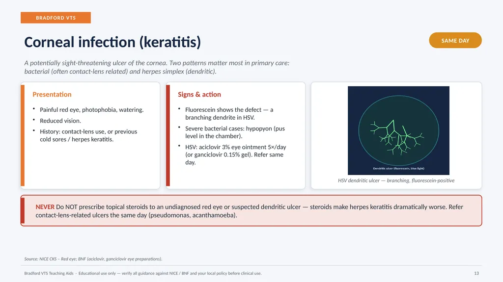
History: contact-lens use, or previous cold sores / herpes keratitis. Signs & action Fluorescein shows the defect — a branching dendrite in HSV.
Severe bacterial cases: hypopyon (pus level in the chamber).
HSV: aciclovir 3% eye ointment 5×/day (or ganciclovir 0.15% gel). Refer same day. HSV dendritic ulcer — branching, fluorescein-positive NEVER Do NOT prescribe topical steroids to an undiagnosed red eye or suspected dendritic ulcer — steroids make herpes keratitis dramatically worse. Refer contact-lens-related ulcers the same day (pseudomonas, acanthamoeba). Source: NICE CKS – Red eye; BNF (aciclovir, ganciclovir eye preparations). Bradford VTS Teaching Aids · Educational use only — verify all guidance against NICE / BNF and your local policy before clinical use. 13<br>
14
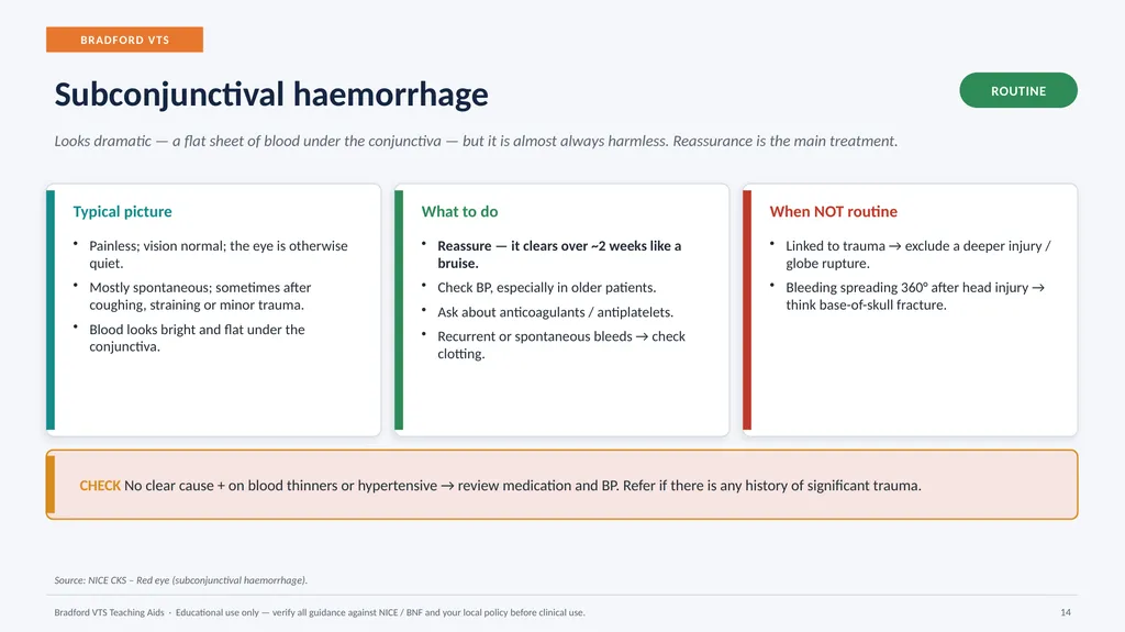
BRADFORD VTS Subconjunctival haemorrhage ROUTINE Looks dramatic — a flat sheet of blood under the conjunctiva — but it is almost always harmless. Reassurance is the main treatment. Typical picture Painless; vision normal; the eye is otherwise quiet.
Mostly spontaneous; sometimes after coughing, straining or minor trauma.
Blood looks bright and flat under the conjunctiva. What to do Reassure — it clears over ~2 weeks like a bruise.
Check BP, especially in older patients.
Ask about anticoagulants / antiplatelets.
Recurrent or spontaneous bleeds → check clotting. When NOT routine Linked to trauma → exclude a deeper injury / globe rupture.
Bleeding spreading 360° after head injury → think base-of-skull fracture. CHECK No clear cause + on blood thinners or hypertensive → review medication and BP. Refer if there is any history of significant trauma. Source: NICE CKS – Red eye (subconjunctival haemorrhage). Bradford VTS Teaching Aids · Educational use only — verify all guidance against NICE / BNF and your local policy before clinical use. 14<br>
15
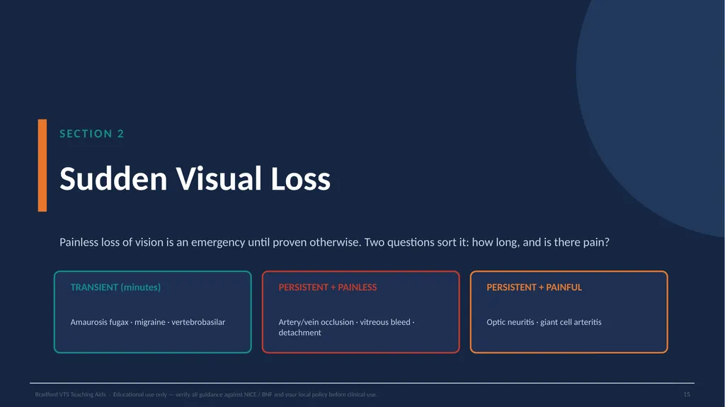
SECTION 2 Sudden Visual Loss Painless loss of vision is an emergency until proven otherwise. Two questions sort it: how long, and is there pain? TRANSIENT (minutes) Amaurosis fugax · migraine · vertebrobasilar PERSISTENT + PAINLESS Artery/vein occlusion · vitreous bleed · detachment PERSISTENT + PAINFUL Optic neuritis · giant cell arteritis Bradford VTS Teaching Aids · Educational use only — verify all guidance against NICE / BNF and your local policy before clinical use. 15<br>
16
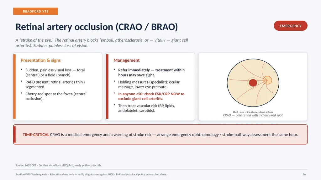
BRADFORD VTS Retinal artery occlusion (CRAO / BRAO) EMERGENCY A “stroke of the eye.” The retinal artery blocks (emboli, atherosclerosis, or — vitally — giant cell arteritis). Sudden, painless loss of vision. Presentation & signs Sudden, painless visual loss — total (central) or a field (branch).
RAPD present; retinal arteries thin / segmented.
Cherry-red spot at the fovea (central occlusion). Management Refer immediately — treatment within hours may save sight.
Holding measures (specialist): ocular massage, lower eye pressure.
In anyone ≥50: check ESR/CRP NOW to exclude giant cell arteritis.
Then treat vascular risk (BP, lipids, antiplatelet, carotids). CRAO — pale retina with a cherry-red spot TIME-CRITICAL CRAO is a medical emergency and a warning of stroke risk — arrange emergency ophthalmology / stroke-pathway assessment the same hour. Source: NICE CKS – Sudden visual loss; RCOphth; verify pathway locally. Bradford VTS Teaching Aids · Educational use only — verify all guidance against NICE / BNF and your local policy before clinical use. 16<br>
17
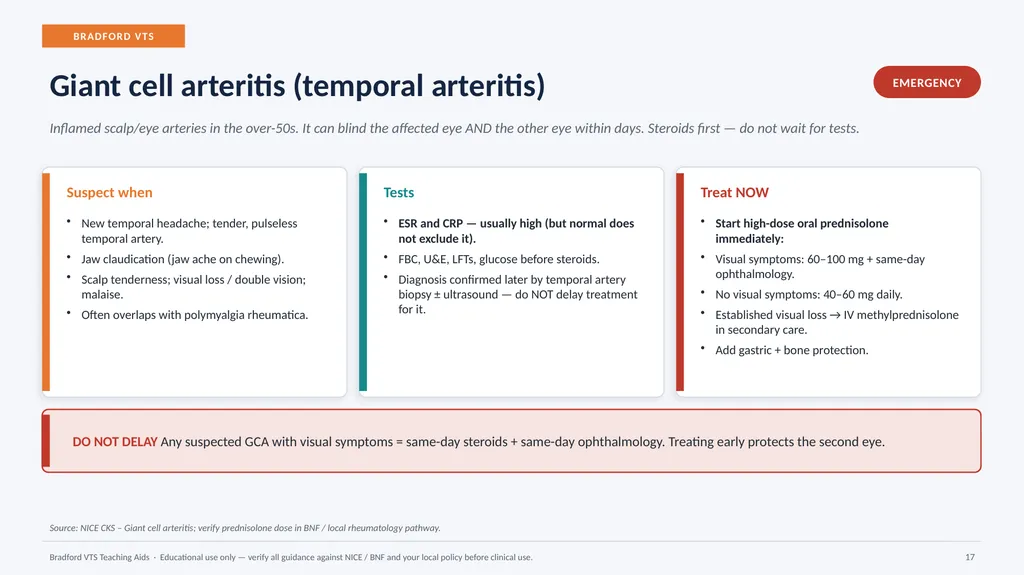
BRADFORD VTS Giant cell arteritis (temporal arteritis) EMERGENCY Inflamed scalp/eye arteries in the over-50s. It can blind the affected eye AND the other eye within days. Steroids first — do not wait for tests. Suspect when New temporal headache; tender, pulseless temporal artery.
Jaw claudication (jaw ache on chewing).
Scalp tenderness; visual loss / double vision; malaise.
Often overlaps with polymyalgia rheumatica. Tests ESR and CRP — usually high (but normal does not exclude it).
FBC, U&E, LFTs, glucose before steroids.
Diagnosis confirmed later by temporal artery biopsy ± ultrasound — do NOT delay treatment for it. Treat NOW Start high-dose oral prednisolone immediately:
Visual symptoms: 60–100 mg + same-day ophthalmology.
No visual symptoms: 40–60 mg daily.
Established visual loss → IV methylprednisolone in secondary care.
Add gastric + bone protection. DO NOT DELAY Any suspected GCA with visual symptoms = same-day steroids + same-day ophthalmology. Treating early protects the second eye. Source: NICE CKS – Giant cell arteritis; verify prednisolone dose in BNF / local rheumatology pathway. Bradford VTS Teaching Aids · Educational use only — verify all guidance against NICE / BNF and your local policy before clinical use. 17<br>
18
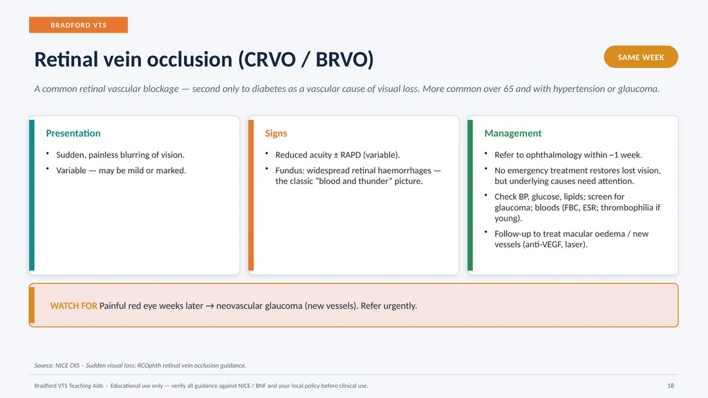
BRADFORD VTS Retinal vein occlusion (CRVO / BRVO) SAME WEEK A common retinal vascular blockage — second only to diabetes as a vascular cause of visual loss. More common over 65 and with hypertension or glaucoma. Presentation Sudden, painless blurring of vision.
Variable — may be mild or marked. Signs Reduced acuity ± RAPD (variable).
Fundus: widespread retinal haemorrhages — the classic “blood and thunder” picture. Management Refer to ophthalmology within ~1 week.
No emergency treatment restores lost vision, but underlying causes need attention.
Check BP, glucose, lipids; screen for glaucoma; bloods (FBC, ESR; thrombophilia if young).
Follow-up to treat macular oedema / new vessels (anti-VEGF, laser). WATCH FOR Painful red eye weeks later → neovascular glaucoma (new vessels). Refer urgently. Source: NICE CKS – Sudden visual loss; RCOphth retinal vein occlusion guidance. Bradford VTS Teaching Aids · Educational use only — verify all guidance against NICE / BNF and your local policy before clinical use. 18<br>
19
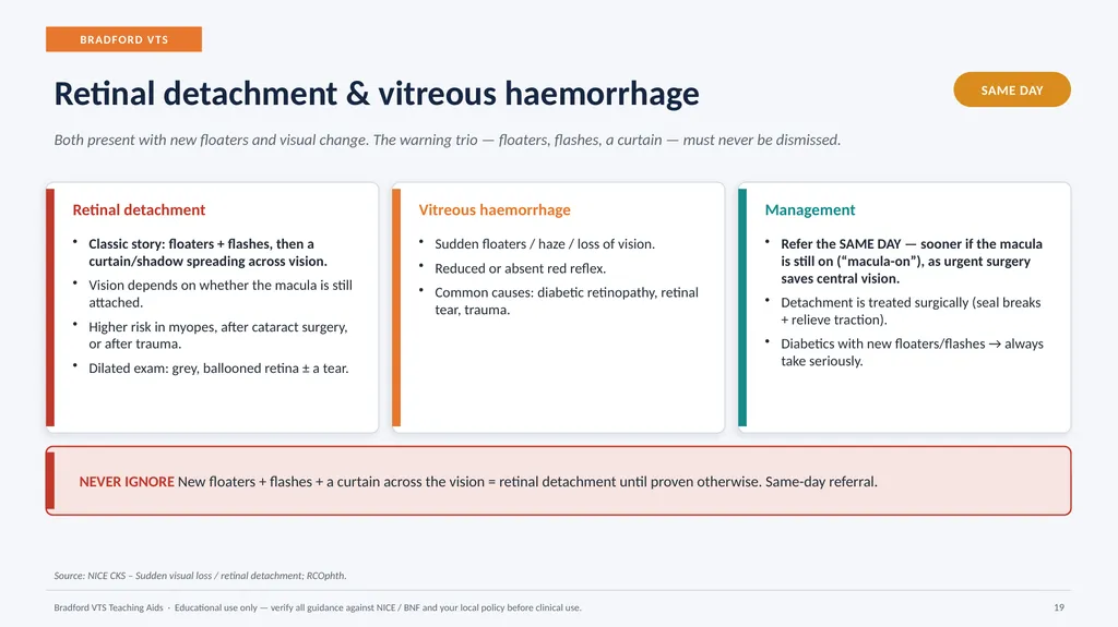
BRADFORD VTS Retinal detachment & vitreous haemorrhage SAME DAY Both present with new floaters and visual change. The warning trio — floaters, flashes, a curtain — must never be dismissed. Retinal detachment Classic story: floaters + flashes, then a curtain/shadow spreading across vision.
Vision depends on whether the macula is still attached.
Higher risk in myopes, after cataract surgery, or after trauma.
Dilated exam: grey, ballooned retina ± a tear. Vitreous haemorrhage Sudden floaters / haze / loss of vision.
Reduced or absent red reflex.
Common causes: diabetic retinopathy, retinal tear, trauma. Management Refer the SAME DAY — sooner if the macula is still on (“macula-on”), as urgent surgery saves central vision.
Detachment is treated surgically (seal breaks + relieve traction).
Diabetics with new floaters/flashes → always take seriously. NEVER IGNORE New floaters + flashes + a curtain across the vision = retinal detachment until proven otherwise. Same-day referral. Source: NICE CKS – Sudden visual loss / retinal detachment; RCOphth. Bradford VTS Teaching Aids · Educational use only — verify all guidance against NICE / BNF and your local policy before clinical use. 19<br>
20
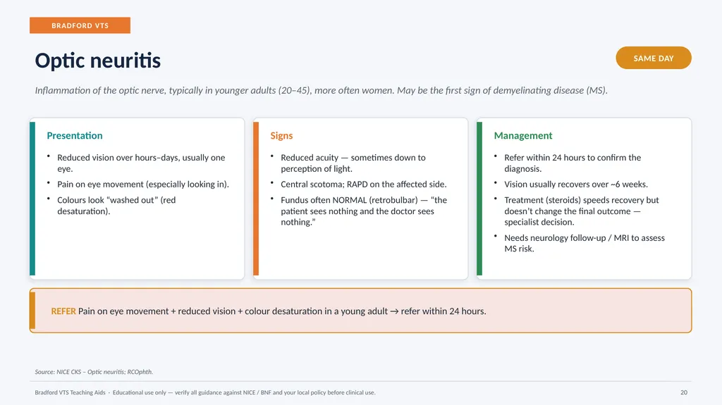
BRADFORD VTS Optic neuritis SAME DAY Inflammation of the optic nerve, typically in younger adults (20–45), more often women. May be the first sign of demyelinating disease (MS). Presentation Reduced vision over hours–days, usually one eye.
Pain on eye movement (especially looking in).
Colours look “washed out” (red desaturation). Signs Reduced acuity — sometimes down to perception of light.
Central scotoma; RAPD on the affected side.
Fundus often NORMAL (retrobulbar) — “the patient sees nothing and the doctor sees nothing.” Management Refer within 24 hours to confirm the diagnosis.
Vision usually recovers over ~6 weeks.
Treatment (steroids) speeds recovery but doesn’t change the final outcome — specialist decision.
Needs neurology follow-up / MRI to assess MS risk. REFER Pain on eye movement + reduced vision + colour desaturation in a young adult → refer within 24 hours. Source: NICE CKS – Optic neuritis; RCOphth. Bradford VTS Teaching Aids · Educational use only — verify all guidance against NICE / BNF and your local policy before clinical use. 20<br>
21
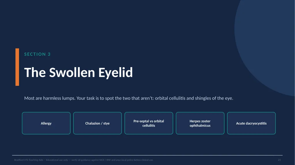
SECTION 3 The Swollen Eyelid Most are harmless lumps. Your task is to spot the two that aren’t: orbital cellulitis and shingles of the eye. Allergy Chalazion / stye Pre-septal vs orbital cellulitis Herpes zoster ophthalmicus Acute dacryocystitis Bradford VTS Teaching Aids · Educational use only — verify all guidance against NICE / BNF and your local policy before clinical use. 21<br>
22
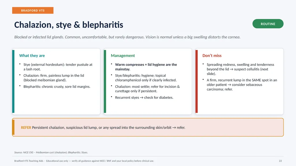
BRADFORD VTS Chalazion, stye & blepharitis ROUTINE Blocked or infected lid glands. Common, uncomfortable, but rarely dangerous. Vision is normal unless a big swelling distorts the cornea. What they are Stye (external hordeolum): tender pustule at a lash root.
Chalazion: firm, painless lump in the lid (blocked meibomian gland).
Blepharitis: chronic crusty, sore lid margins. Management Warm compresses + lid hygiene are the mainstay.
Stye/blepharitis: hygiene; topical chloramphenicol only if clearly infected.
Chalazion: most settle; refer for incision & curettage only if persistent.
Recurrent styes → check for diabetes. Don’t miss Spreading redness, swelling and tenderness beyond the lid → suspect cellulitis (next slide).
A firm, recurrent lump in the SAME spot in an older patient → consider sebaceous carcinoma; refer. REFER Persistent chalazion, suspicious lid lump, or any spread into the surrounding skin/orbit → refer. Source: NICE CKS – Meibomian cyst (chalazion); Blepharitis; Styes. Bradford VTS Teaching Aids · Educational use only — verify all guidance against NICE / BNF and your local policy before clinical use. 22<br>
23
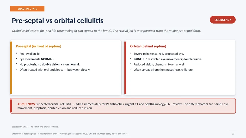
BRADFORD VTS Pre-septal vs orbital cellulitis EMERGENCY Orbital cellulitis is sight- and life-threatening (it can spread to the brain). The crucial job is to separate it from the milder pre-septal form. Pre-septal (in front of septum) Red, swollen lid.
Eye movements NORMAL.
No proptosis, no double vision, vision normal.
Often treated with oral antibiotics — but watch closely. Orbital (behind septum) Severe pain; tense, red, proptosed eye.
PAINFUL / restricted eye movements; double vision.
Reduced vision; chemosis; fever, unwell.
Often spreads from the sinuses (esp. children). ADMIT NOW Suspected orbital cellulitis → admit immediately for IV antibiotics, urgent CT and ophthalmology/ENT review. The differentiators are painful eye movement, proptosis, double vision and reduced vision. Source: NICE CKS – Pre-septal and orbital cellulitis. Bradford VTS Teaching Aids · Educational use only — verify all guidance against NICE / BNF and your local policy before clinical use. 23<br>
24
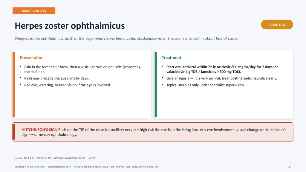
BRADFORD VTS Herpes zoster ophthalmicus SAME DAY Shingles in the ophthalmic branch of the trigeminal nerve. Reactivated chickenpox virus. The eye is involved in about half of cases. Presentation Pain in the forehead / brow, then a vesicular rash on one side (respecting the midline).
Rash may precede the eye signs by days.
Red eye, watering, blurred vision if the eye is involved. Treatment Start oral antiviral within 72 h: aciclovir 800 mg 5×/day for 7 days (or valaciclovir 1 g TDS / famciclovir 500 mg TDS).
Give analgesia — it is very painful; treat post-herpetic neuralgia early.
Topical steroids only under specialist supervision. HUTCHINSON’S SIGN Rash on the TIP of the nose (nasociliary nerve) = high risk the eye is in the firing line. Any eye involvement, visual change or Hutchinson’s sign → same-day ophthalmology. Source: NICE CKS – Shingles; BNF (aciclovir/valaciclovir doses — verify). Bradford VTS Teaching Aids · Educational use only — verify all guidance against NICE / BNF and your local policy before clinical use. 24<br>
25
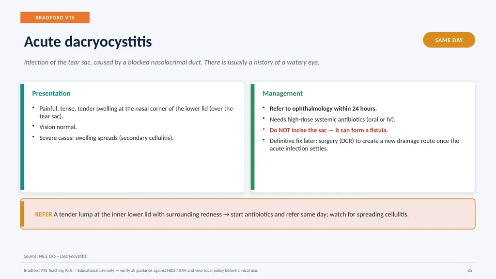
BRADFORD VTS Acute dacryocystitis SAME DAY Infection of the tear sac, caused by a blocked nasolacrimal duct. There is usually a history of a watery eye. Presentation Painful, tense, tender swelling at the nasal corner of the lower lid (over the tear sac).
Vision normal.
Severe cases: swelling spreads (secondary cellulitis). Management Refer to ophthalmology within 24 hours.
Needs high-dose systemic antibiotics (oral or IV).
Do NOT incise the sac — it can form a fistula.
Definitive fix later: surgery (DCR) to create a new drainage route once the acute infection settles. REFER A tender lump at the inner lower lid with surrounding redness → start antibiotics and refer same day; watch for spreading cellulitis. Source: NICE CKS – Dacryocystitis. Bradford VTS Teaching Aids · Educational use only — verify all guidance against NICE / BNF and your local policy before clinical use. 25<br>
26
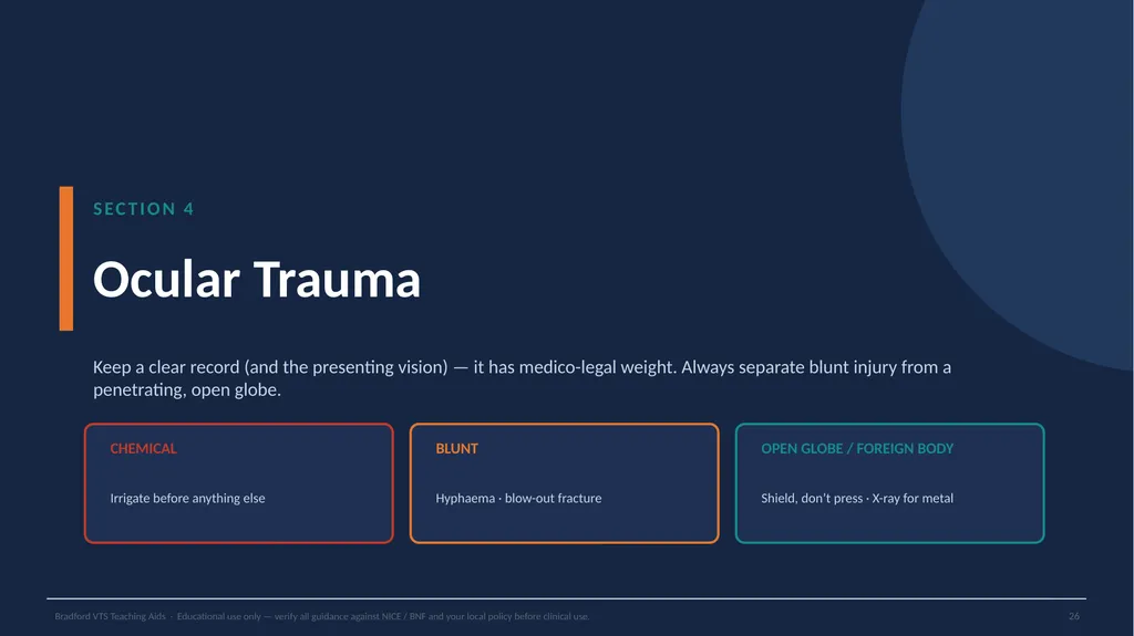
SECTION 4 Ocular Trauma Keep a clear record (and the presenting vision) — it has medico-legal weight. Always separate blunt injury from a penetrating, open globe. CHEMICAL Irrigate before anything else BLUNT Hyphaema · blow-out fracture OPEN GLOBE / FOREIGN BODY Shield, don’t press · X-ray for metal Bradford VTS Teaching Aids · Educational use only — verify all guidance against NICE / BNF and your local policy before clinical use. 26<br>
27
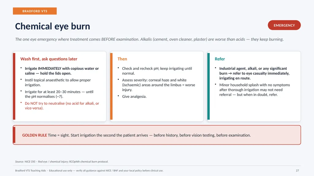
BRADFORD VTS Chemical eye burn EMERGENCY The one eye emergency where treatment comes BEFORE examination. Alkalis (cement, oven cleaner, plaster) are worse than acids — they keep burning. Wash first, ask questions later Irrigate IMMEDIATELY with copious water or saline — hold the lids open.
Instil topical anaesthetic to allow proper irrigation.
Irrigate for at least 20–30 minutes — until the pH normalises (~7).
Do NOT try to neutralise (no acid for alkali, or vice-versa). Then Check and recheck pH; keep irrigating until normal.
Assess severity: corneal haze and white (ischaemic) areas around the limbus = worse injury.
Give analgesia. Refer Industrial agent, alkali, or any significant burn → refer to eye casualty immediately, irrigating en route.
Minor household splash with no symptoms after thorough irrigation may not need referral — but when in doubt, refer. GOLDEN RULE Time = sight. Start irrigation the second the patient arrives — before history, before vision testing, before examination. Source: NICE CKS – Red eye / chemical injury; RCOphth chemical burn protocol. Bradford VTS Teaching Aids · Educational use only — verify all guidance against NICE / BNF and your local policy before clinical use. 27<br>
28
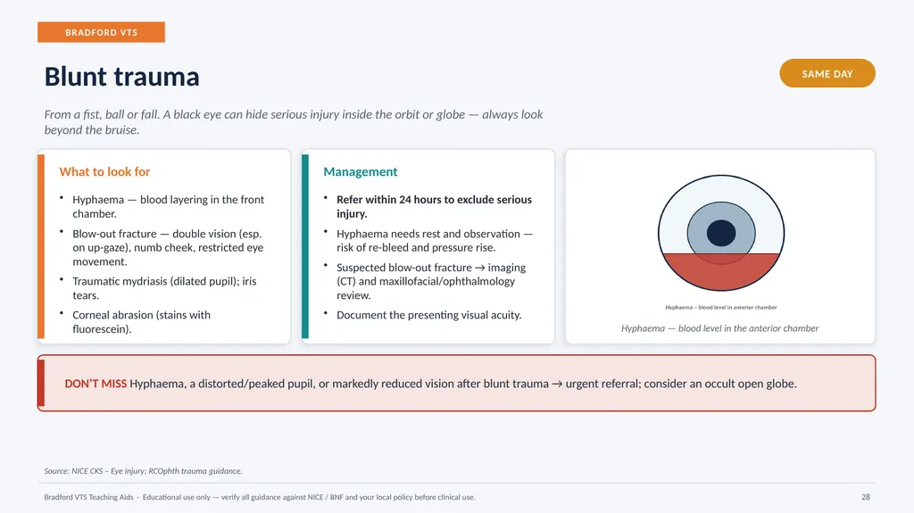
BRADFORD VTS Blunt trauma SAME DAY From a fist, ball or fall. A black eye can hide serious injury inside the orbit or globe — always look beyond the bruise. What to look for Hyphaema — blood layering in the front chamber.
Blow-out fracture — double vision (esp. on up-gaze), numb cheek, restricted eye movement.
Traumatic mydriasis (dilated pupil); iris tears.
Corneal abrasion (stains with fluorescein). Management Refer within 24 hours to exclude serious injury.
Hyphaema needs rest and observation — risk of re-bleed and pressure rise.
Suspected blow-out fracture → imaging (CT) and maxillofacial/ophthalmology review.
Document the presenting visual acuity. Hyphaema — blood level in the anterior chamber DON’T MISS Hyphaema, a distorted/peaked pupil, or markedly reduced vision after blunt trauma → urgent referral; consider an occult open globe. Source: NICE CKS – Eye injury; RCOphth trauma guidance. Bradford VTS Teaching Aids · Educational use only — verify all guidance against NICE / BNF and your local policy before clinical use. 28<br>
29
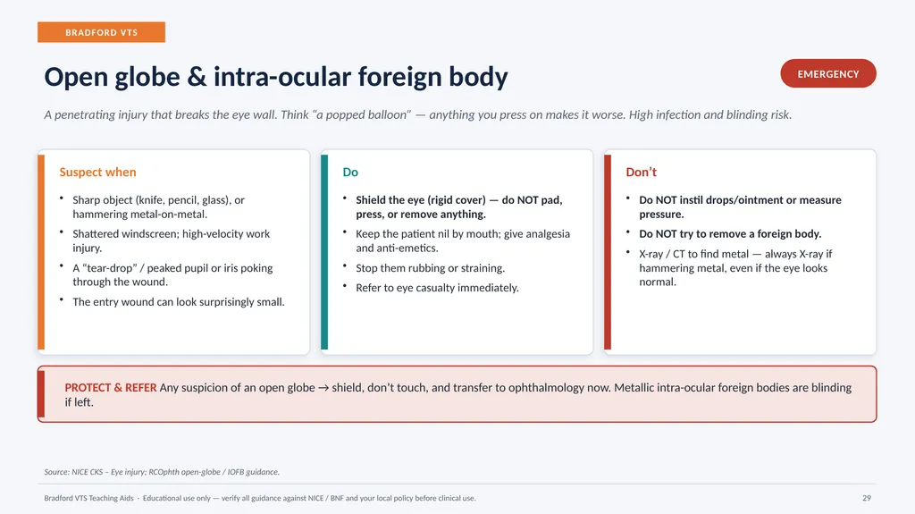
BRADFORD VTS Open globe & intra-ocular foreign body EMERGENCY A penetrating injury that breaks the eye wall. Think “a popped balloon” — anything you press on makes it worse. High infection and blinding risk. Suspect when Sharp object (knife, pencil, glass), or hammering metal-on-metal.
Shattered windscreen; high-velocity work injury.
A “tear-drop” / peaked pupil or iris poking through the wound.
The entry wound can look surprisingly small. Do Shield the eye (rigid cover) — do NOT pad, press, or remove anything.
Keep the patient nil by mouth; give analgesia and anti-emetics.
Stop them rubbing or straining.
Refer to eye casualty immediately. Don’t Do NOT instil drops/ointment or measure pressure.
Do NOT try to remove a foreign body.
X-ray / CT to find metal — always X-ray if hammering metal, even if the eye looks normal. PROTECT & REFER Any suspicion of an open globe → shield, don’t touch, and transfer to ophthalmology now. Metallic intra-ocular foreign bodies are blinding if left. Source: NICE CKS – Eye injury; RCOphth open-globe / IOFB guidance. Bradford VTS Teaching Aids · Educational use only — verify all guidance against NICE / BNF and your local policy before clinical use. 29<br>
30
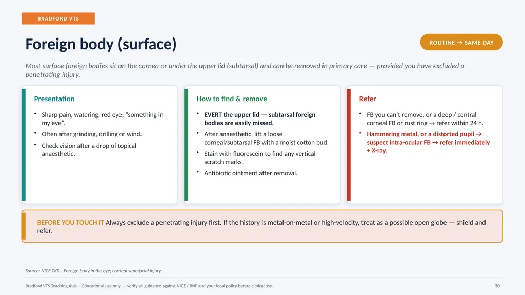
BRADFORD VTS Foreign body (surface) ROUTINE → SAME DAY Most surface foreign bodies sit on the cornea or under the upper lid (subtarsal) and can be removed in primary care — provided you have excluded a penetrating injury. Presentation Sharp pain, watering, red eye; “something in my eye”.
Often after grinding, drilling or wind.
Check vision after a drop of topical anaesthetic. How to find & remove EVERT the upper lid — subtarsal foreign bodies are easily missed.
After anaesthetic, lift a loose corneal/subtarsal FB with a moist cotton bud.
Stain with fluorescein to find any vertical scratch marks.
Antibiotic ointment after removal. Refer FB you can’t remove, or a deep / central corneal FB or rust ring → refer within 24 h.
Hammering metal, or a distorted pupil → suspect intra-ocular FB → refer immediately + X-ray. BEFORE YOU TOUCH IT Always exclude a penetrating injury first. If the history is metal-on-metal or high-velocity, treat as a possible open globe — shield and refer. Source: NICE CKS – Foreign body in the eye; corneal superficial injury. Bradford VTS Teaching Aids · Educational use only — verify all guidance against NICE / BNF and your local policy before clinical use. 30<br>
31
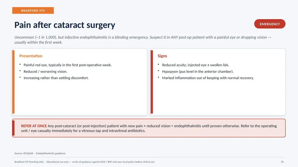
BRADFORD VTS Pain after cataract surgery EMERGENCY Uncommon (~1 in 1,000), but infective endophthalmitis is a blinding emergency. Suspect it in ANY post-op patient with a painful eye or dropping vision — usually within the first week. Presentation Painful red eye, typically in the first post-operative week.
Reduced / worsening vision.
Increasing rather than settling discomfort. Signs Reduced acuity; injected eye ± swollen lids.
Hypopyon (pus level in the anterior chamber).
Marked inflammation out of keeping with normal recovery. REFER AT ONCE Any post-cataract (or post-injection) patient with new pain + reduced vision = endophthalmitis until proven otherwise. Refer to the operating unit / eye casualty immediately for a vitreous tap and intravitreal antibiotics. Source: RCOphth – Endophthalmitis guidance. Bradford VTS Teaching Aids · Educational use only — verify all guidance against NICE / BNF and your local policy before clinical use. 31<br>
32
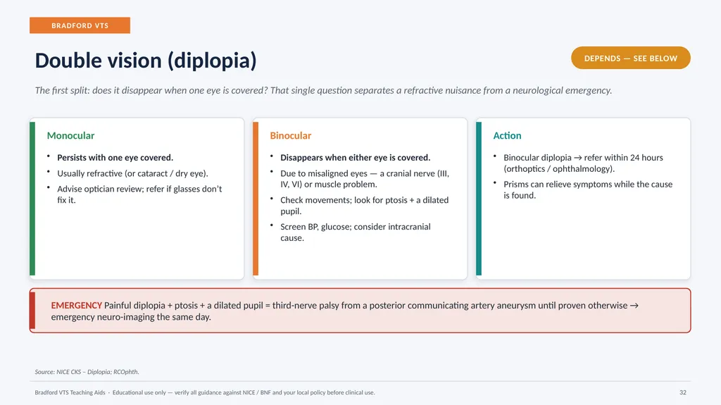
BRADFORD VTS Double vision (diplopia) DEPENDS — SEE BELOW The first split: does it disappear when one eye is covered? That single question separates a refractive nuisance from a neurological emergency. Monocular Persists with one eye covered.
Usually refractive (or cataract / dry eye).
Advise optician review; refer if glasses don’t fix it. Binocular Disappears when either eye is covered.
Due to misaligned eyes — a cranial nerve (III, IV, VI) or muscle problem.
Check movements; look for ptosis + a dilated pupil.
Screen BP, glucose; consider intracranial cause. Action Binocular diplopia → refer within 24 hours (orthoptics / ophthalmology).
Prisms can relieve symptoms while the cause is found. EMERGENCY Painful diplopia + ptosis + a dilated pupil = third-nerve palsy from a posterior communicating artery aneurysm until proven otherwise → emergency neuro-imaging the same day. Source: NICE CKS – Diplopia; RCOphth. Bradford VTS Teaching Aids · Educational use only — verify all guidance against NICE / BNF and your local policy before clinical use. 32<br>
33
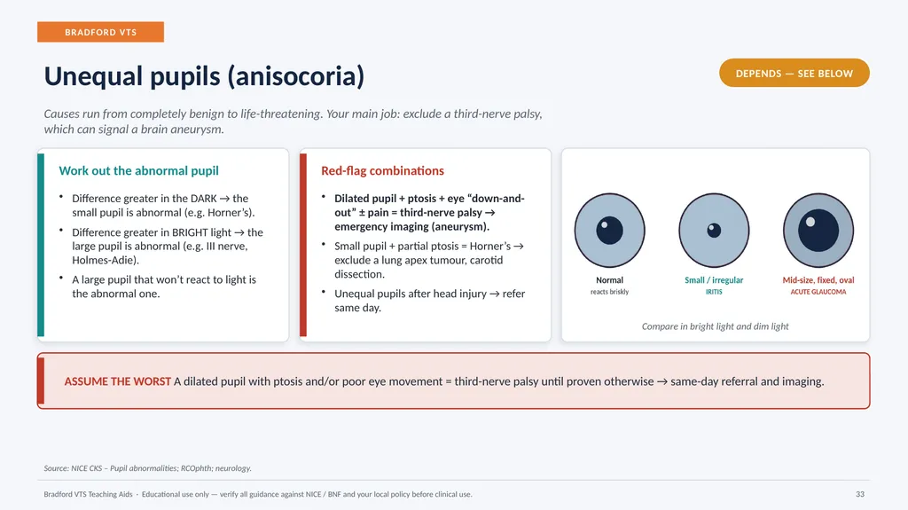
BRADFORD VTS Unequal pupils (anisocoria) DEPENDS — SEE BELOW Causes run from completely benign to life-threatening. Your main job: exclude a third-nerve palsy, which can signal a brain aneurysm. Work out the abnormal pupil Difference greater in the DARK → the small pupil is abnormal (e.g. Horner’s).
Difference greater in BRIGHT light → the large pupil is abnormal (e.g. III nerve, Holmes-Adie).
A large pupil that won’t react to light is the abnormal one. Red-flag combinations Dilated pupil + ptosis + eye “down-and-out” ± pain = third-nerve palsy → emergency imaging (aneurysm).
Small pupil + partial ptosis = Horner’s → exclude a lung apex tumour, carotid dissection.
Unequal pupils after head injury → refer same day. Compare in bright light and dim light ASSUME THE WORST A dilated pupil with ptosis and/or poor eye movement = third-nerve palsy until proven otherwise → same-day referral and imaging. Source: NICE CKS – Pupil abnormalities; RCOphth; neurology. Bradford VTS Teaching Aids · Educational use only — verify all guidance against NICE / BNF and your local policy before clinical use. 33<br>
34
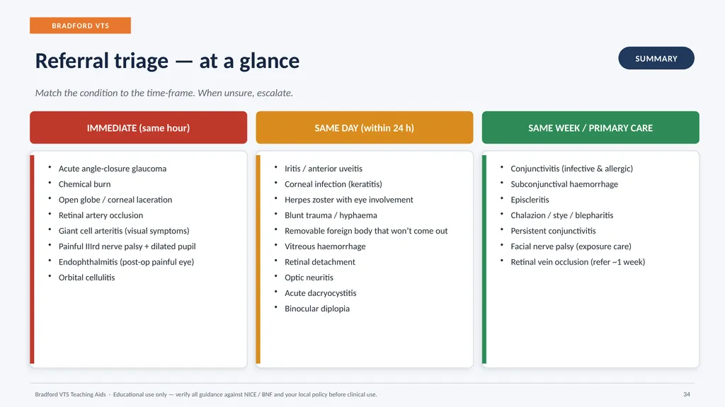
BRADFORD VTS Referral triage — at a glance SUMMARY Match the condition to the time-frame. When unsure, escalate. IMMEDIATE (same hour) Acute angle-closure glaucoma
Chemical burn
Open globe / corneal laceration
Retinal artery occlusion
Giant cell arteritis (visual symptoms)
Painful IIIrd nerve palsy + dilated pupil
Endophthalmitis (post-op painful eye)
Orbital cellulitis SAME DAY (within 24 h) Iritis / anterior uveitis
Corneal infection (keratitis)
Herpes zoster with eye involvement
Blunt trauma / hyphaema
Removable foreign body that won’t come out
Vitreous haemorrhage
Retinal detachment
Optic neuritis
Acute dacryocystitis
Binocular diplopia SAME WEEK / PRIMARY CARE Conjunctivitis (infective & allergic)
Subconjunctival haemorrhage
Episcleritis
Chalazion / stye / blepharitis
Persistent conjunctivitis
Facial nerve palsy (exposure care)
Retinal vein occlusion (refer ~1 week) Bradford VTS Teaching Aids · Educational use only — verify all guidance against NICE / BNF and your local policy before clinical use. 34<br>
35
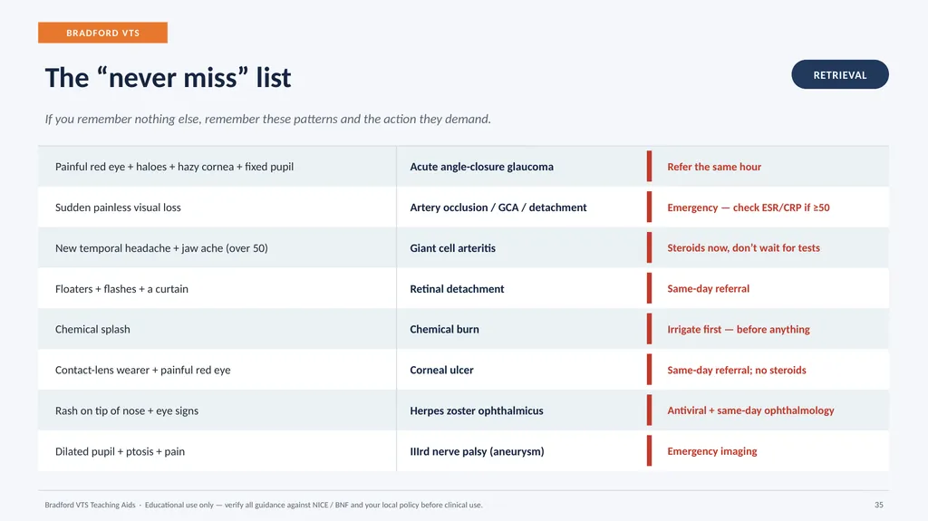
BRADFORD VTS The “never miss” list RETRIEVAL If you remember nothing else, remember these patterns and the action they demand. Painful red eye + haloes + hazy cornea + fixed pupil Acute angle-closure glaucoma Refer the same hour Sudden painless visual loss Artery occlusion / GCA / detachment Emergency — check ESR/CRP if ≥50 New temporal headache + jaw ache (over 50) Giant cell arteritis Steroids now, don’t wait for tests Floaters + flashes + a curtain Retinal detachment Same-day referral Chemical splash Chemical burn Irrigate first — before anything Contact-lens wearer + painful red eye Corneal ulcer Same-day referral; no steroids Rash on tip of nose + eye signs Herpes zoster ophthalmicus Antiviral + same-day ophthalmology Dilated pupil + ptosis + pain IIIrd nerve palsy (aneurysm) Emergency imaging Bradford VTS Teaching Aids · Educational use only — verify all guidance against NICE / BNF and your local policy before clinical use. 35<br>
36
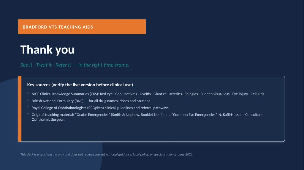
BRADFORD VTS TEACHING AIDS Thank you See it · Treat it · Refer it — in the right time-frame. Key sources (verify the live version before clinical use) NICE Clinical Knowledge Summaries (CKS): Red eye · Conjunctivitis · Uveitis · Giant cell arteritis · Shingles · Sudden visual loss · Eye injury · Cellulitis.
British National Formulary (BNF) — for all drug names, doses and cautions.
Royal College of Ophthalmologists (RCOphth) clinical guidelines and referral pathways.
Original teaching material: “Ocular Emergencies” (Smith & Nephew, Booklet No. 4) and “Common Eye Emergencies”, N. Kafil-Hussain, Consultant Ophthalmic Surgeon. This deck is a teaching aid only and does not replace current national guidance, local policy, or specialist advice. June 2026.<br>