
Candida spp. Showing Pseudohyphae in a Direct Wet

Published  . 0 views
↓ Download
Candida spp. Showing Pseudohyphae in a Direct Wet
1 / 1
Candida spp. Showing Pseudohyphae in a Direct Wet - slide 1 of 4 Candida spp. Showing Pseudohyphae in a Direct Wet - slide 2 of 4 Candida spp. Showing Pseudohyphae in a Direct Wet - slide 3 of 4 Candida spp. Showing Pseudohyphae in a Direct Wet - slide 4 of 4
Description: Candida spp. Showing Pseudohyphae in a Direct Wet Mount of a Vaginal Discharge Authors: Guadalupe del Rocio Dominguez-Figueroa, Javier Gutierrez Jimenez, Ulises Alberto Santiago-Vazquez Citation: Guadalupe del Rocio Dominguez-Figueroa,

Related Topics

Download Presentation

"Candida spp. Showing Pseudohyphae in a Direct Wet" is the property of its rightful owner. Permission is granted to download and print the materials on this website for personal, non-commercial use only, and to display it on your personal computer provided you do not modify the materials and that you retain all copyright notices contained in the materials. By downloading content from our website, you accept the terms of this agreement.

Presentation Transcript

slide1. Candida spp. Showing Pseudohyphae in a Direct Wet Mount of a Vaginal Discharge Authors: Guadalupe del Rocio Dominguez-Figueroa, Javier Gutierrez Jimenez, Ulises Alberto Santiago-Vazquez
Citation: Guadalupe del Rocio Dominguez-Figueroa, Javier Gutierrez Jimenez, Ulises Alberto Santiago-Vazquez. 2009. Candida spp. showing pseudohyphae in a direct wet mount of a vaginal discharge.
Publication Date : August 2009<br>
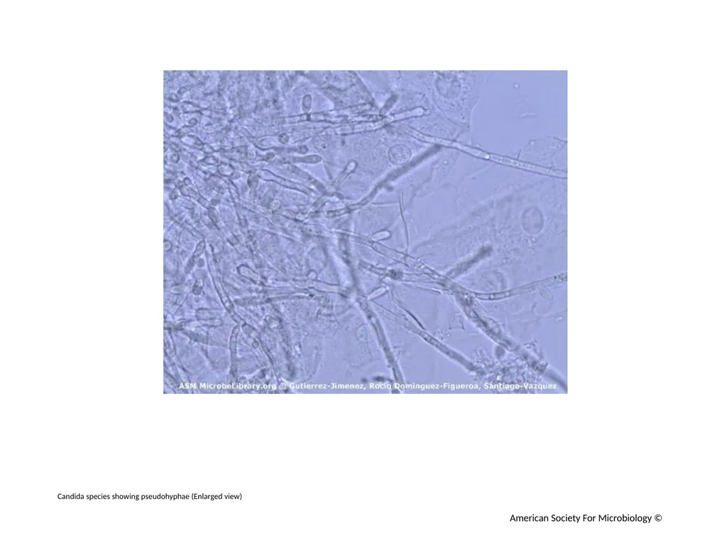
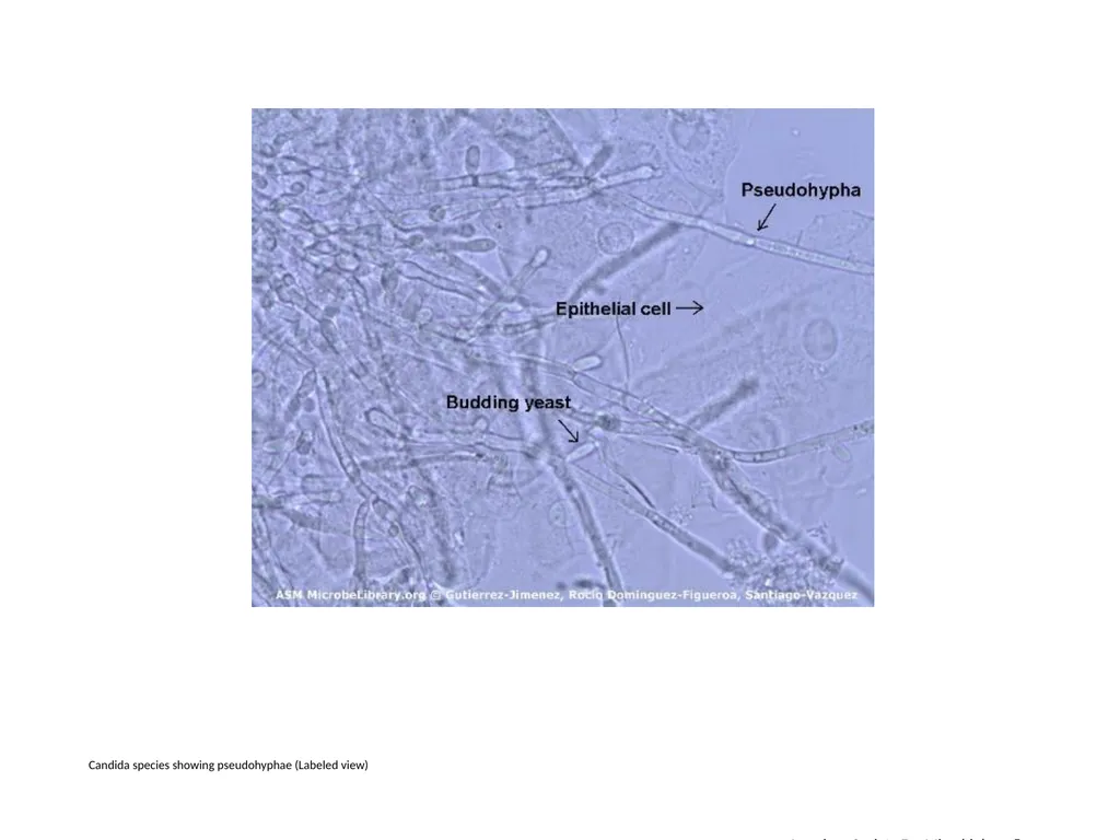
slide2. Introduction The image shows a direct wet mount prepared from a white vaginal discharge showing pseudohyphae, budding yeast, and human epithelial cells.  Examination of a fresh vaginal discharge provides the simplest and most rapid diagnostic test in aiding presumptive identification of Candida species.  
Methods
The vaginal discharge was collected from a 37-year-old female with dysuria and vulvovaginal pruritus.  The sample was diluted with equal parts of physiologic sterile saline and applied directly to the surface of a slide. A coverslip was placed over the surface of the sample, and excess liquid was blotted from around the edges.
References
1.  Buxton, R. 2007.  Examination of Gram stains of vaginal secretions.  ASM Microbe Library.
2.  Casasone, A., F. De Berrnardis,  and G. Santoni. 2007. Anticandidal immunity and vaginitis: novel opportunities for immune intervention. Infec. Immun. 75:4675–4686.
3.  Gilligan, P. H., M. L. Smiley, and D. S. Shapiro. 1997. Cases in medical microbiology and infectious diseases. p. 2–4. ASM Press, Washington, DC.
4. Jo Baron, E., and S. M. Finegold. 1990. Bailey & Scott´s diagnostic microbiology, p. 66, 263–269. Mosby, St. Louis, MO.<br>
slide3. Candida species showing pseudohyphae (Enlarged view) American Society For Microbiology ©<br>
slide4. Candida species showing pseudohyphae (Labeled view) American Society For Microbiology ©<br>