
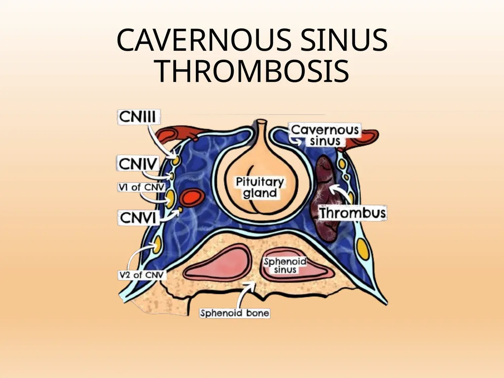
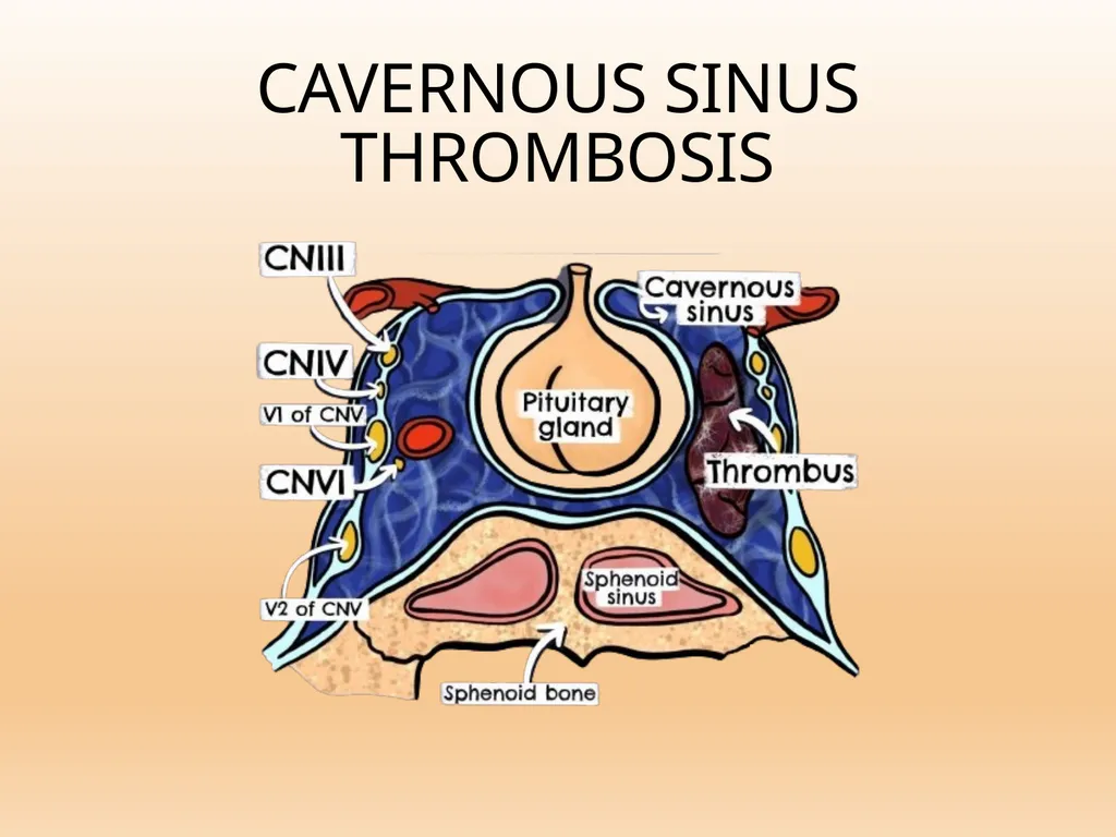
CAVERNOUS SINUS THROMBOSIS Cavernous Sinus Anatomy
PC
Published · 29 slides · 0 views
1 / 1


Description
CAVERNOUS SINUS THROMBOSIS Cavernous Sinus Anatomy Large venous space situated in the middle cranial fossa, on either side of body of the sphenoid bone. Each sinus is about 2 cm long and 1 cm wide. Interior is divided into a number of
Related Topics
Share
Embed code
Download this presentation From Below
"CAVERNOUS SINUS THROMBOSIS Cavernous Sinus Anatomy" is the property of its rightful owner. Permission is granted to download and print the materials on this website for personal, non-commercial use only, and to display it on your personal computer provided you do not modify the materials and that you retain all copyright notices contained in the materials. By downloading content from our website, you accept the terms of this agreement.
Presentation Transcript
01
CAVERNOUS SINUS THROMBOSIS<br>
02
Cavernous Sinus Anatomy Large venous space situated in the middle cranial fossa, on either side of body of the sphenoid bone.
Each sinus is about 2 cm long and 1 cm wide.
Interior is divided into a number of spaces or caverns by trabeculae.<br>
Each sinus is about 2 cm long and 1 cm wide.
Interior is divided into a number of spaces or caverns by trabeculae.<br>
03
Cavernous Sinus Anatomy
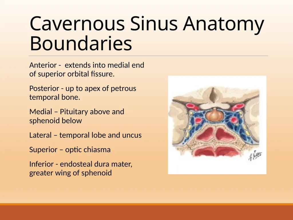
Boundaries Anterior - extends into medial end of superior orbital fissure.
Posterior - up to apex of petrous temporal bone.
Medial – Pituitary above and sphenoid below
Lateral – temporal lobe and uncus
Superior – optic chiasma
Inferior - endosteal dura mater, greater wing of sphenoid<br>
Posterior - up to apex of petrous temporal bone.
Medial – Pituitary above and sphenoid below
Lateral – temporal lobe and uncus
Superior – optic chiasma
Inferior - endosteal dura mater, greater wing of sphenoid<br>
04
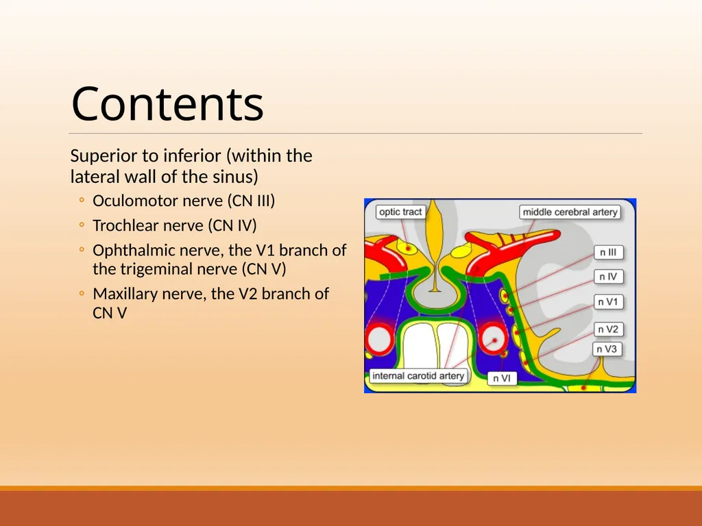
Contents Superior to inferior (within the lateral wall of the sinus)
Oculomotor nerve (CN III)
Trochlear nerve (CN IV)
Ophthalmic nerve, the V1 branch of the trigeminal nerve (CN V)
Maxillary nerve, the V2 branch of CN V<br>
Oculomotor nerve (CN III)
Trochlear nerve (CN IV)
Ophthalmic nerve, the V1 branch of the trigeminal nerve (CN V)
Maxillary nerve, the V2 branch of CN V<br>
05
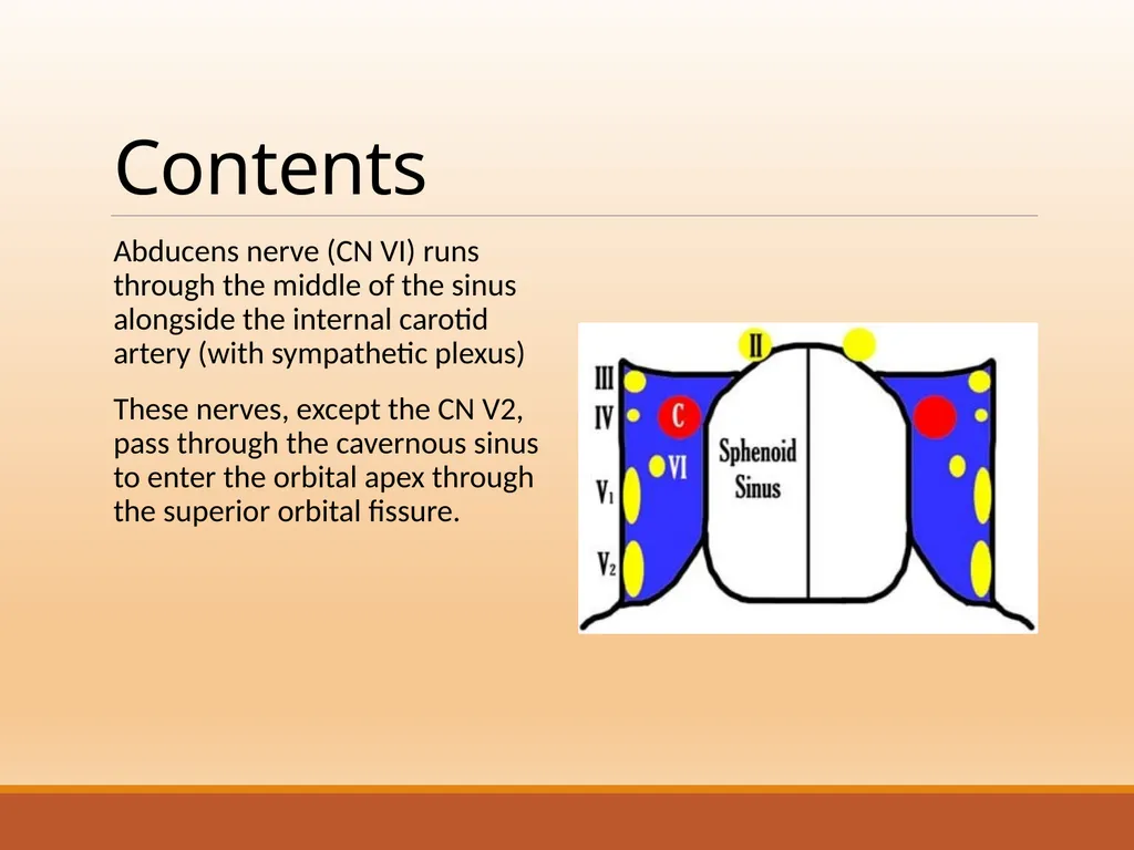
Contents Abducens nerve (CN VI) runs through the middle of the sinus alongside the internal carotid artery (with sympathetic plexus)
These nerves, except the CN V2, pass through the cavernous sinus to enter the orbital apex through the superior orbital fissure.<br>
These nerves, except the CN V2, pass through the cavernous sinus to enter the orbital apex through the superior orbital fissure.<br>
06
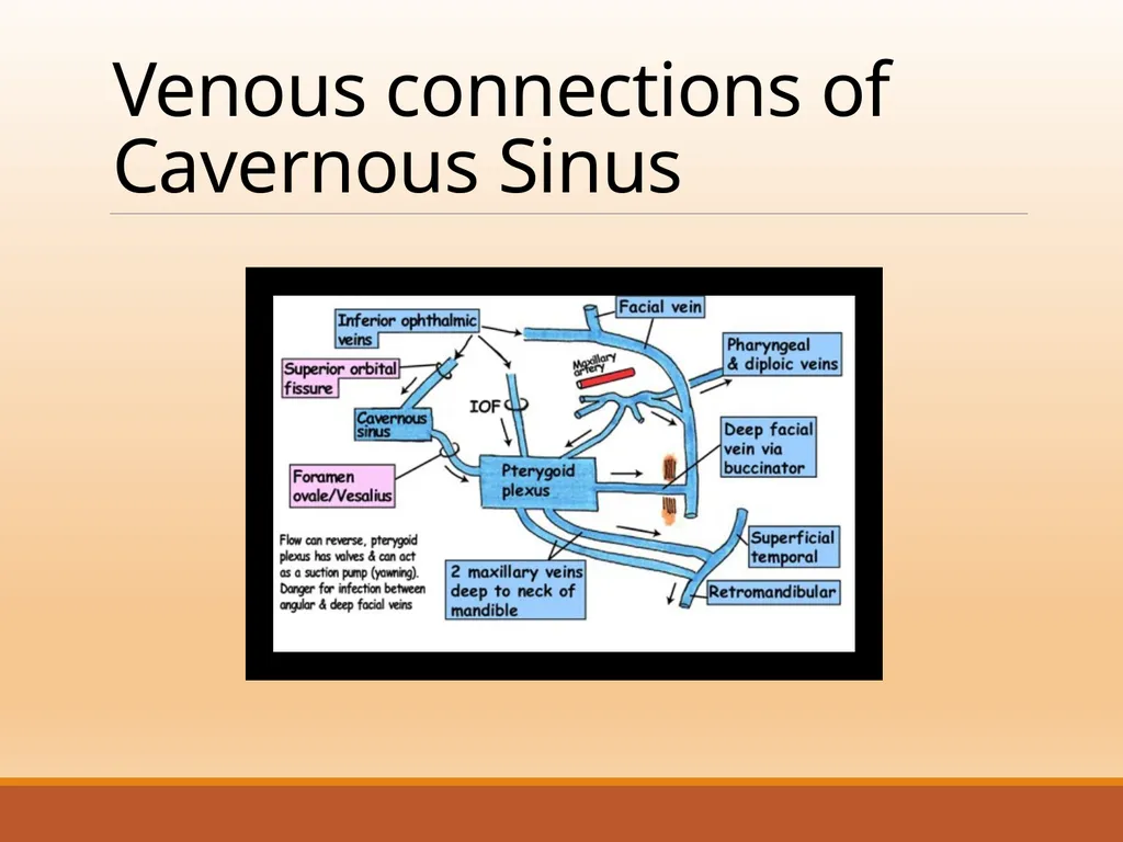
Venous connections of Cavernous Sinus<br>
07
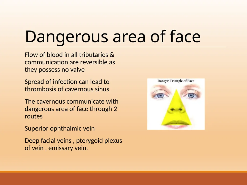
Dangerous area of face Flow of blood in all tributaries & communication are reversible as they possess no valve
Spread of infection can lead to thrombosis of cavernous sinus
The cavernous communicate with dangerous area of face through 2 routes
Superior ophthalmic vein
Deep facial veins , pterygoid plexus of vein , emissary vein.<br>
Spread of infection can lead to thrombosis of cavernous sinus
The cavernous communicate with dangerous area of face through 2 routes
Superior ophthalmic vein
Deep facial veins , pterygoid plexus of vein , emissary vein.<br>
08
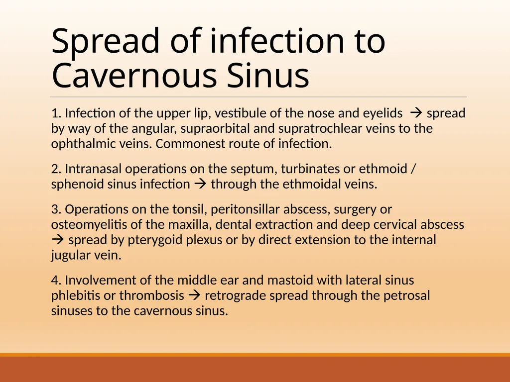
Spread of infection to Cavernous Sinus 1. Infection of the upper lip, vestibule of the nose and eyelids spread by way of the angular, supraorbital and supratrochlear veins to the ophthalmic veins. Commonest route of infection.
2. Intranasal operations on the septum, turbinates or ethmoid / sphenoid sinus infection through the ethmoidal veins.
3. Operations on the tonsil, peritonsillar abscess, surgery or osteomyelitis of the maxilla, dental extraction and deep cervical abscess spread by pterygoid plexus or by direct extension to the internal jugular vein.
4. Involvement of the middle ear and mastoid with lateral sinus phlebitis or thrombosis retrograde spread through the petrosal sinuses to the cavernous sinus.<br>
2. Intranasal operations on the septum, turbinates or ethmoid / sphenoid sinus infection through the ethmoidal veins.
3. Operations on the tonsil, peritonsillar abscess, surgery or osteomyelitis of the maxilla, dental extraction and deep cervical abscess spread by pterygoid plexus or by direct extension to the internal jugular vein.
4. Involvement of the middle ear and mastoid with lateral sinus phlebitis or thrombosis retrograde spread through the petrosal sinuses to the cavernous sinus.<br>
09
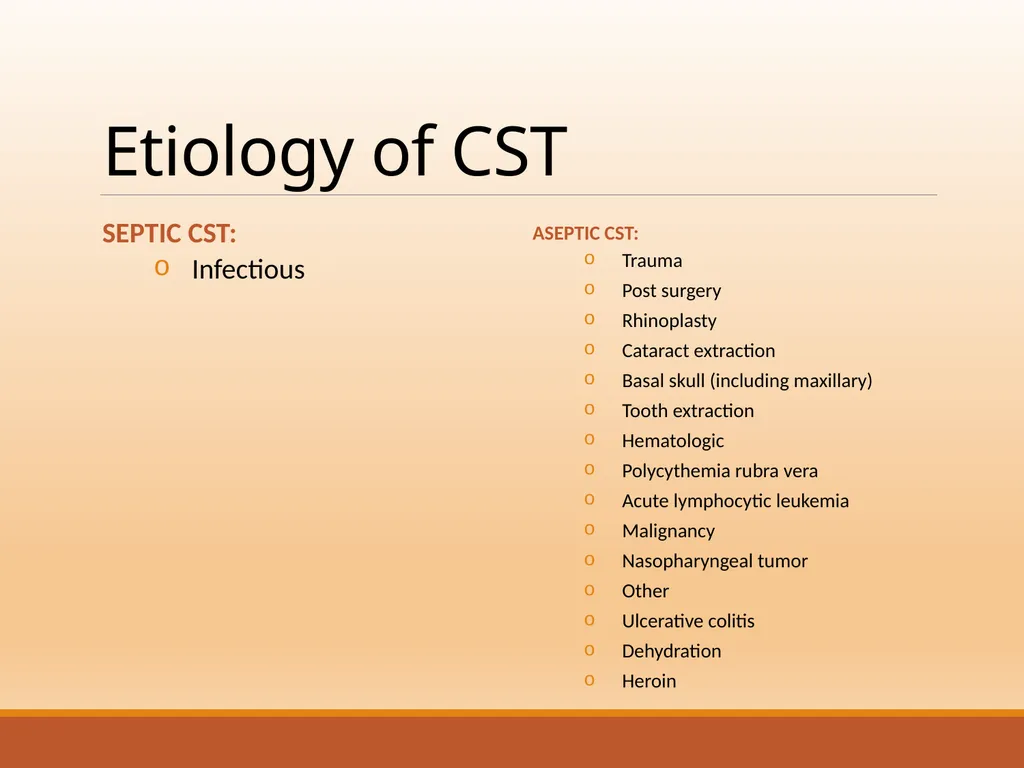
Etiology of CST Septic CST:
Infectious Aseptic CST:
Trauma
Post surgery
Rhinoplasty
Cataract extraction
Basal skull (including maxillary)
Tooth extraction
Hematologic
Polycythemia rubra vera
Acute lymphocytic leukemia
Malignancy
Nasopharyngeal tumor
Other
Ulcerative colitis
Dehydration
Heroin<br>
Infectious Aseptic CST:
Trauma
Post surgery
Rhinoplasty
Cataract extraction
Basal skull (including maxillary)
Tooth extraction
Hematologic
Polycythemia rubra vera
Acute lymphocytic leukemia
Malignancy
Nasopharyngeal tumor
Other
Ulcerative colitis
Dehydration
Heroin<br>
10
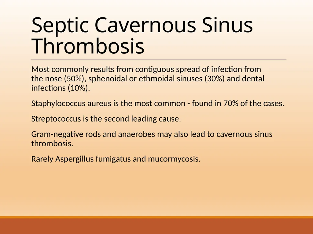
Septic Cavernous Sinus Thrombosis Most commonly results from contiguous spread of infection from the nose (50%), sphenoidal or ethmoidal sinuses (30%) and dental infections (10%).
Staphylococcus aureus is the most common - found in 70% of the cases.
Streptococcus is the second leading cause.
Gram-negative rods and anaerobes may also lead to cavernous sinus thrombosis.
Rarely Aspergillus fumigatus and mucormycosis.<br>
Staphylococcus aureus is the most common - found in 70% of the cases.
Streptococcus is the second leading cause.
Gram-negative rods and anaerobes may also lead to cavernous sinus thrombosis.
Rarely Aspergillus fumigatus and mucormycosis.<br>
11
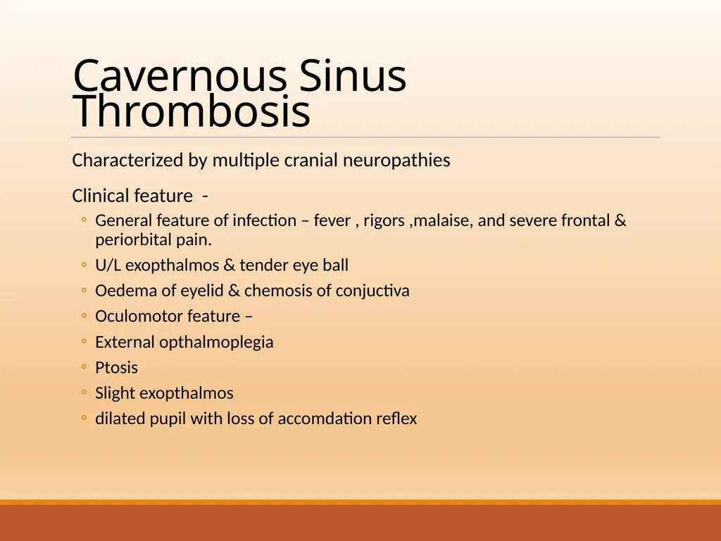
Cavernous Sinus Thrombosis Characterized by multiple cranial neuropathies
Clinical feature -
General feature of infection – fever , rigors ,malaise, and severe frontal & periorbital pain.
U/L exopthalmos & tender eye ball
Oedema of eyelid & chemosis of conjuctiva
Oculomotor feature –
External opthalmoplegia
Ptosis
Slight exopthalmos
dilated pupil with loss of accomdation reflex<br>
Clinical feature -
General feature of infection – fever , rigors ,malaise, and severe frontal & periorbital pain.
U/L exopthalmos & tender eye ball
Oedema of eyelid & chemosis of conjuctiva
Oculomotor feature –
External opthalmoplegia
Ptosis
Slight exopthalmos
dilated pupil with loss of accomdation reflex<br>
12
Cavernous Sinus thrombosis Impairment of ocular motor nerves, Horner’s syndrome and sensory loss of the first or second divisions of the trigeminal nerve in various combination
The pupil may be involved or spared or may appear spared with concomitant oculosympathetic involvement.<br>
The pupil may be involved or spared or may appear spared with concomitant oculosympathetic involvement.<br>
13
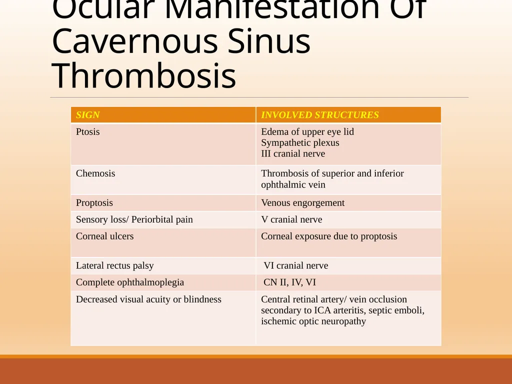
Ocular Manifestation Of Cavernous Sinus Thrombosis<br>
14
Complication of Cavernous Sinus Thrombosis Intracranial extension of infection may result in meningitis, encephalitis, brain abscess, pituitary infection, and epidural and subdural empyema.
Cortical vein thrombosis can result in hemorrhagic infarction.
Extension of the thrombus to other sinuses can occur.<br>
Cortical vein thrombosis can result in hemorrhagic infarction.
Extension of the thrombus to other sinuses can occur.<br>
15
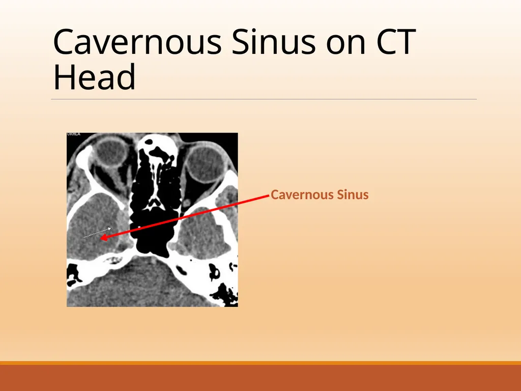
Cavernous Sinus on CT Head Cavernous Sinus<br>
16
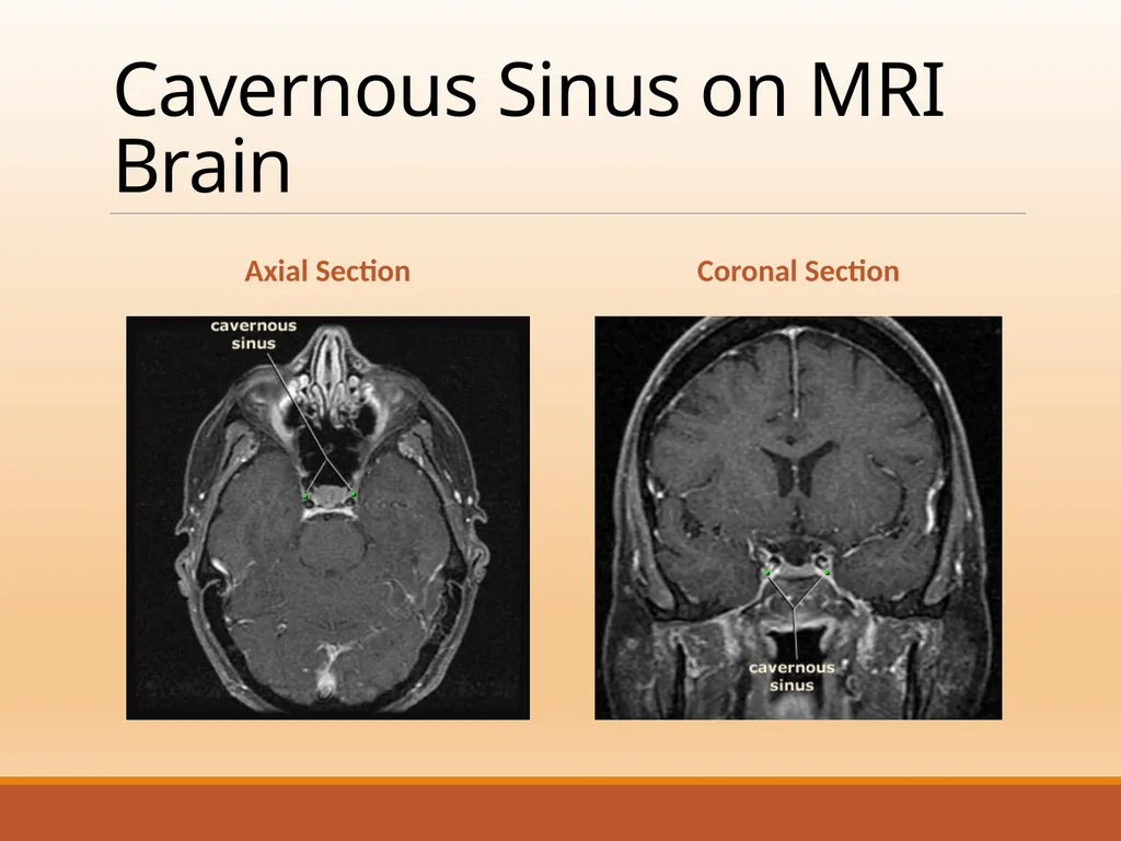
Cavernous Sinus on MRI Brain Axial Section Coronal Section<br>
17
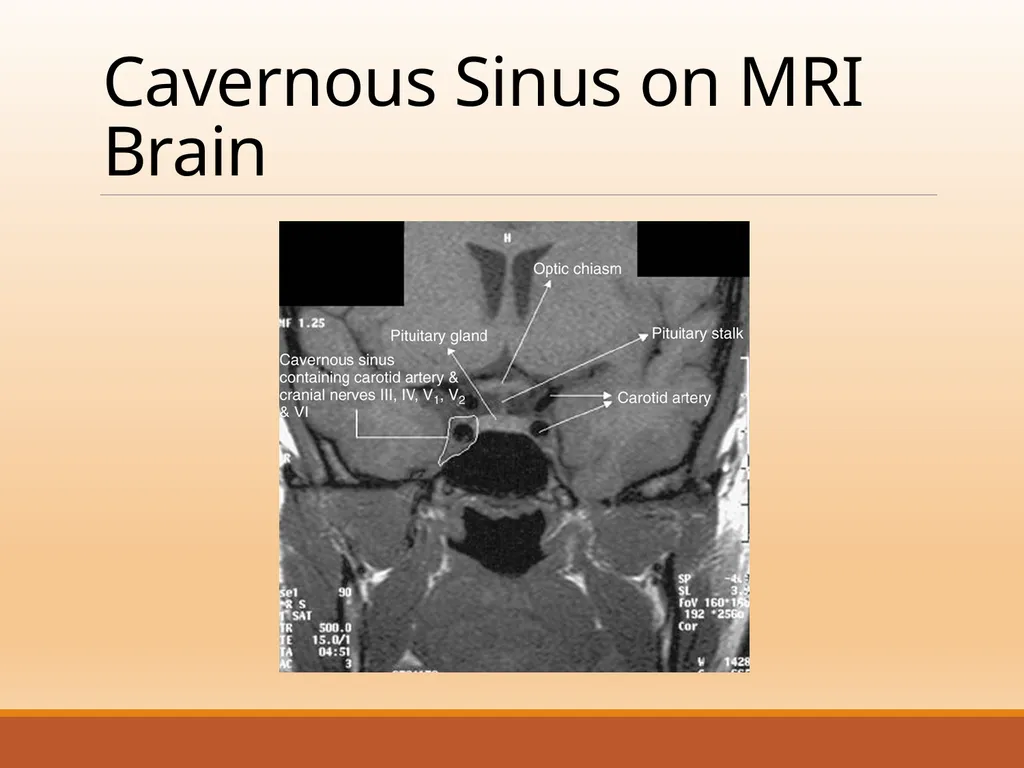
Cavernous Sinus on MRI Brain<br>
18
TREATMENT OF CAVERNOUS SINUS THROMBOSIS Septic cavernous sinus thrombosis –
The mainstay of therapy is early and aggressive antibiotic administration.
Although S aureus is the usual cause, broad-spectrum coverage for gram-positive, gram-negative, and anaerobic organisms should be instituted pending the outcome of cultures.
Empiric antibiotic therapy should include a penicillinase-resistant penicillin plus a third generation cephalosporin.
Vancomycin may be added for MRSA.
IV antibiotics are recommended for a minimum of 3-4 weeks.<br>
The mainstay of therapy is early and aggressive antibiotic administration.
Although S aureus is the usual cause, broad-spectrum coverage for gram-positive, gram-negative, and anaerobic organisms should be instituted pending the outcome of cultures.
Empiric antibiotic therapy should include a penicillinase-resistant penicillin plus a third generation cephalosporin.
Vancomycin may be added for MRSA.
IV antibiotics are recommended for a minimum of 3-4 weeks.<br>
19
TREATMENT OF CAVERNOUS SINUS THROMBOSIS The indication of anticoagulation is still debated because of possible bleeding complications and an eventual suppressive role of the thrombus on the extension of the infectious thrombophlebitis.
Although, no randomized controlled studies have been conducted, early anticoagulant therapy may have a beneficial effect on mortality and morbidity, reducing oculomotor sequelae, blindness, and motor sequelae as well as the risk of hypopituitarism. (studied in only 7 cases)
(Levine SR, Twyman RE, Gilman S: The role of anticoagulation in cavernous sinus thrombosis. Neurology, 1988; 38: 517–22)<br>
Although, no randomized controlled studies have been conducted, early anticoagulant therapy may have a beneficial effect on mortality and morbidity, reducing oculomotor sequelae, blindness, and motor sequelae as well as the risk of hypopituitarism. (studied in only 7 cases)
(Levine SR, Twyman RE, Gilman S: The role of anticoagulation in cavernous sinus thrombosis. Neurology, 1988; 38: 517–22)<br>
20
TREATMENT OF CAVERNOUS SINUS THROMBOSIS A Cochrane review found 2 small trials involving 79 patients who were treated with anticoagulants.
Limited evidence suggests anticoagulant drugs are probably safe and may be beneficial for people with sinus thrombosis.
Anticoagulation carries a significant risk of hemorrhage if cortical venous infarction or necrosis of intracavernous portions of the carotid artery are present.
Anticoagulant is contraindicated in the presence of intracerebral hemorrhage or other bleeding diathesis.<br>
Limited evidence suggests anticoagulant drugs are probably safe and may be beneficial for people with sinus thrombosis.
Anticoagulation carries a significant risk of hemorrhage if cortical venous infarction or necrosis of intracavernous portions of the carotid artery are present.
Anticoagulant is contraindicated in the presence of intracerebral hemorrhage or other bleeding diathesis.<br>
21
Prognosis 100% mortality prior to effective antimicrobials
Typically, death is due to sepsis or central nervous system (CNS) infection.
With aggressive management, the mortality rate is now less than 30%.
Morbidity, however, remains high, and complete recovery is rare.
Roughly one sixth of patients are left with some degree of visual impairment, and one half (50 %) have cranial nerve deficits.<br>
Typically, death is due to sepsis or central nervous system (CNS) infection.
With aggressive management, the mortality rate is now less than 30%.
Morbidity, however, remains high, and complete recovery is rare.
Roughly one sixth of patients are left with some degree of visual impairment, and one half (50 %) have cranial nerve deficits.<br>
22
Fungal Infection Intracranial extension is the most dreaded complication of fungal sinusitis with high mortality rates.
Aspergillus is the most common.
Mucor, rhizopus, cladosporium, candida, cryptococcus are amongst the others.
Mode of spread:
Hematogenous spread
Direct extension<br>
Aspergillus is the most common.
Mucor, rhizopus, cladosporium, candida, cryptococcus are amongst the others.
Mode of spread:
Hematogenous spread
Direct extension<br>
23
Fungal Infection Uncontrolled diabetics are more susceptible to mucormycosis .
The clinical signs of Mucormycosis are commonly opthalmoplegia, proptosis, blindness, palatal ulcer, coma and stupor.
Nasal examination shows black areas along the inferior turbinates, which on biopsy appear as non-septate hyphae.<br>
The clinical signs of Mucormycosis are commonly opthalmoplegia, proptosis, blindness, palatal ulcer, coma and stupor.
Nasal examination shows black areas along the inferior turbinates, which on biopsy appear as non-septate hyphae.<br>
24
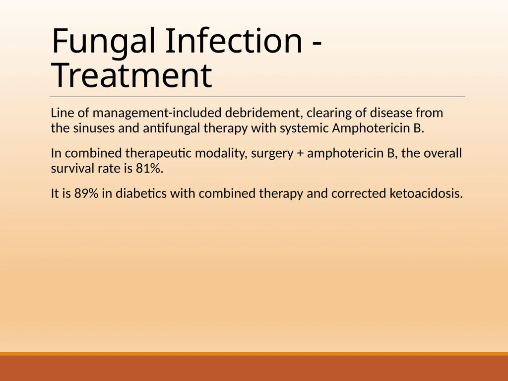
Fungal Infection - Treatment Line of management-included debridement, clearing of disease from the sinuses and antifungal therapy with systemic Amphotericin B.
In combined therapeutic modality, surgery + amphotericin B, the overall survival rate is 81%.
It is 89% in diabetics with combined therapy and corrected ketoacidosis.<br>
In combined therapeutic modality, surgery + amphotericin B, the overall survival rate is 81%.
It is 89% in diabetics with combined therapy and corrected ketoacidosis.<br>
25
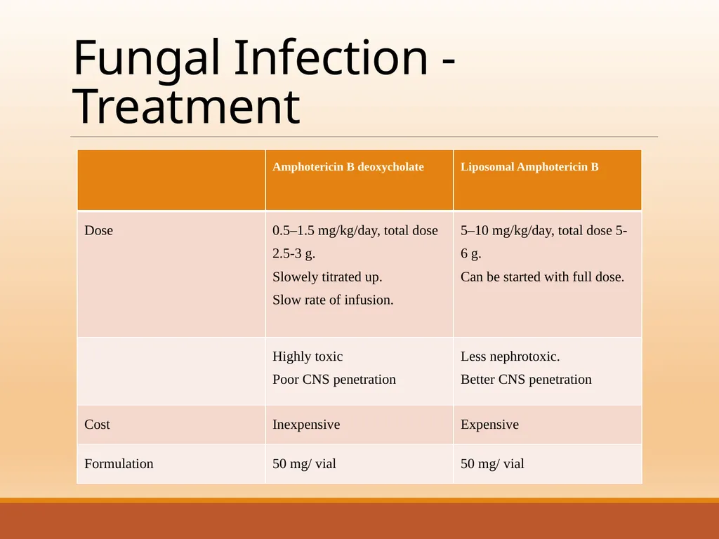
Fungal infection - treatment Amphotericin B deoxycholate (AmB) remains the only licensed antifungal agent for the treatment of mucormycosis.
However, lipid formulations of AmB (LFABs) are significantly less nephrotoxic and can be safely administered at higher doses for a longer period of time than AmB.
Patients who respond to a parenteral lipid amphotericin B-based treatment, given for at least 3 weeks, are transitioned to oral posaconazole as maintenance/secondary prophylaxis.<br>
However, lipid formulations of AmB (LFABs) are significantly less nephrotoxic and can be safely administered at higher doses for a longer period of time than AmB.
Patients who respond to a parenteral lipid amphotericin B-based treatment, given for at least 3 weeks, are transitioned to oral posaconazole as maintenance/secondary prophylaxis.<br>
26
Fungal Infection - Treatment<br>
27
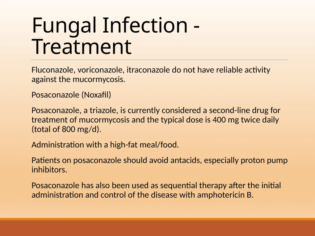
Fungal Infection - Treatment Fluconazole, voriconazole, itraconazole do not have reliable activity against the mucormycosis.
Posaconazole (Noxafil)
Posaconazole, a triazole, is currently considered a second-line drug for treatment of mucormycosis and the typical dose is 400 mg twice daily (total of 800 mg/d).
Administration with a high-fat meal/food.
Patients on posaconazole should avoid antacids, especially proton pump inhibitors.
Posaconazole has also been used as sequential therapy after the initial administration and control of the disease with amphotericin B.<br>
Posaconazole (Noxafil)
Posaconazole, a triazole, is currently considered a second-line drug for treatment of mucormycosis and the typical dose is 400 mg twice daily (total of 800 mg/d).
Administration with a high-fat meal/food.
Patients on posaconazole should avoid antacids, especially proton pump inhibitors.
Posaconazole has also been used as sequential therapy after the initial administration and control of the disease with amphotericin B.<br>
28
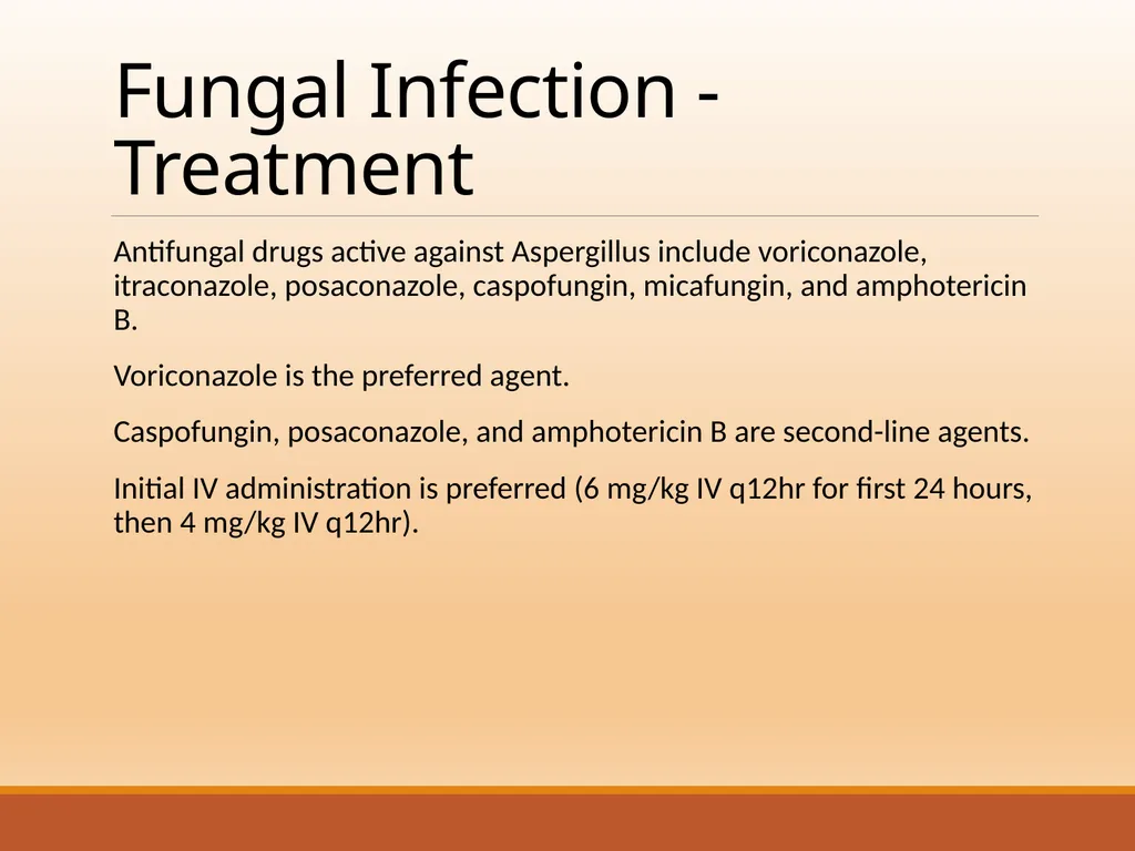
Fungal Infection - Treatment Antifungal drugs active against Aspergillus include voriconazole, itraconazole, posaconazole, caspofungin, micafungin, and amphotericin B.
Voriconazole is the preferred agent.
Caspofungin, posaconazole, and amphotericin B are second-line agents.
Initial IV administration is preferred (6 mg/kg IV q12hr for first 24 hours, then 4 mg/kg IV q12hr).<br>
Voriconazole is the preferred agent.
Caspofungin, posaconazole, and amphotericin B are second-line agents.
Initial IV administration is preferred (6 mg/kg IV q12hr for first 24 hours, then 4 mg/kg IV q12hr).<br>
29
THANK YOU!<br>