
CELL WALL: STRUCTURE AND DYNAMICS INTRODUCTION
CB
Published · 41 slides · 0 views
1 / 1


Description
CELL WALL: STRUCTURE AND DYNAMICS INTRODUCTION Cell wall was first observed and named simply as a wall by Robert Hooke in 1665. In 1804, Karl Rudolphi and J.H.F. Link proved that cells have independent cell walls. A cell wall is a
Related Topics
Share
Embed code
Download this presentation From Below
"CELL WALL: STRUCTURE AND DYNAMICS INTRODUCTION" is the property of its rightful owner. Permission is granted to download and print the materials on this website for personal, non-commercial use only, and to display it on your personal computer provided you do not modify the materials and that you retain all copyright notices contained in the materials. By downloading content from our website, you accept the terms of this agreement.
Presentation Transcript
01
CELL WALL: STRUCTURE AND DYNAMICS<br>
02
INTRODUCTION Cell wall was first observed and named simply as a “wall” by Robert Hooke in 1665.
In 1804, Karl Rudolphi and J.H.F. Link proved that cells have independent cell walls.
A cell wall is a structural layer that surrounds some types of cells, situated outside the cell membrane.
It can be tough, flexible and rigid which provides cell with both structural support and protection.<br>
In 1804, Karl Rudolphi and J.H.F. Link proved that cells have independent cell walls.
A cell wall is a structural layer that surrounds some types of cells, situated outside the cell membrane.
It can be tough, flexible and rigid which provides cell with both structural support and protection.<br>
03
INTRODUCTION On the basis of chemical composition of cell wall there are three types of cell wall:
Bacterial Cell Wall : made up of Mucopeptide and Muramic acid.
Cell wall of Fungi: made up of Chitin.
Plant Cell wall: made up of Cellulose.<br>
Bacterial Cell Wall : made up of Mucopeptide and Muramic acid.
Cell wall of Fungi: made up of Chitin.
Plant Cell wall: made up of Cellulose.<br>
04
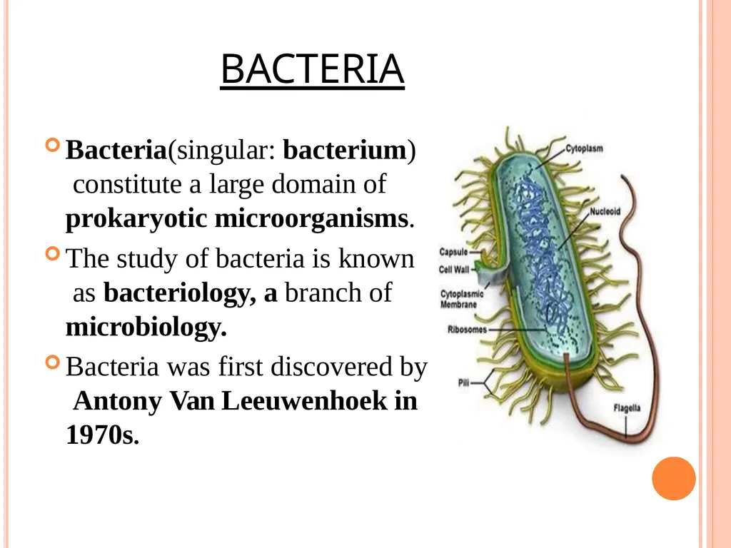
BACTERIA Bacteria(singular: bacterium) constitute a large domain of prokaryotic microorganisms.
The study of bacteria is known as bacteriology, a branch of microbiology.
Bacteria was first discovered by Antony Van Leeuwenhoek in 1970s.<br>
The study of bacteria is known as bacteriology, a branch of microbiology.
Bacteria was first discovered by Antony Van Leeuwenhoek in 1970s.<br>
05
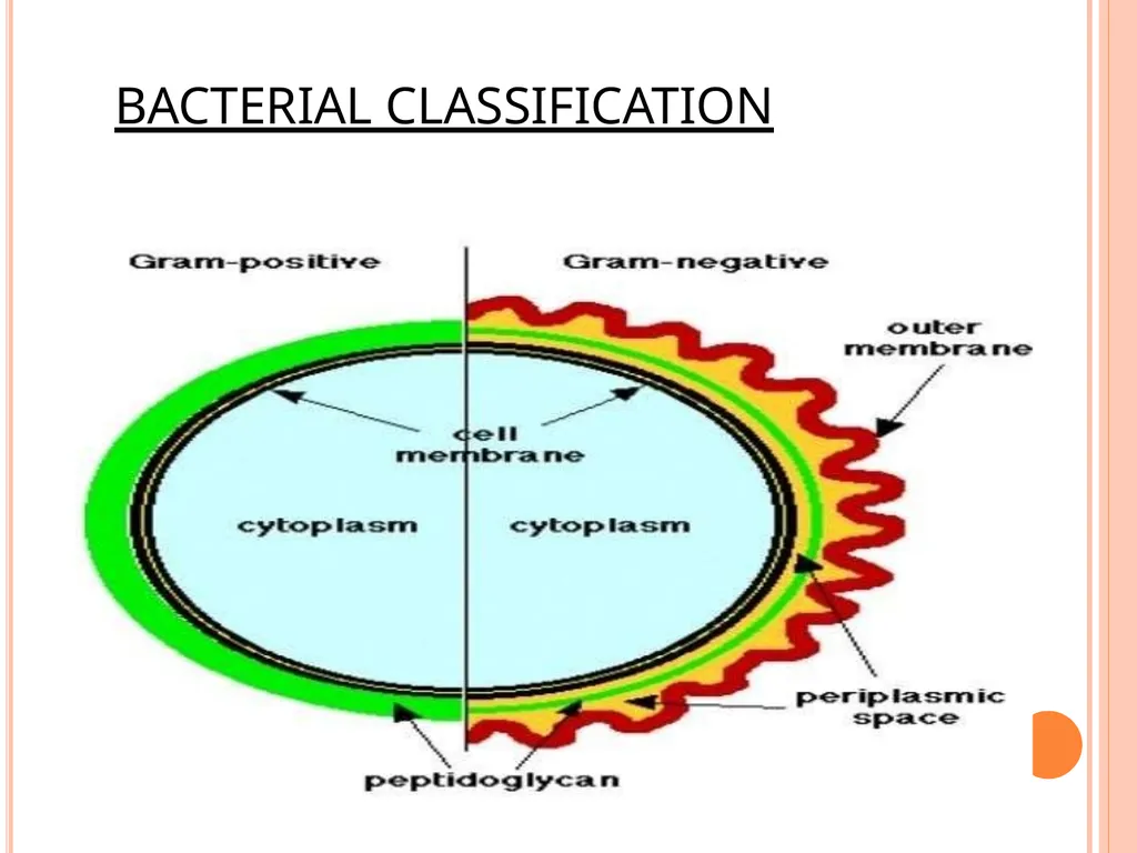
BACTERIAL CLASSIFICATION<br>
06
PEPTIDOGLYCAN Peptidoglycan ,also known as murein, is a polymer consisting of sugars and amino acids that forms a mesh – like layer outside the cell membrane of most bacteria forming cell wall.
The sugars component consist of alternating residues of ᵦ- (1,4) linked N- acetylglucosamine and N- acetylmuramic acid.
These subunits which are related to glucose in their structure are covalently joined to one another to form glycan chains.<br>
The sugars component consist of alternating residues of ᵦ- (1,4) linked N- acetylglucosamine and N- acetylmuramic acid.
These subunits which are related to glucose in their structure are covalently joined to one another to form glycan chains.<br>
07
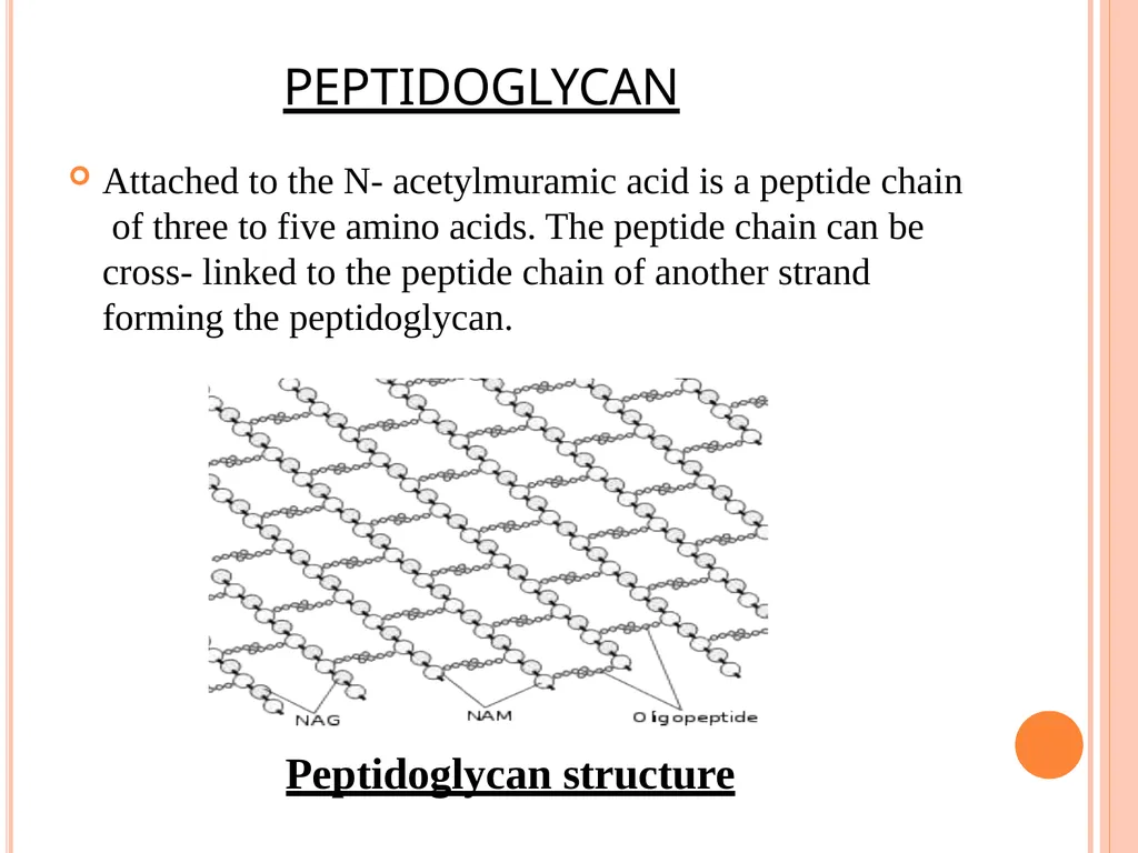
PEPTIDOGLYCAN Attached to the N- acetylmuramic acid is a peptide chain of three to five amino acids. The peptide chain can be cross- linked to the peptide chain of another strand forming the peptidoglycan. Peptidoglycan structure<br>
08
TYPES OF BACTERIAL CELL WALL On the based structure of the cell wall and to their response to stain, bacteria have been classified into two types:
Gram Positive Bacteria
Gram Negative Bacteria<br>
Gram Positive Bacteria
Gram Negative Bacteria<br>
09
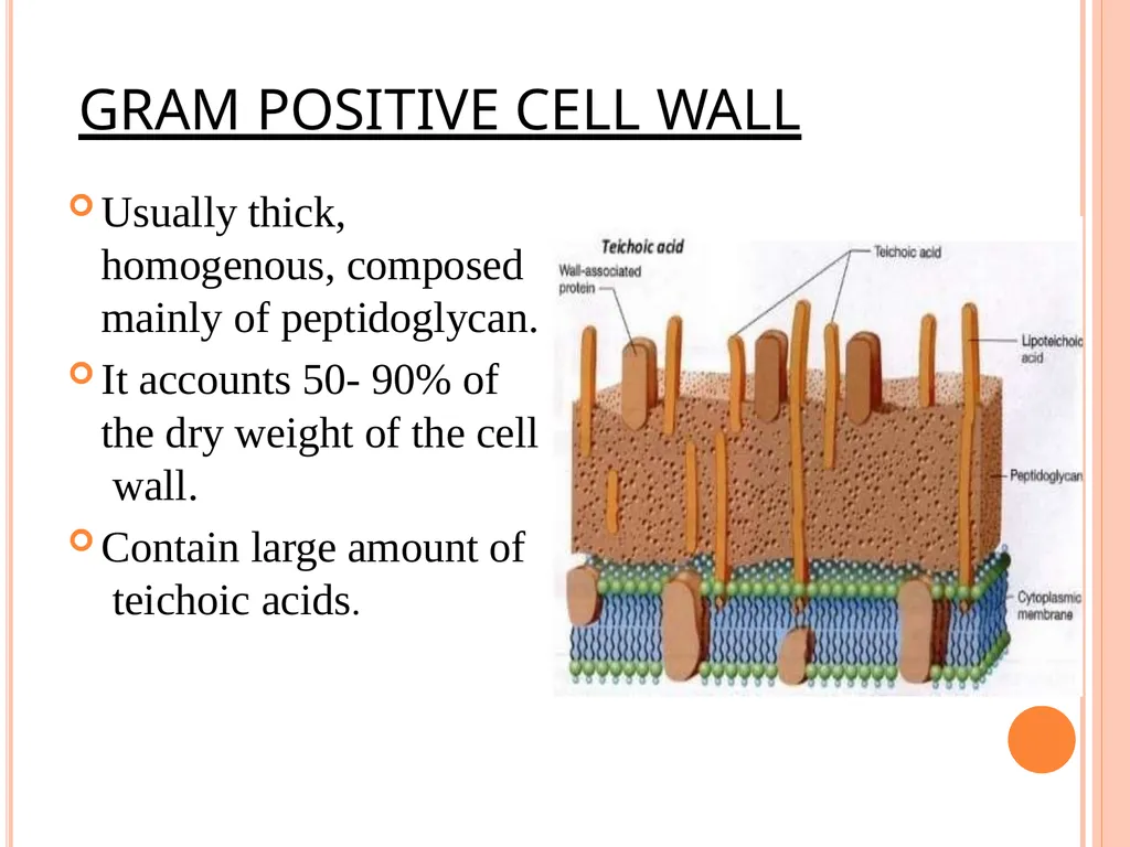
GRAM POSITIVE CELL WALL Usually thick, homogenous, composed mainly of peptidoglycan.
It accounts 50- 90% of the dry weight of the cell wall.
Contain large amount of teichoic acids.<br>
It accounts 50- 90% of the dry weight of the cell wall.
Contain large amount of teichoic acids.<br>
10
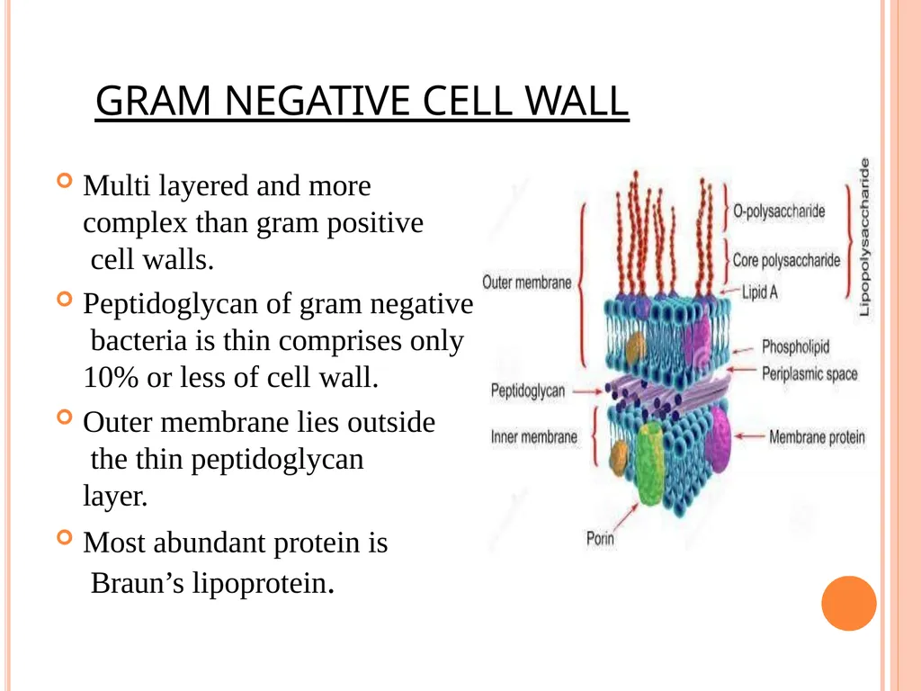
GRAM NEGATIVE CELL WALL Multi layered and more complex than gram positive cell walls.
Peptidoglycan of gram negative bacteria is thin comprises only 10% or less of cell wall.
Outer membrane lies outside the thin peptidoglycan layer.
Most abundant protein is Braun’s lipoprotein.<br>
Peptidoglycan of gram negative bacteria is thin comprises only 10% or less of cell wall.
Outer membrane lies outside the thin peptidoglycan layer.
Most abundant protein is Braun’s lipoprotein.<br>
11
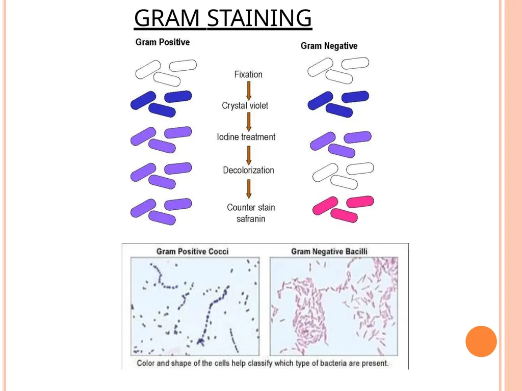
GRAM STAINING<br>
12
FUNCTIONS OF BACTERIAL CELL WALL Protect cells against osmotic shock and physical damage.
Regulation of substance transport into and out of cells.
Contain supplemental genetic information such as resistance to antibiotics, production of toxins and tolerance to toxic environment.
Take part in protein synthesis.
Movement of cells.
Mineral storage of cells.<br>
Regulation of substance transport into and out of cells.
Contain supplemental genetic information such as resistance to antibiotics, production of toxins and tolerance to toxic environment.
Take part in protein synthesis.
Movement of cells.
Mineral storage of cells.<br>
13
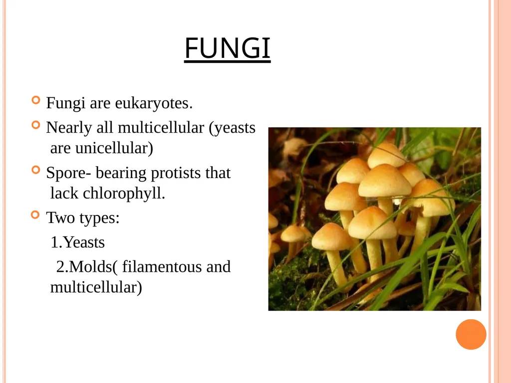
FUNGI Fungi are eukaryotes.
Nearly all multicellular (yeasts are unicellular)
Spore- bearing protists that lack chlorophyll.
Two types: 1.Yeasts
2.Molds( filamentous and multicellular)<br>
Nearly all multicellular (yeasts are unicellular)
Spore- bearing protists that lack chlorophyll.
Two types: 1.Yeasts
2.Molds( filamentous and multicellular)<br>
14
FUNGAL CELL WALL The cell wall is made up of :
Chitin (polymers of acetylated amino sugar N-acetyl- glucosamine)
2.Glucans 3.Proteins
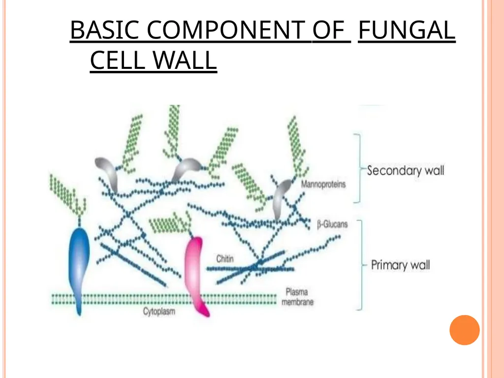
Glucan and Chitin are components of the primary wall.
Proteins are components of the secondary wall.
Other components include chitosan, melanins and lipids.<br>
Chitin (polymers of acetylated amino sugar N-acetyl- glucosamine)
2.Glucans 3.Proteins
Glucan and Chitin are components of the primary wall.
Proteins are components of the secondary wall.
Other components include chitosan, melanins and lipids.<br>
15
BASIC COMPONENT OF FUNGAL CELL WALL<br>
16
FUNCTION OF FUNGAL CELL WALL Protects against osmotic lysis.
Acts as a molecular sieve.
Contains pigments for protection.
Binding sites for enzymes.
The outermost surface of the cell wall
provides a medium between the cell and the environment.
a site where antigen and agglutinin gets attached to the substrate, host and other cells.<br>
Acts as a molecular sieve.
Contains pigments for protection.
Binding sites for enzymes.
The outermost surface of the cell wall
provides a medium between the cell and the environment.
a site where antigen and agglutinin gets attached to the substrate, host and other cells.<br>
17
PLANT CELL WALL The plant cell wall is a remarkable structure. It provides the most significant difference between plant cells and other eukaryotic cells.
The wall is rigid(up to many micrometers in thickness) and gives plant cells a very defined shape.
While most cells have a outer membrane , none is comparable in strength to the plant cell wall. The cell wall is the reason for the difference between plant and animal cell functions. Because the plant has evolved this rigid structure.<br>
The wall is rigid(up to many micrometers in thickness) and gives plant cells a very defined shape.
While most cells have a outer membrane , none is comparable in strength to the plant cell wall. The cell wall is the reason for the difference between plant and animal cell functions. Because the plant has evolved this rigid structure.<br>
18
COMPONENTS OF PLANT CELL WALL The plant cell wall composed of :
The Middle Lamella
The Primary Cell Wall 3.The Secondary Cell Wall
4. The Tertiary Cell Wall<br>
The Middle Lamella
The Primary Cell Wall 3.The Secondary Cell Wall
4. The Tertiary Cell Wall<br>
19
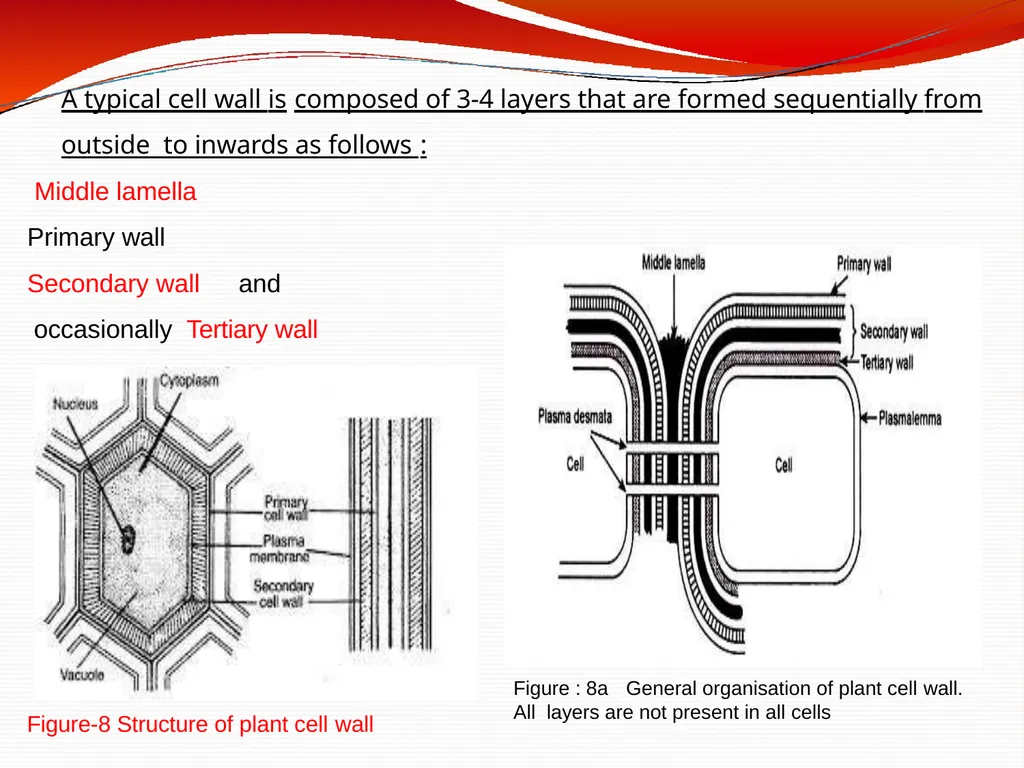
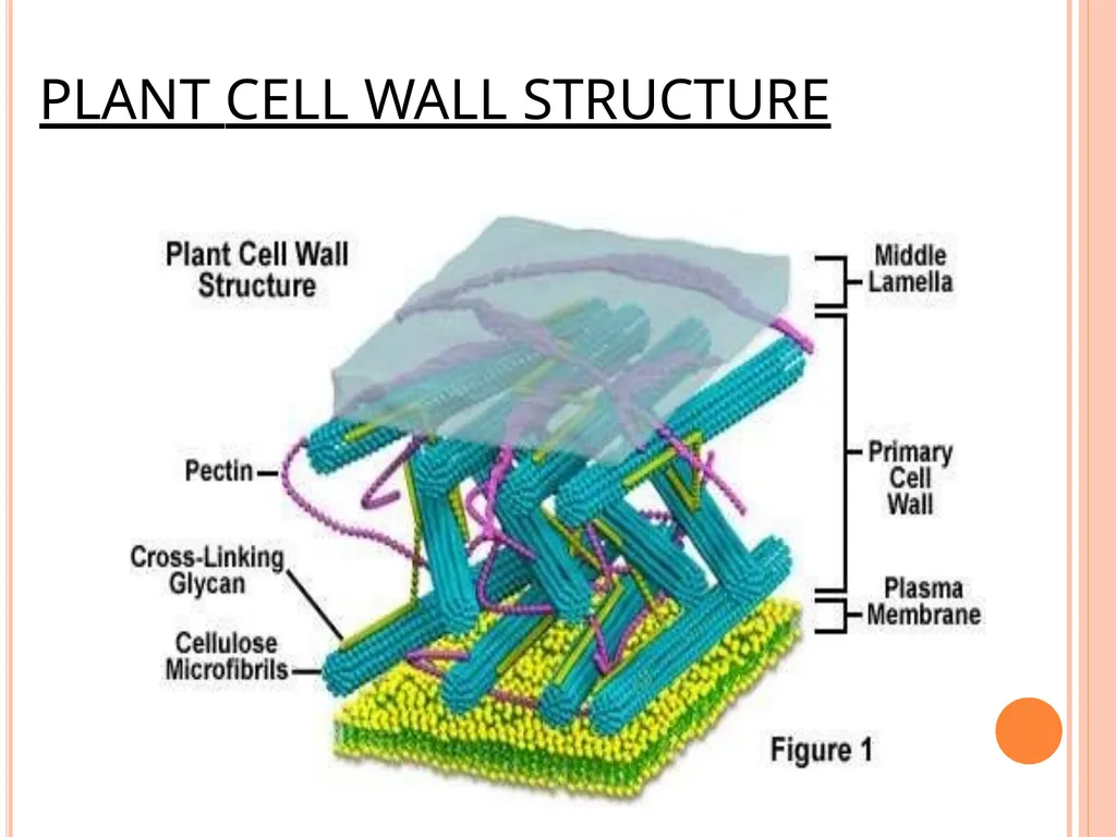
Figure : 8a General organisation of plant cell wall. All layers are not present in all cells Figure-8 Structure of plant cell wall A typical cell wall is composed of 3-4 layers that are formed sequentially from outside to inwards as follows : Middle lamella
Primary wall
Secondary wall and occasionally Tertiary wall<br>
Primary wall
Secondary wall and occasionally Tertiary wall<br>
20
PLANT CELL WALL STRUCTURE<br>
21
Middle lamella is the first layer to be formed when a cell divides .
It is an amorphous intercellular layer between primary walls of adjacent cells .
It is cementing layer made up of Ca and Mg pectinate/pectate which joins or glues two neighbouring plant cells.
It is absent on the free surface of plant cells and in plasmadesmata region. Middle lamella dissolves in ripe fruits which results in softening.
Middle lamella can be dissolved artificially by means of acid treatment ( during root tip cytological preparation ) The cell walls and the middle lamella of plants never occur in the form of continuous layers but have many minute apertures through which the cells of a tissue maintain cytoplasmic relations with each other.<br>
It is an amorphous intercellular layer between primary walls of adjacent cells .
It is cementing layer made up of Ca and Mg pectinate/pectate which joins or glues two neighbouring plant cells.
It is absent on the free surface of plant cells and in plasmadesmata region. Middle lamella dissolves in ripe fruits which results in softening.
Middle lamella can be dissolved artificially by means of acid treatment ( during root tip cytological preparation ) The cell walls and the middle lamella of plants never occur in the form of continuous layers but have many minute apertures through which the cells of a tissue maintain cytoplasmic relations with each other.<br>
22
Primary cell wall
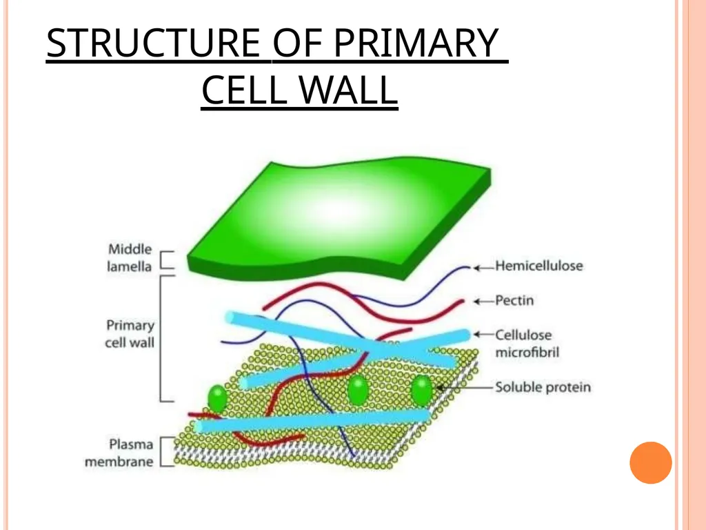
The primary cell wall, generally a thin, flexible and extensible layer formed while the cell is growing.
It is the first deposition product of protoplasm outside plasma membrane and is present inner to middle lamella.
Primary cell wall is delicate,flexible and thin (0.1-3.0µm) and capable of further extension.
Its thickness increases with the growth of the plant cell. It grows by deposition of wall material into the existing primary wall. Parenchymatous,meristematic cells and cell involved in photosynthesis,
respiration and secretion and unicellular plants have only primary cell wall.<br>
The primary cell wall, generally a thin, flexible and extensible layer formed while the cell is growing.
It is the first deposition product of protoplasm outside plasma membrane and is present inner to middle lamella.
Primary cell wall is delicate,flexible and thin (0.1-3.0µm) and capable of further extension.
Its thickness increases with the growth of the plant cell. It grows by deposition of wall material into the existing primary wall. Parenchymatous,meristematic cells and cell involved in photosynthesis,
respiration and secretion and unicellular plants have only primary cell wall.<br>
23
Both the structure and function of cell walls change as plant cells develop.
The thickness, as well as the composition and organization of cell walls can vary significantly.
The walls of growing plant cells (called primary cell walls) are relatively thin and
flexible, allowing the cell to expand in size.
The microfibrils of the cellulose are deposited on both sides of the middle lamella and form the primary cell wall
Once cells have ceased growth, they frequently lay down secondary cell walls between the plasma membrane and the primary cell wall
Such secondary cell walls, which are both thicker and more rigid than primary walls, are particularly important in cell types responsible for conducting water and providing mechanical strength to the plant.<br>
The thickness, as well as the composition and organization of cell walls can vary significantly.
The walls of growing plant cells (called primary cell walls) are relatively thin and
flexible, allowing the cell to expand in size.
The microfibrils of the cellulose are deposited on both sides of the middle lamella and form the primary cell wall
Once cells have ceased growth, they frequently lay down secondary cell walls between the plasma membrane and the primary cell wall
Such secondary cell walls, which are both thicker and more rigid than primary walls, are particularly important in cell types responsible for conducting water and providing mechanical strength to the plant.<br>
24
STRUCTURE OF PRIMARY CELL WALL<br>
25
Secondary wall Secondary cell wall – it is often deposited on inner side of primary walls after the growth of cell stops ( cell matures).
It is 4-10 µm thick, rigid, non-elastic, permeable and made up of cellulose and is not lignin deposits.
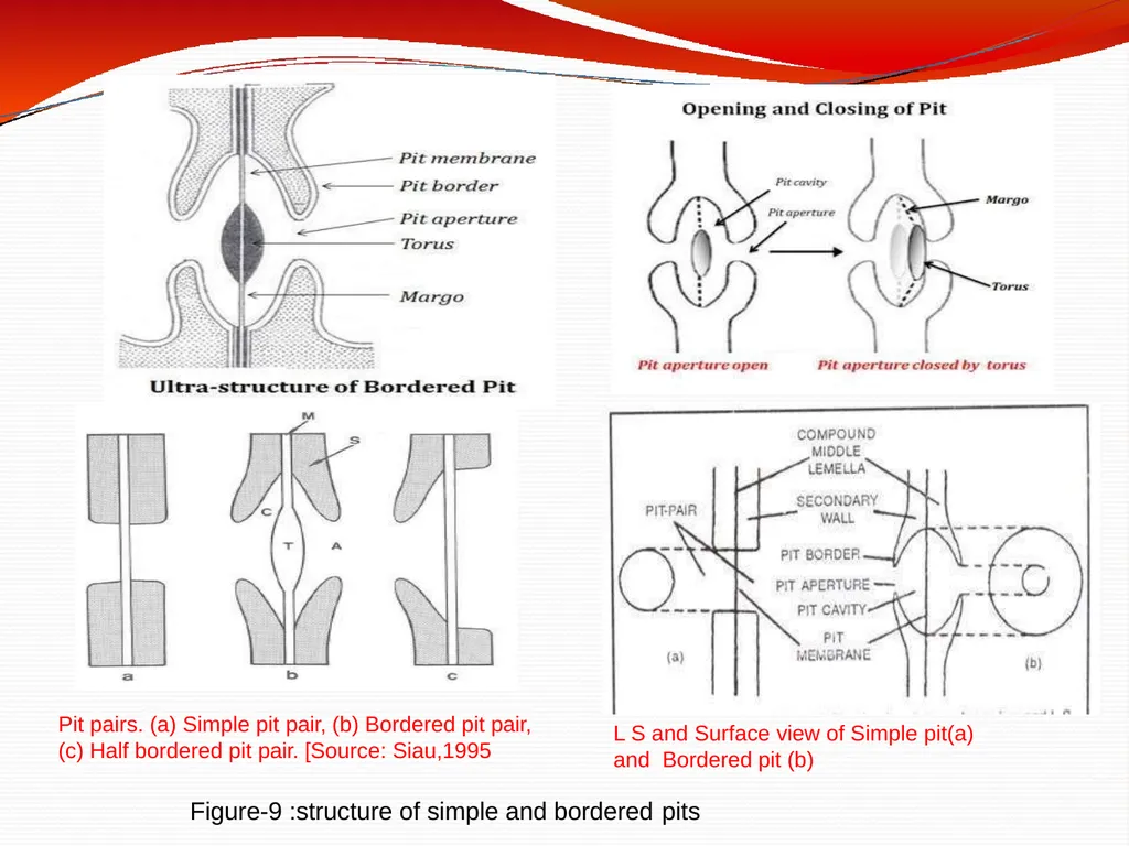
Secondary wall deposition is not uniform. At some places secondary wall laid down. Such unthickened areas are called pits .Pits of two neighbouring cell form pit pair. Pits can be of two types – simple or bordered , pit chamber becomes flask shaped due to deposition in the form of border. (Figure-9)
During the development of pits, the secondary cell wall may over arch the pit
cavity forming a border, leaving an inner opening called pit-aperture. Such pits
with borders are called bordered pits. Two opposite bordered pits are called bordered pit pair.<br>
It is 4-10 µm thick, rigid, non-elastic, permeable and made up of cellulose and is not lignin deposits.
Secondary wall deposition is not uniform. At some places secondary wall laid down. Such unthickened areas are called pits .Pits of two neighbouring cell form pit pair. Pits can be of two types – simple or bordered , pit chamber becomes flask shaped due to deposition in the form of border. (Figure-9)
During the development of pits, the secondary cell wall may over arch the pit
cavity forming a border, leaving an inner opening called pit-aperture. Such pits
with borders are called bordered pits. Two opposite bordered pits are called bordered pit pair.<br>
26
through border pit shows two circles , one of pit and other of A transverse section through simple pit shows only one circle and transverse section border.
Bordered pits are present in tracheids of Gymnosperms and vessels of angiosperms. Pits which lack the borders are called simple pits. Two opposite simple pits on
adjacent cells are called simple pit pair.
Plasmodesmata (Singular Plasmodesma ) A number of fine cytoplasmic strands (20-40 nm in diameter) pass through pits from one cell to other and make connection between the cytoplasm of two cells.
Endoplasmic reticulum plays a significant role in origin of plasmodesmata . Plasmodesmata were studied in details by Strasburger (1901).
Plasmodesmata help in transfer of nutrients, stimuli and other material between
adjacent cells and thus Produce a protoplasmic continuum called symplast.<br>
Bordered pits are present in tracheids of Gymnosperms and vessels of angiosperms. Pits which lack the borders are called simple pits. Two opposite simple pits on
adjacent cells are called simple pit pair.
Plasmodesmata (Singular Plasmodesma ) A number of fine cytoplasmic strands (20-40 nm in diameter) pass through pits from one cell to other and make connection between the cytoplasm of two cells.
Endoplasmic reticulum plays a significant role in origin of plasmodesmata . Plasmodesmata were studied in details by Strasburger (1901).
Plasmodesmata help in transfer of nutrients, stimuli and other material between
adjacent cells and thus Produce a protoplasmic continuum called symplast.<br>
27
Pit pairs. (a) Simple pit pair, (b) Bordered pit pair,
(c) Half bordered pit pair. [Source: Siau,1995 L S and Surface view of Simple pit(a) and Bordered pit (b) Figure-9 :structure of simple and bordered pits<br>
(c) Half bordered pit pair. [Source: Siau,1995 L S and Surface view of Simple pit(a) and Bordered pit (b) Figure-9 :structure of simple and bordered pits<br>
28
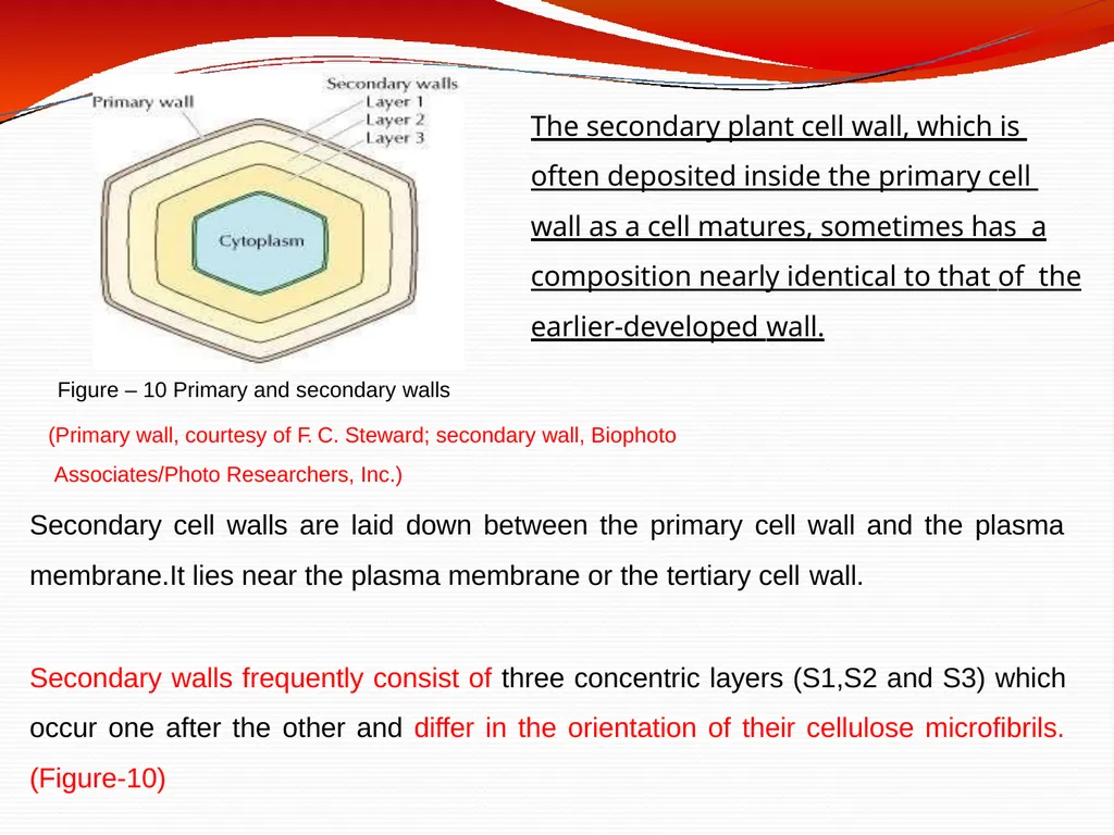
Figure – 10 Primary and secondary walls
(Primary wall, courtesy of F. C. Steward; secondary wall, Biophoto Associates/Photo Researchers, Inc.)
Secondary cell walls are laid down between the primary cell wall and the plasma membrane.It lies near the plasma membrane or the tertiary cell wall.
Secondary walls frequently consist of three concentric layers (S1,S2 and S3) which occur one after the other and differ in the orientation of their cellulose microfibrils. (Figure-10) The secondary plant cell wall, which is often deposited inside the primary cell wall as a cell matures, sometimes has a composition nearly identical to that of the earlier-developed wall.<br>
(Primary wall, courtesy of F. C. Steward; secondary wall, Biophoto Associates/Photo Researchers, Inc.)
Secondary cell walls are laid down between the primary cell wall and the plasma membrane.It lies near the plasma membrane or the tertiary cell wall.
Secondary walls frequently consist of three concentric layers (S1,S2 and S3) which occur one after the other and differ in the orientation of their cellulose microfibrils. (Figure-10) The secondary plant cell wall, which is often deposited inside the primary cell wall as a cell matures, sometimes has a composition nearly identical to that of the earlier-developed wall.<br>
29
Primary and secondary cell walls differ in composition as well as in thickness. Primary cell walls contain approximately equal amounts of cellulose, hemicelluloses, and pectins.
In contrast, the more rigid secondary walls generally lack pectin and contain 50 to 80% cellulose.
Many secondary walls are further strengthened by lignin, a complex polymer of phenolic residues(aromatic alcohols ) that is responsible for much of the strength and density of wood. (Figure-11)
The orientation of cellulose microfibrils also differs in primary and secondary cell walls. In primary wall microfibrils are short, wavy and loosely scattered.
In secondary wall microfibrils are long, straight, close and parallely arranged
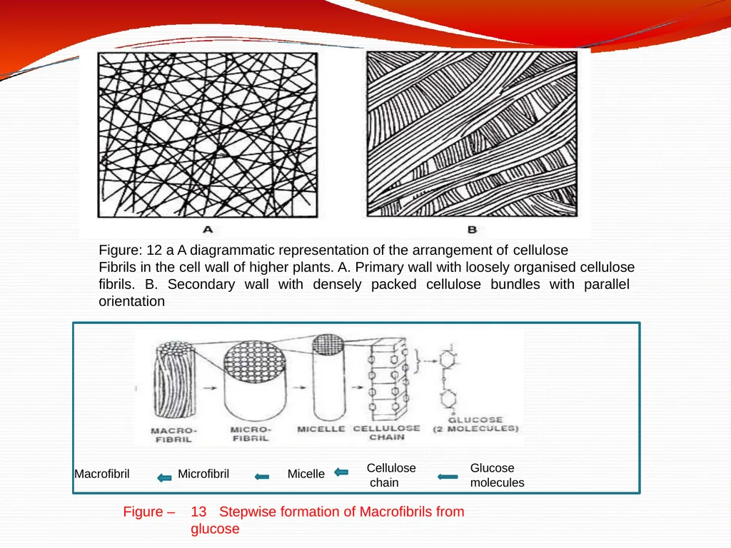
The cellulose fibers of primary walls appear to be randomly arranged, whereas
those of secondary walls are highly ordered (Figure -12 a )<br>
In contrast, the more rigid secondary walls generally lack pectin and contain 50 to 80% cellulose.
Many secondary walls are further strengthened by lignin, a complex polymer of phenolic residues(aromatic alcohols ) that is responsible for much of the strength and density of wood. (Figure-11)
The orientation of cellulose microfibrils also differs in primary and secondary cell walls. In primary wall microfibrils are short, wavy and loosely scattered.
In secondary wall microfibrils are long, straight, close and parallely arranged
The cellulose fibers of primary walls appear to be randomly arranged, whereas
those of secondary walls are highly ordered (Figure -12 a )<br>
30
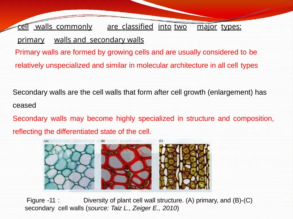
Figure -11 : Diversity of plant cell wall structure. (A) primary, and (B)-(C) secondary cell walls (source: Taiz L., Zeiger E., 2010) cell walls commonly are classified into two major types: primary walls and secondary walls Primary walls are formed by growing cells and are usually considered to be
relatively unspecialized and similar in molecular architecture in all cell types
Secondary walls are the cell walls that form after cell growth (enlargement) has ceased
Secondary walls may become highly specialized in structure and composition,
reflecting the differentiated state of the cell.<br>
relatively unspecialized and similar in molecular architecture in all cell types
Secondary walls are the cell walls that form after cell growth (enlargement) has ceased
Secondary walls may become highly specialized in structure and composition,
reflecting the differentiated state of the cell.<br>
31
Secondary walls are frequently laid down in layers in which the cellulose fibers differ in orientation, forming a laminated structure that greatly increases cell wall strength.
In certain plant cells, there occurs another cell wall beneath the secondary cell
wall which is known as tertiary cell wall. It differs from the primary and secondary cell wall in terms of its morphology, chemistry and staining properties.
Tertiary wall –In some tissue a tertiary cell wall is formed on the inner surface of the secondary cell wall. This layer is very thin and is found in the xylem tracheids of gymnosperms. Taxus – Tertiary spiral thickening makes the wood elastic and strong, so is used for making bows. It is composed mainly of xylan, instead of cellulose. It is not found in all the cells. Typically, it does not contain any cellulose micro-fibrils.<br>
In certain plant cells, there occurs another cell wall beneath the secondary cell
wall which is known as tertiary cell wall. It differs from the primary and secondary cell wall in terms of its morphology, chemistry and staining properties.
Tertiary wall –In some tissue a tertiary cell wall is formed on the inner surface of the secondary cell wall. This layer is very thin and is found in the xylem tracheids of gymnosperms. Taxus – Tertiary spiral thickening makes the wood elastic and strong, so is used for making bows. It is composed mainly of xylan, instead of cellulose. It is not found in all the cells. Typically, it does not contain any cellulose micro-fibrils.<br>
32
Ultra-structure of plant cell wall The primary wall and the secondary wall have the same basic structure.
In both cases cellulose micro-fibrils (chief constituent) are found embedded in an
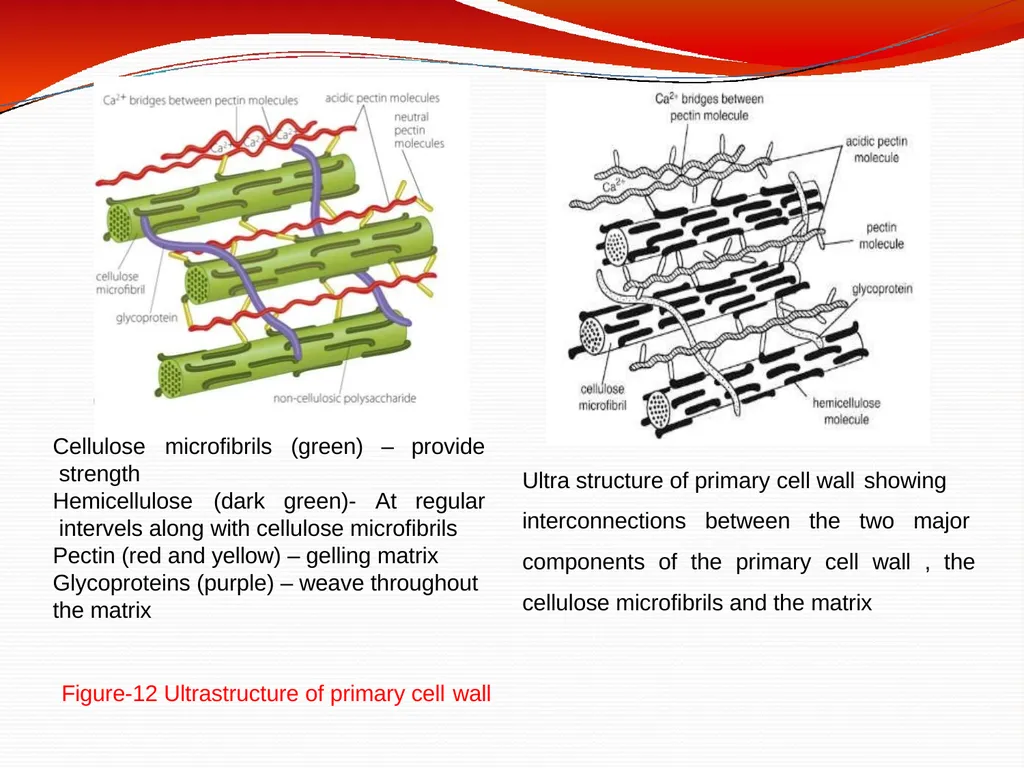
amorphous gel-like matrix (consisting of proteins and two polysaccharides : hemicelluloses and pectins) (Figure-12)
Each cellulose chain (1 -5 µm long) consists of about 2000-25000 glucose units. Nearly 100 cellulose chains arranged parallel to form minute bundle called crystalline domain or micelle (1.0 nm thick).
Micelle is the smallest structural unit of cell wall. About 20-40 micelles assemble in the matrix to form a microfibril (2.6 nm thick).
Nearly 250 microfibrils aggregate in bigger bundles called macrofibrils (~ 0.5 µm in diameter, may reach, 4µm in length). (Figure- 13)
A cotton fibre has 1500 macro fibrils.<br>
In both cases cellulose micro-fibrils (chief constituent) are found embedded in an
amorphous gel-like matrix (consisting of proteins and two polysaccharides : hemicelluloses and pectins) (Figure-12)
Each cellulose chain (1 -5 µm long) consists of about 2000-25000 glucose units. Nearly 100 cellulose chains arranged parallel to form minute bundle called crystalline domain or micelle (1.0 nm thick).
Micelle is the smallest structural unit of cell wall. About 20-40 micelles assemble in the matrix to form a microfibril (2.6 nm thick).
Nearly 250 microfibrils aggregate in bigger bundles called macrofibrils (~ 0.5 µm in diameter, may reach, 4µm in length). (Figure- 13)
A cotton fibre has 1500 macro fibrils.<br>
33
Ultra structure of primary cell wall showing interconnections between the two major components of the primary cell wall , the cellulose microfibrils and the matrix Cellulose microfibrils (green) – provide strength Hemicellulose (dark green)- At regular intervels along with cellulose microfibrils Pectin (red and yellow) – gelling matrix Glycoproteins (purple) – weave throughout the matrix Figure-12 Ultrastructure of primary cell wall<br>
34
Figure: 12 a A diagrammatic representation of the arrangement of cellulose
Fibrils in the cell wall of higher plants. A. Primary wall with loosely organised cellulose fibrils. B. Secondary wall with densely packed cellulose bundles with parallel orientation Glucose molecules Cellulose chain Microfibril Micelle Macrofibril Figure – 13 Stepwise formation of Macrofibrils from glucose<br>
Fibrils in the cell wall of higher plants. A. Primary wall with loosely organised cellulose fibrils. B. Secondary wall with densely packed cellulose bundles with parallel orientation Glucose molecules Cellulose chain Microfibril Micelle Macrofibril Figure – 13 Stepwise formation of Macrofibrils from glucose<br>
35
Plant cell walls encase the plant cells and provide many structural and functional roles.
A major role of the cell wall is to form a framework for the cell to prevent over
expansion.
Cellulose fibers, structural proteins, and other polysaccharides help to maintain the shape and form of the cell.
The cell wall provides mechanical strength and support. It also controls the
direction of cell growth.
Cell wall protects the cell (protoplast) against loss of water, excessive heat and foreign attacks(plant virus and other pathgens ).
It bestows definite shape ,supporting frame work and rigidity to cell.
Provides mechanical strength in higher plants having vascular system It protects the cell from mechanical injury. Functions of cell wall<br>
A major role of the cell wall is to form a framework for the cell to prevent over
expansion.
Cellulose fibers, structural proteins, and other polysaccharides help to maintain the shape and form of the cell.
The cell wall provides mechanical strength and support. It also controls the
direction of cell growth.
Cell wall protects the cell (protoplast) against loss of water, excessive heat and foreign attacks(plant virus and other pathgens ).
It bestows definite shape ,supporting frame work and rigidity to cell.
Provides mechanical strength in higher plants having vascular system It protects the cell from mechanical injury. Functions of cell wall<br>
36
Cells communicate with one another via plasmodesmata ( fine cytoplasmic strands) between plant cell walls and form a which pass through pores or channels present system of interconnected protoplasts
(two adjacent cells )called the symplast. It provides a porous medium for circulation (movement) of water, minerals and other nutrients from between adjacent cells
Cuticle present on outer surface of epidermal cells (leaves) and suberin present in periderm ( bark) prevents water loss.
The cell wall has an important function in regulating how plant cells achieve their final size and shape and consequently have an essential role in regulating plant growth.
Some of the proteins of cell wall possess catalytic activity by acting as enzymes to
polymerize wall monomers, enzymes that cross-link polymers and enzymes that cleave polymers.<br>
(two adjacent cells )called the symplast. It provides a porous medium for circulation (movement) of water, minerals and other nutrients from between adjacent cells
Cuticle present on outer surface of epidermal cells (leaves) and suberin present in periderm ( bark) prevents water loss.
The cell wall has an important function in regulating how plant cells achieve their final size and shape and consequently have an essential role in regulating plant growth.
Some of the proteins of cell wall possess catalytic activity by acting as enzymes to
polymerize wall monomers, enzymes that cross-link polymers and enzymes that cleave polymers.<br>
37
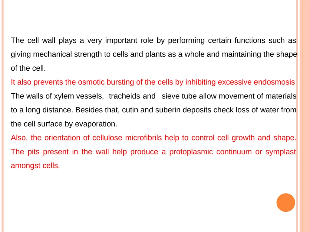
The cell wall plays a very important role by performing certain functions such as giving mechanical strength to cells and plants as a whole and maintaining the shape of the cell.
It also prevents the osmotic bursting of the cells by inhibiting excessive endosmosis The walls of xylem vessels, tracheids and sieve tube allow movement of materials to a long distance. Besides that, cutin and suberin deposits check loss of water from the cell surface by evaporation.
Also, the orientation of cellulose microfibrils help to control cell growth and shape. The pits present in the wall help produce a protoplasmic continuum or symplast amongst cells.<br>
It also prevents the osmotic bursting of the cells by inhibiting excessive endosmosis The walls of xylem vessels, tracheids and sieve tube allow movement of materials to a long distance. Besides that, cutin and suberin deposits check loss of water from the cell surface by evaporation.
Also, the orientation of cellulose microfibrils help to control cell growth and shape. The pits present in the wall help produce a protoplasmic continuum or symplast amongst cells.<br>
38
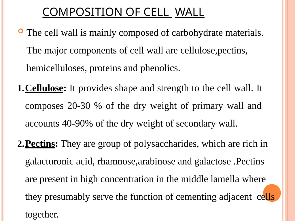
COMPOSITION OF CELL WALL The cell wall is mainly composed of carbohydrate materials. The major components of cell wall are cellulose,pectins, hemicelluloses, proteins and phenolics.
Cellulose: It provides shape and strength to the cell wall. It composes 20-30 % of the dry weight of primary wall and accounts 40-90% of the dry weight of secondary wall.
Pectins: They are group of polysaccharides, which are rich in galacturonic acid, rhamnose,arabinose and galactose .Pectins are present in high concentration in the middle lamella where they presumably serve the function of cementing adjacent cells together.<br>
Cellulose: It provides shape and strength to the cell wall. It composes 20-30 % of the dry weight of primary wall and accounts 40-90% of the dry weight of secondary wall.
Pectins: They are group of polysaccharides, which are rich in galacturonic acid, rhamnose,arabinose and galactose .Pectins are present in high concentration in the middle lamella where they presumably serve the function of cementing adjacent cells together.<br>
39
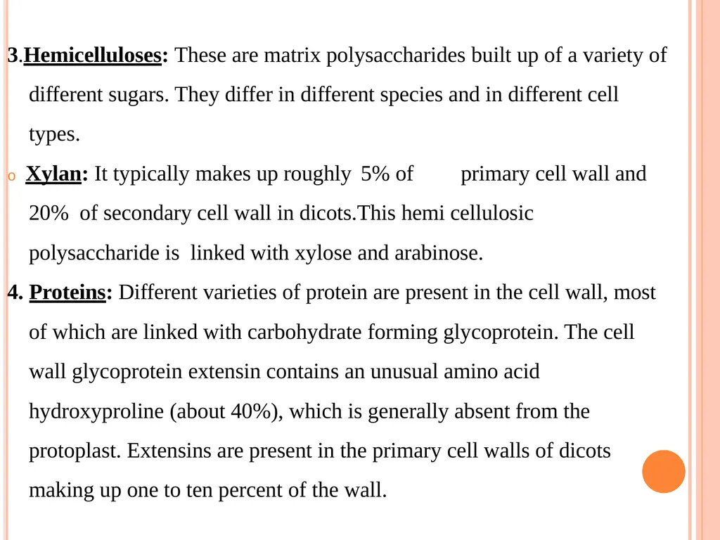
3.Hemicelluloses: These are matrix polysaccharides built up of a variety of different sugars. They differ in different species and in different cell types.
o Xylan: It typically makes up roughly 5% of primary cell wall and 20% of secondary cell wall in dicots.This hemi cellulosic polysaccharide is linked with xylose and arabinose.
4. Proteins: Different varieties of protein are present in the cell wall, most of which are linked with carbohydrate forming glycoprotein. The cell wall glycoprotein extensin contains an unusual amino acid hydroxyproline (about 40%), which is generally absent from the protoplast. Extensins are present in the primary cell walls of dicots making up one to ten percent of the wall.<br>
o Xylan: It typically makes up roughly 5% of primary cell wall and 20% of secondary cell wall in dicots.This hemi cellulosic polysaccharide is linked with xylose and arabinose.
4. Proteins: Different varieties of protein are present in the cell wall, most of which are linked with carbohydrate forming glycoprotein. The cell wall glycoprotein extensin contains an unusual amino acid hydroxyproline (about 40%), which is generally absent from the protoplast. Extensins are present in the primary cell walls of dicots making up one to ten percent of the wall.<br>
40
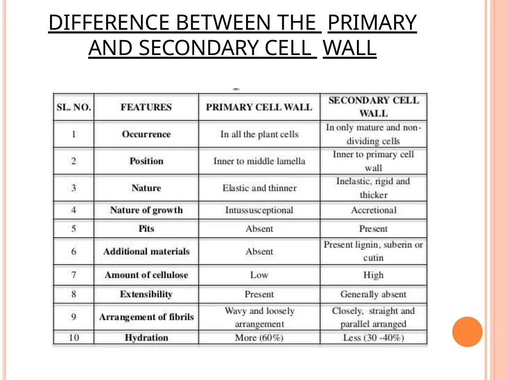
DIFFERENCE BETWEEN THE PRIMARY AND SECONDARY CELL WALL<br>