
CHAPTER 18 Neurologic Emergencies National EMS






Description: CHAPTER 18 Neurologic Emergencies National EMS Education Standard Competencies (1 of 3) Medicine Applies fundamental knowledge to provide basic emergency care and transportation based on assessment findings for an acutely ill patient.
Related Topics
Download Presentation
"CHAPTER 18 Neurologic Emergencies National EMS" is the property of its rightful owner. Permission is granted to download and print the materials on this website for personal, non-commercial use only, and to display it on your personal computer provided you do not modify the materials and that you retain all copyright notices contained in the materials. By downloading content from our website, you accept the terms of this agreement.
Presentation Transcript
slide1. CHAPTER 18 Neurologic Emergencies<br>
slide2. National EMS Education Standard Competencies (1 of 3) Medicine
Applies fundamental knowledge to provide basic emergency care and transportation based on assessment findings for an acutely ill patient.<br>
slide3. National EMS Education Standard Competencies (2 of 3) Neurology
Anatomy, presentations, and management of
Decreased level of responsiveness
Seizure
Stroke<br>
slide4. National EMS Education Standard Competencies (3 of 3) Neurology (cont’d)
Anatomy, physiology, pathophysiology, assessment, and management of
Stroke/transient ischemic attack
Seizure
Status epilepticus
Headache<br>
slide5. Introduction (1 of 3) Stroke is the fifth-leading cause of death and the leading cause of adult disability in the United States.
Common in geriatric patients
Contributing factors for stroke include family history and race.
New treatments are available.<br>
slide6. Introduction (2 of 3) Seizures and altered mental status may also occur.
Seizures may occur as a result of:
Recent or prior head injury
A brain tumor
Metabolic problems
Fever
A genetic disposition<br>
slide7. Introduction (3 of 3) Possible causes of altered mental status include:
Intoxication
Head injury
Hypoxia
Stroke
Metabolic disturbances
Treatment varies widely.<br>
slide8. Anatomy and Physiology (1 of 7) The brain is the body’s computer.
Controls breathing, speech, and all body functions
Three major parts: brainstem, cerebellum, and cerebrum
The cerebrum is the largest part.<br>
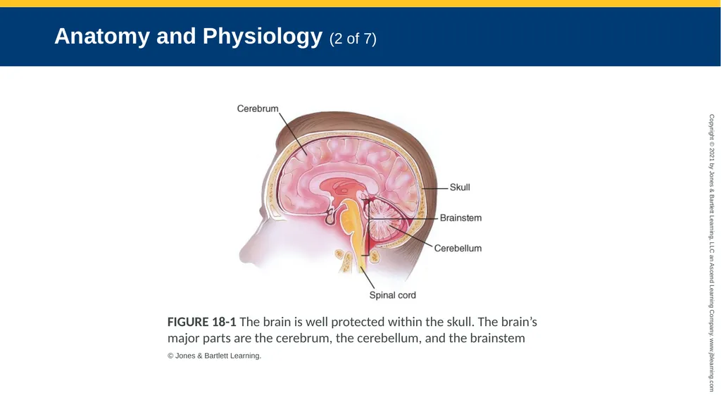
slide9. Anatomy and Physiology (2 of 7) FIGURE 18-1 The brain is well protected within the skull. The brain’s major parts are the cerebrum, the cerebellum, and the brainstem © Jones & Bartlett Learning.<br>
slide10. Anatomy and Physiology (3 of 7) The brainstem controls the most basic functions.
Breathing, blood pressure, swallowing, pupil constriction
The cerebellum controls muscle and body coordination.<br>
slide11. Anatomy and Physiology (4 of 7) The cerebrum is divided into right and left hemispheres.
Each controls activities on the opposite side of the body.
The front of the cerebrum controls emotion and thought.
The middle controls sensation and movement.
The back processes sight.<br>
slide12. Anatomy and Physiology (5 of 7) In most people, speech is controlled on the left side of the brain near the middle of the cerebrum.
Messages sent to and from the brain travel through nerves.
Twelve cranial nerves run directly from the brain to parts of the head.<br>
slide13. Anatomy and Physiology (6 of 7) The rest of the nerves join in the spinal cord and exit the brain through a large opening in the base of the skull called the foramen magnum.
At each vertebra in the neck and back, two nerves branch out (spinal nerves).
These carry signals to and from the body.<br>
slide14. Anatomy and Physiology (7 of 7) FIGURE 18-2 The spinal cord is the continuation of the
brainstem. It exits the skull at the foramen magnum and
extends down to the level of the second lumbar vertebra. © Jones & Bartlett Learning.<br>
slide15. Pathophysiology Many different disorders may cause brain dysfunction.
May affect the patient’s level of consciousness, speech, and voluntary muscle control
The brain is sensitive to changes in oxygen, glucose, and temperature.<br>
slide16. Headache (1 of 5) One of the most common complaints
Can be a symptom of another condition or a neurologic condition on its own
Only a small percentage of headaches are caused by a serious medical condition.
Tension headaches, migraines, and sinus headaches are the most common.<br>
slide17. Headache (2 of 5) Tension headaches
Caused by muscle contractions in the head and neck
Attributed to stress
Pain is usually described as squeezing, dull, or as an ache.
Usually do not require medical attention<br>
slide18. Headache (3 of 5) Migraine headaches
Thought to be caused by changes in blood vessel size in the base of the brain.
Pain is usually described as pounding, throbbing, and pulsating.
Often associated with nausea and vomiting, and may be preceded by visual changes
Can last for several hours or days<br>
slide19. Headache (4 of 5) Sinus headaches
Caused by pressure that is the result of fluid accumulation in the sinus cavities
Patients may also have cold-like symptoms of nasal congestion, cough, and fever.
Prehospital emergency care is not required.<br>
slide20. Headache (5 of 5) Serious conditions that include headache as a symptom are hemorrhagic stroke, brain tumor, and meningitis.<br>
slide21. Stroke Also called a cerebrovascular accident (CVA)
Interruption of blood flow to an area within the brain
Results in the loss of brain function
There are two main types of stroke: ischemic and hemorrhagic.<br>
slide22. Ischemic Stroke (1 of 2) Most common, accounting for 87% of strokes
Results from thrombosis or an embolus
Symptoms may range from nothing at all to complete paralysis.
Atherosclerosis in the blood vessels is often the cause.<br>
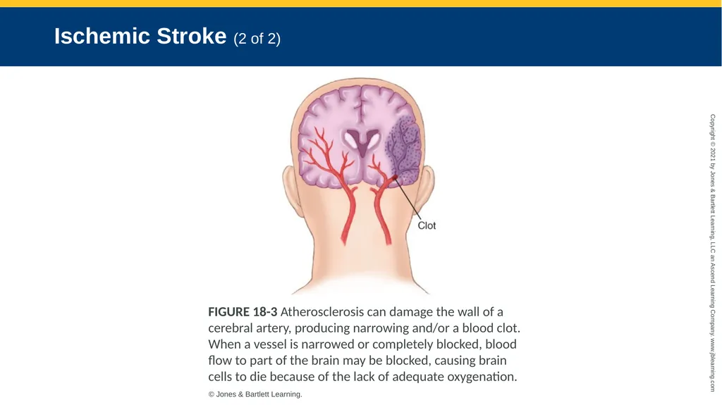
slide23. Ischemic Stroke (2 of 2) FIGURE 18-3 Atherosclerosis can damage the wall of a
cerebral artery, producing narrowing and/or a blood clot.
When a vessel is narrowed or completely blocked, blood
flow to part of the brain may be blocked, causing brain
cells to die because of the lack of adequate oxygenation. © Jones & Bartlett Learning.<br>
slide24. Hemorrhagic Stroke (1 of 2) Accounts for 13% of strokes
Results from bleeding inside the brain
Cerebral hemorrhages are often fatal.
People at high risk include those experiencing stress or exertion.
People at highest risk are those who have very high blood pressure.<br>
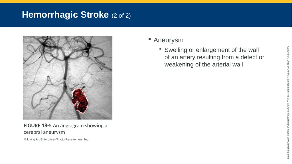
slide25. Hemorrhagic Stroke (2 of 2) Aneurysm
Swelling or enlargement of the wall of an artery resulting from a defect or weakening of the arterial wall FIGURE 18-5 An angiogram showing a cerebral aneurysm © Living Art Enterprises/Photo Researchers, Inc.<br>
slide26. Transient Ischemic Attack (TIA) Stroke-like symptoms go away on their own in less than 24 hours.
May be a warning sign of a larger stroke to come
About one-third of patients who have a TIA will experience a stroke.<br>
slide27. Signs and Symptoms of Stroke (1 of 4) Facial drooping
Sudden weakness or numbness in the face, arm, leg, or one side of body
Decreased or absent movement and sensation on one side of the body
Lack of muscle coordination (ataxia) or loss of balance<br>
slide28. Signs and Symptoms of Stroke (2 of 4) Sudden vision loss in one eye
Blurred and double vision
Difficulty swallowing
Decreased level of responsiveness
Speech disorders<br>
slide29. Signs and Symptoms of Stroke (3 of 4) Aphasia
Slurred speech (dysarthria)
Sudden and severe headache
Confusion
Dizziness<br>
slide30. Signs and Symptoms of Stroke (4 of 4) Weakness
Combativeness
Restlessness
Tongue deviation
Coma<br>
slide31. Left Hemisphere Stroke in the left cerebral hemisphere may cause aphasia.
Inability to produce or understand speech
Speech problems can vary widely.
May also cause paralysis of the right side of the body<br>
slide32. Right Hemisphere Stroke may cause paralysis of the left side of the body.
Usually, patients can understand language and are able to speak.
Patients may be oblivious to their problem (neglect).
Neglect and lack of pain cause many patients to delay seeking help.<br>
slide33. Bleeding in the Brain Patients may have high blood pressure.
May be the cause of the bleeding
May be caused by the bleeding, as a compensatory response
Increasing blood pressure is an important sign.
Significant drops in blood pressure may occur as the patient’s condition worsens.<br>
slide34. Hypoglycemia
Postictal state
Subdural or epidural bleeding Conditions That May Mimic Stroke (1 of 2)<br>
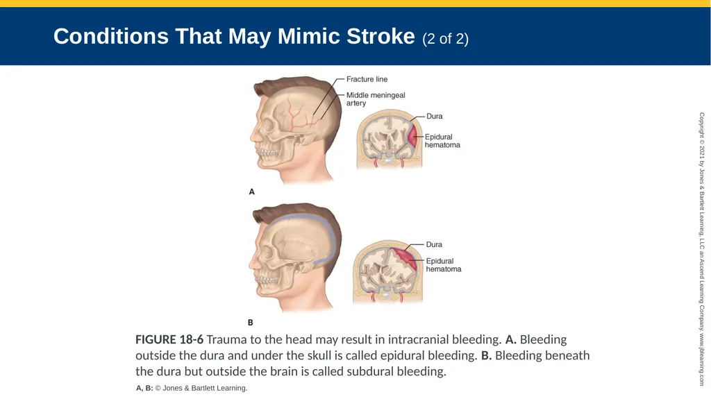
slide35. Conditions That May Mimic Stroke (2 of 2) FIGURE 18-6 Trauma to the head may result in intracranial bleeding. A. Bleeding outside the dura and under the skull is called epidural bleeding. B. Bleeding beneath the dura but outside the brain is called subdural bleeding. A, B: © Jones & Bartlett Learning.<br>
slide36. Seizures A neurologic episode caused by a surge of electrical activity in the brain
Can take the form of a convulsion and/or can be associated with a temporary alteration in consciousness.
Two basic groups: generalized and partial (focal)<br>
slide37. Results from abnormal electrical discharges from large areas of the brain
Typically characterized by unconsciousness and a generalized severe twitching of all muscles lasting several minutes or longer Generalized Seizure<br>
slide38. Does not involve any changes in motor activity
Characterized by a brief lapse of consciousness in which the patient seems to stare and not respond Absence Seizure<br>
slide39. Partial (Focal) Seizure (1 of 2) Focal-onset aware seizure
No change in the patient’s level of consciousness
May have numbness, weakness, dizziness, visual changes, or unusual smells/tastes
May have some twitching or brief paralysis<br>
slide40. Partial (Focal) Seizure (2 of 2) Focal-onset, impaired awareness seizure
Altered mental status
Results from abnormal discharges from the temporal lobe of the brain
Lip smacking, eye blinking, isolated jerking
Unpleasant smells, visual hallucinations, uncontrollable fear, repetitive physical behavior<br>
slide41. Aura Patients may experience an aura prior to a seizure.
Can include visual changes or hallucinations
People with a history of seizures recognize their auras and usually take steps to minimize injury.
Auras do not occur prior to every seizure, and not all patients with a seizure disorder experience an aura.<br>
slide42. Generalized Seizure Characterized by sudden loss of consciousness, chaotic muscle movement and tone, and apnea.
May exhibit bilateral muscle movement characterized by a cycle of muscle rigidity and relaxation
Typically lasts less than 5 minutes
Followed by a postictal state<br>
slide43. Absence Seizure Formerly called petit mal
May last for seconds
Patient fully recovers with a brief lapse of memory<br>
slide44. Status Epilepticus Seizures lasting more than 5 minutes are likely to progress to status epilepticus.
Seizures that continue every few minutes without the person regaining consciousness or last longer than 30 minutes<br>
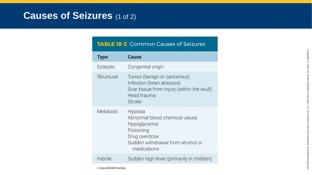
slide45. Causes of Seizures (1 of 2)<br>
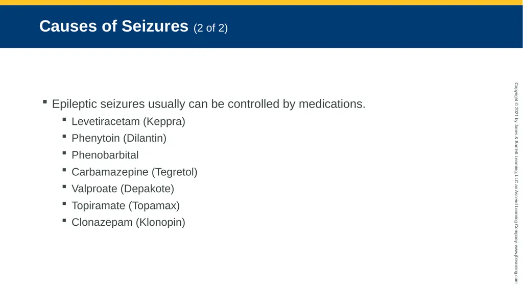
slide46. Causes of Seizures (2 of 2) Epileptic seizures usually can be controlled by medications.
Levetiracetam (Keppra)
Phenytoin (Dilantin)
Phenobarbital
Carbamazepine (Tegretol)
Valproate (Depakote)
Topiramate (Topamax)
Clonazepam (Klonopin)<br>
slide47. The Importance of Recognizing Seizures Recognize when a seizure is occurring and whether this episode differs from previous ones.
Recognize the postictal state and complications of seizures.
Identify other problems associated with seizures.<br>
slide48. The Postictal State After a seizure, the muscles relax and breathing becomes labored.
May be characterized by hemiparesis
Most commonly characterized by lethargy and confusion
If the patient does not improve, consider other possible underlying conditions.<br>
slide49. Syncope Seizures are often mistaken for syncope, or fainting.
Fainting typically occurs while the patient is standing.
Seizures may occur in any position.
Fainting is not associated with a postictal state.<br>
slide50. Altered Mental Status Aside from stroke and seizures, the most common neurologic emergency
Patient is not thinking clearly or is incapable of being aroused.
In some cases, the patient will be unconscious; in others, the patient may be alert but confused.<br>
slide51. Causes of AMS (1 of 2) Hypoglycemia
Hypoxemia
Intoxication
Delirium
Drug overdose
Unrecognized head injury<br>
slide52. Causes of AMS (2 of 2) Brain infection
Body temperature abnormality
Brain tumor
Overdose and/or poisoning<br>
slide53. Scene Size-up Make an early determination whether the cause is medical or trauma.
Look for threats to safety.
Follow standard precautions.
Consider the need for spinal motion restriction.
Call for additional resources early.<br>
slide54. Primary Assessment Look for and treat life-threatening conditions.
Perform a rapid exam.
Establish priorities of care based on assessment of the patient’s LOC and XABCs.<br>
slide55. History Taking Investigate the chief complaint.
For unresponsive patients, gather any history from family or bystanders.
If no one is around, quickly look for explanations for the AMS.
Try to determine the events leading up to the incident.
Obtain a SAMPLE history.<br>
slide56. Secondary Assessment (1 of 8) Vital signs
Significant intracranial bleeding leads to a great deal of pressure in the skull, compressing the brain.
Slow pulse and erratic respirations
High blood pressure
Changes in pupil size
If the patient has an AMS, check the blood glucose level.<br>
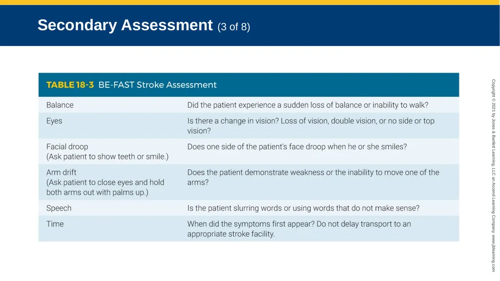
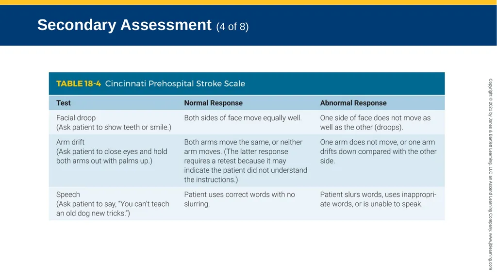
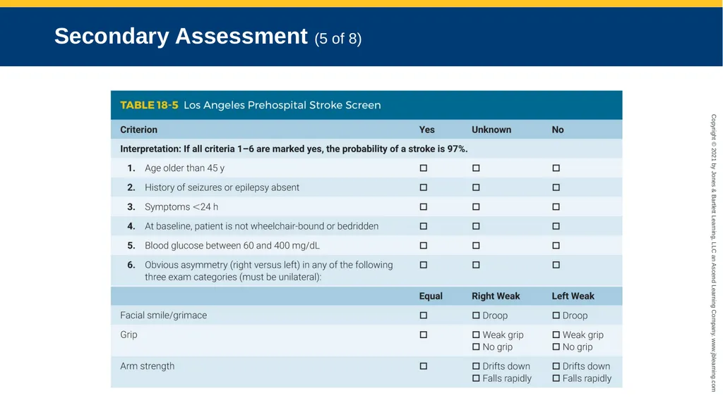
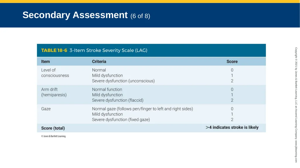
slide57. Secondary Assessment (2 of 8) Stroke assessment
Stroke scales evaluate the face, arms, and speech.
BE-FAST mnemonic
Cincinnati Prehospital Stroke Scale
Los Angeles Prehospital Stroke Screen
3-Item Stroke Severity Scale (LAG)
Glasgow Coma Scale (GCS) score<br>
slide58. Secondary Assessment (3 of 8)<br>
slide59. Secondary Assessment (4 of 8)<br>
slide60. Secondary Assessment (5 of 8)<br>
slide61. Secondary Assessment (6 of 8)<br>
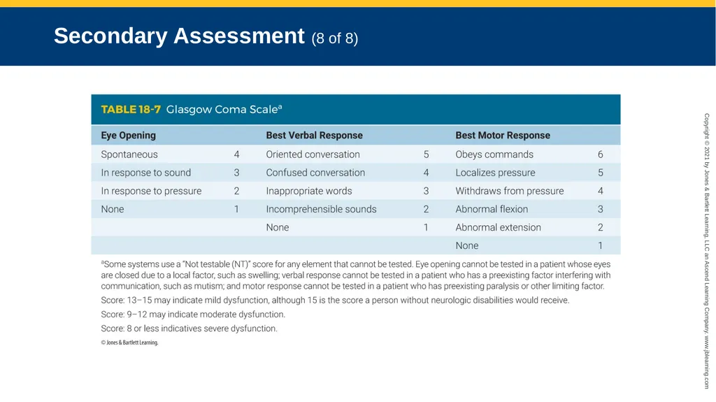
slide62. Secondary Assessment (7 of 8) All patients with an altered mental status should also have a Glasgow Coma Scale (GCS) score calculated.<br>
slide63. Secondary Assessment (8 of 8)<br>
slide64. Reassessment Focus on reassessing the ABCs, vital signs, and interventions.
Compare baseline findings with updated information.
Watch carefully for changes in pulse, blood pressure, respirations, and GCS scores.
Notify the receiving facility of patient’s chief complaint and assessment findings.<br>
slide65. Emergency Medical Care (1 of 3) ED physicians determine if there is bleeding in the brain for patients with a suspected stroke with a CT scan of the head.
If no bleeding is present, the patient may be a candidate for blood clot dissolving medication.
Notify the hospital regarding the last time the patient was known to be without their current signs and symptoms of stroke.<br>
slide66. Emergency Medical Care (2 of 3) Patients who have had a seizure require definitive evaluation and treatment.
Supplemental oxygen is strongly advised.
For patients who are having a seizure:
Protect them from harm.
Maintain a clear airway by suctioning.
Provide oxygen as quickly as possible.
If head or neck trauma is suspected, provide spinal immobilization.<br>
slide67. Emergency Medical Care (3 of 3) For patients who continue to have a seizure, as in status epilepticus:
Suction the airway.
Provide positive pressure ventilations.
Transport quickly to the hospital.
Rendezvous with ALS, if possible.<br>
slide68. Emergency Medical Care: Headache You should be concerned if the patient complains of:
A sudden-onset, severe headache
A sudden headache with fever, seizures, altered mental status, or following trauma<br>
slide69. Emergency Medical Care: Migraine Always assess the patient for other signs and symptoms that might indicate a more serious condition.
Apply high-flow oxygen, if tolerated.
Provide a darkened, quiet environment.
Do not use lights and siren during transport.<br>
slide70. Emergency Medical Care: Stroke Support XABCs and provide rapid transport to a stroke center.
Maintain a SpO2 level of at least 94%.
Oxygen therapy not recommended unless the patient is in respiratory distress or is hypoxic.
If possible, transport to a designated stroke center.<br>
slide71. Emergency Medical Care: Seizure (1 of 2) The patient may be in a postictal state upon your arrival.
The patient may still be having a seizure:
Continue to assess and treat XABCs.
Protect the patient from harm.<br>
slide72. Emergency Medical Care: Seizure (2 of 2) If the patient refuses transport after a seizure:
Contact medical control.
Follow local protocols.<br>
slide73. Emergency Medical Care: Altered Mental Status Determine the cause.
Provide spinal motion restriction.
Provide airway and ventilation support.
Transport to the appropriate facility.<br>
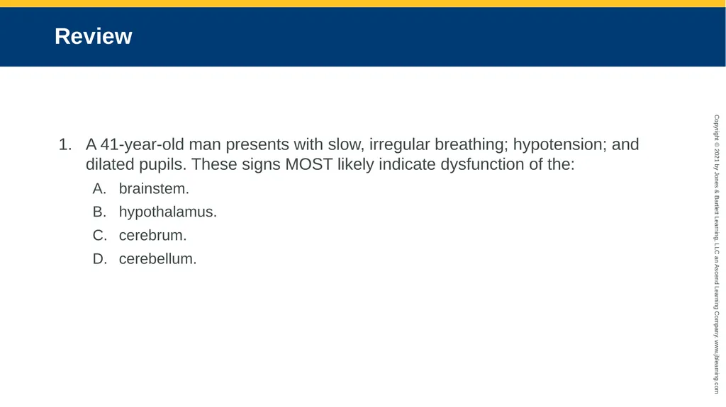
slide74. Review A 41-year-old man presents with slow, irregular breathing; hypotension; and dilated pupils. These signs MOST likely indicate dysfunction of the:
brainstem.
hypothalamus.
cerebrum.
cerebellum.<br>
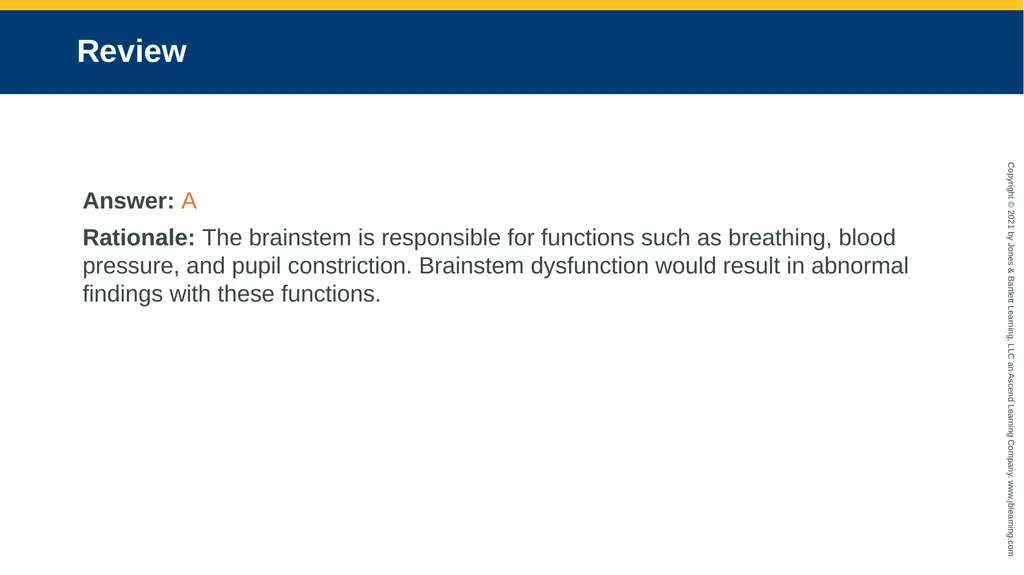
slide75. Review Answer: A
Rationale: The brainstem is responsible for functions such as breathing, blood pressure, and pupil constriction. Brainstem dysfunction would result in abnormal findings with these functions.<br>
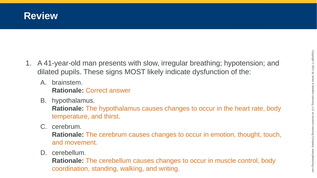
slide76. Review A 41-year-old man presents with slow, irregular breathing; hypotension; and dilated pupils. These signs MOST likely indicate dysfunction of the:
brainstem. Rationale: Correct answer
hypothalamus. Rationale: The hypothalamus causes changes to occur in the heart rate, body temperature, and thirst.
cerebrum. Rationale: The cerebrum causes changes to occur in emotion, thought, touch, and movement.
cerebellum. Rationale: The cerebellum causes changes to occur in muscle control, body coordination, standing, walking, and writing.<br>
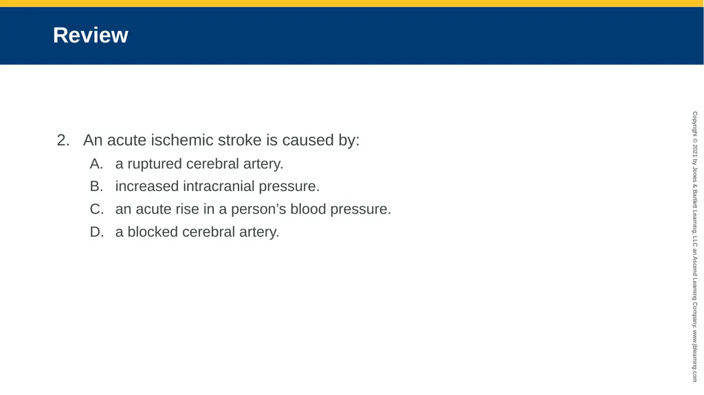
slide77. Review An acute ischemic stroke is caused by:
a ruptured cerebral artery.
increased intracranial pressure.
an acute rise in a person’s blood pressure.
a blocked cerebral artery.<br>
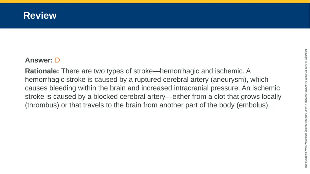
slide78. Review Answer: D
Rationale: There are two types of stroke—hemorrhagic and ischemic. A hemorrhagic stroke is caused by a ruptured cerebral artery (aneurysm), which causes bleeding within the brain and increased intracranial pressure. An ischemic stroke is caused by a blocked cerebral artery—either from a clot that grows locally (thrombus) or that travels to the brain from another part of the body (embolus).<br>
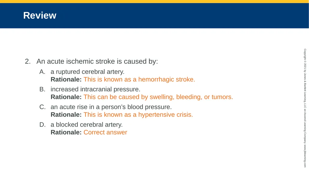
slide79. Review An acute ischemic stroke is caused by:
a ruptured cerebral artery. Rationale: This is known as a hemorrhagic stroke.
increased intracranial pressure. Rationale: This can be caused by swelling, bleeding, or tumors.
an acute rise in a person’s blood pressure. Rationale: This is known as a hypertensive crisis.
a blocked cerebral artery. Rationale: Correct answer<br>
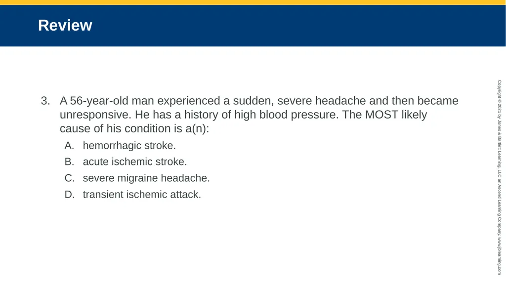
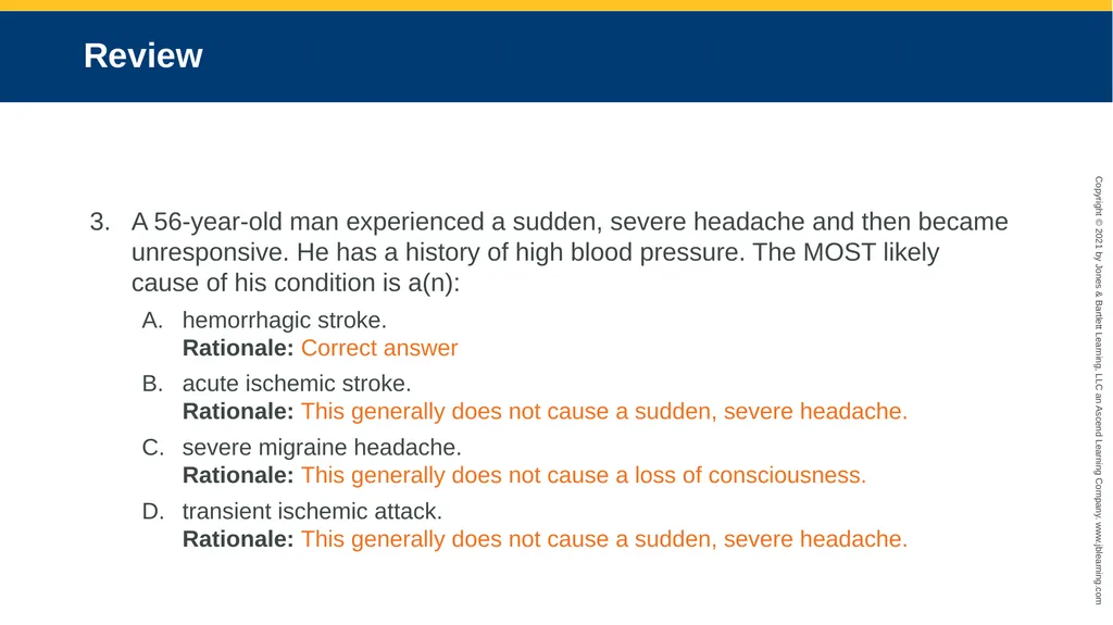
slide80. Review A 56-year-old man experienced a sudden, severe headache and then became unresponsive. He has a history of high blood pressure. The MOST likely cause of his condition is a(n):
hemorrhagic stroke.
acute ischemic stroke.
severe migraine headache.
transient ischemic attack.<br>
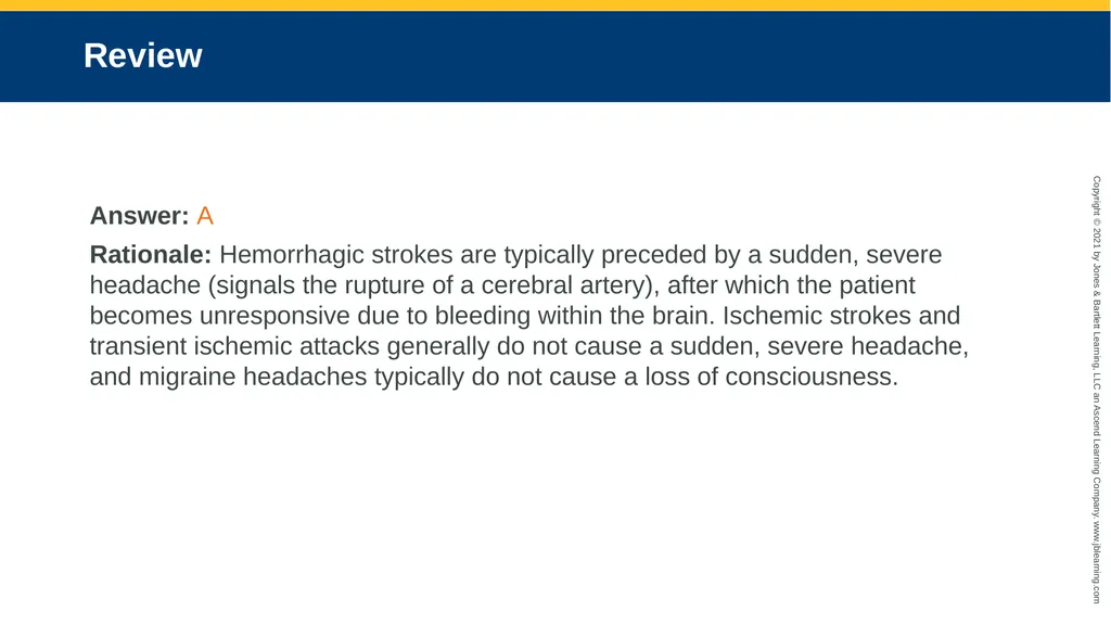
slide81. Review Answer: A
Rationale: Hemorrhagic strokes are typically preceded by a sudden, severe headache (signals the rupture of a cerebral artery), after which the patient becomes unresponsive due to bleeding within the brain. Ischemic strokes and transient ischemic attacks generally do not cause a sudden, severe headache, and migraine headaches typically do not cause a loss of consciousness.<br>
slide82. Review A 56-year-old man experienced a sudden, severe headache and then became unresponsive. He has a history of high blood pressure. The MOST likely cause of his condition is a(n):
hemorrhagic stroke. Rationale: Correct answer
acute ischemic stroke. Rationale: This generally does not cause a sudden, severe headache.
severe migraine headache. Rationale: This generally does not cause a loss of consciousness.
transient ischemic attack. Rationale: This generally does not cause a sudden, severe headache.<br>
slide83. Review Unlike an ischemic stroke, a transient ischemic attack is characterized by all of the following, EXCEPT:
symptoms that resolve within 24 hours.
symptoms that persist for longer than 24 hours.
weakness or paralysis to one side of the body.
an acute onset of confusion and slurred speech.<br>
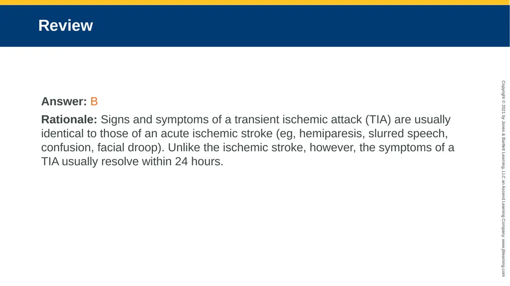
slide84. Review Answer: B
Rationale: Signs and symptoms of a transient ischemic attack (TIA) are usually identical to those of an acute ischemic stroke (eg, hemiparesis, slurred speech, confusion, facial droop). Unlike the ischemic stroke, however, the symptoms of a TIA usually resolve within 24 hours.<br>
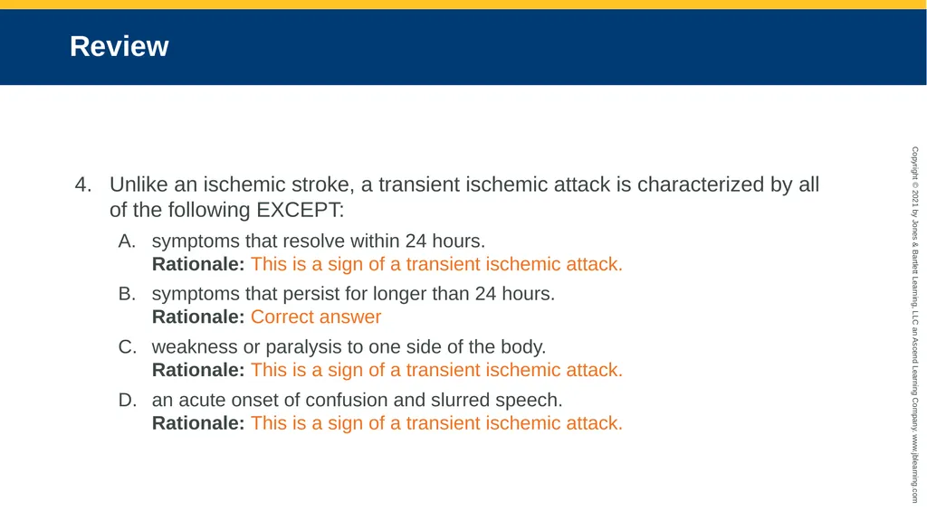
slide85. Review Unlike an ischemic stroke, a transient ischemic attack is characterized by all of the following EXCEPT:
symptoms that resolve within 24 hours. Rationale: This is a sign of a transient ischemic attack.
symptoms that persist for longer than 24 hours. Rationale: Correct answer
weakness or paralysis to one side of the body. Rationale: This is a sign of a transient ischemic attack.
an acute onset of confusion and slurred speech. Rationale: This is a sign of a transient ischemic attack.<br>
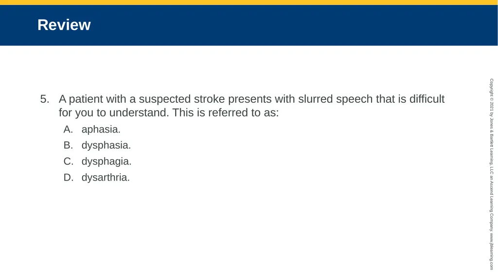
slide86. Review A patient with a suspected stroke presents with slurred speech that is difficult for you to understand. This is referred to as:
aphasia.
dysphasia.
dysphagia.
dysarthria.<br>
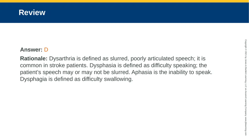
slide87. Review Answer: D
Rationale: Dysarthria is defined as slurred, poorly articulated speech; it is common in stroke patients. Dysphasia is defined as difficulty speaking; the patient’s speech may or may not be slurred. Aphasia is the inability to speak. Dysphagia is defined as difficulty swallowing.<br>
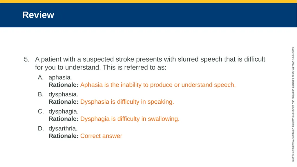
slide88. Review A patient with a suspected stroke presents with slurred speech that is difficult for you to understand. This is referred to as:
aphasia. Rationale: Aphasia is the inability to produce or understand speech.
dysphasia. Rationale: Dysphasia is difficulty in speaking.
dysphagia. Rationale: Dysphagia is difficulty in swallowing.
dysarthria. Rationale: Correct answer<br>
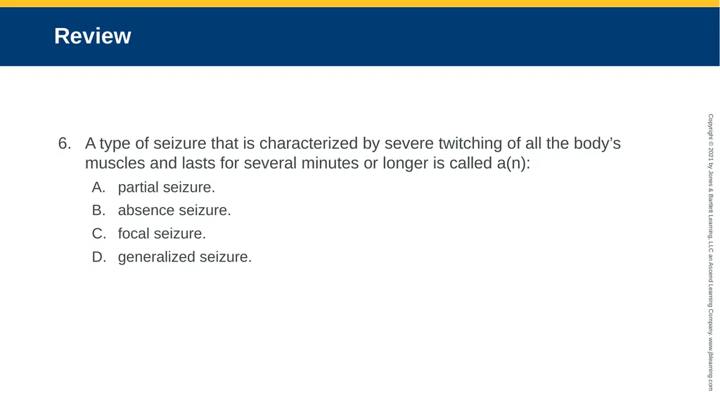
slide89. Review A type of seizure that is characterized by severe twitching of all the body’s muscles and lasts for several minutes or longer is called a(n):
partial seizure.
absence seizure.
focal seizure.
generalized seizure.<br>
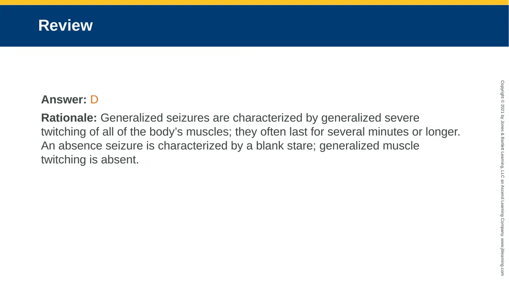
slide90. Review Answer: D
Rationale: Generalized seizures are characterized by generalized severe twitching of all of the body’s muscles; they often last for several minutes or longer. An absence seizure is characterized by a blank stare; generalized muscle twitching is absent.<br>
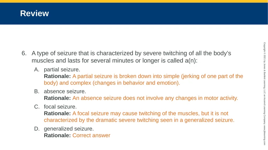
slide91. Review A type of seizure that is characterized by severe twitching of all the body’s muscles and lasts for several minutes or longer is called a(n):
partial seizure. Rationale: A partial seizure is broken down into simple (jerking of one part of the body) and complex (changes in behavior and emotion).
absence seizure. Rationale: An absence seizure does not involve any changes in motor activity.
focal seizure. Rationale: A focal seizure may cause twitching of the muscles, but it is not characterized by the dramatic severe twitching seen in a generalized seizure.
generalized seizure. Rationale: Correct answer<br>
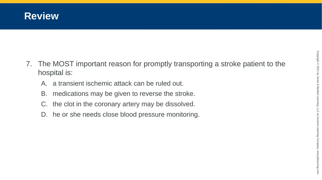
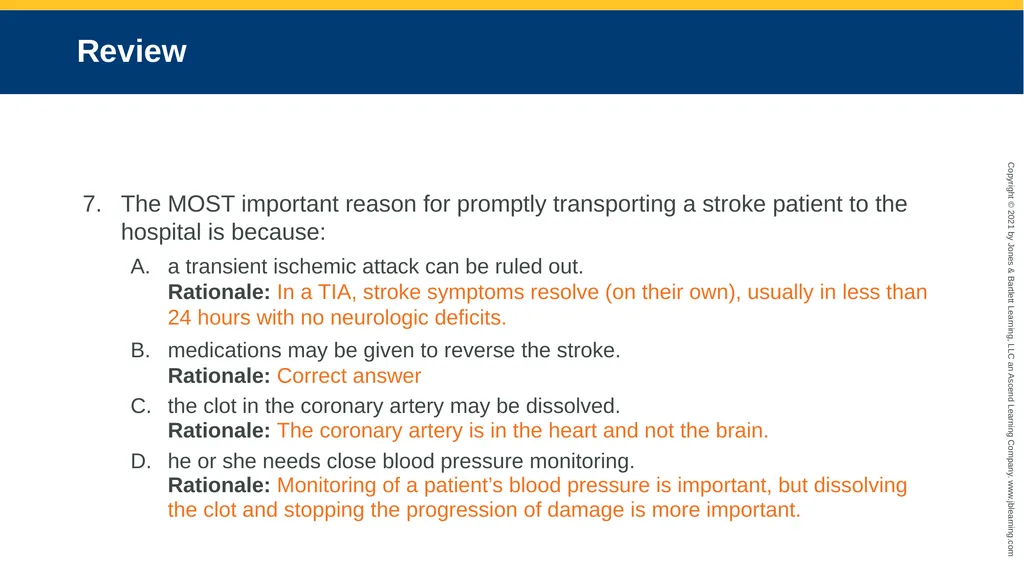
slide92. Review The MOST important reason for promptly transporting a stroke patient to the hospital is:
a transient ischemic attack can be ruled out.
medications may be given to reverse the stroke.
the clot in the coronary artery may be dissolved.
he or she needs close blood pressure monitoring.<br>
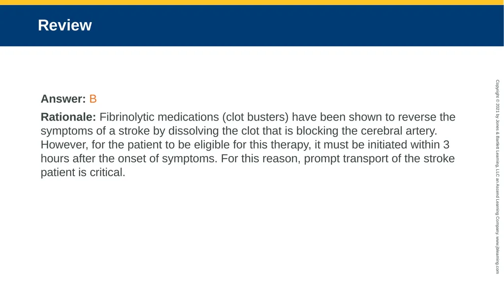
slide93. Review Answer: B
Rationale: Fibrinolytic medications (clot busters) have been shown to reverse the symptoms of a stroke by dissolving the clot that is blocking the cerebral artery. However, for the patient to be eligible for this therapy, it must be initiated within 3 hours after the onset of symptoms. For this reason, prompt transport of the stroke patient is critical.<br>
slide94. Review The MOST important reason for promptly transporting a stroke patient to the hospital is because:
a transient ischemic attack can be ruled out. Rationale: In a TIA, stroke symptoms resolve (on their own), usually in less than 24 hours with no neurologic deficits.
medications may be given to reverse the stroke. Rationale: Correct answer
the clot in the coronary artery may be dissolved. Rationale: The coronary artery is in the heart and not the brain.
he or she needs close blood pressure monitoring. Rationale: Monitoring of a patient’s blood pressure is important, but dissolving the clot and stopping the progression of damage is more important.<br>
slide95. Review Which of the following are components of the Cincinnati Prehospital Stroke Scale?
Arm drift, blood pressure, speech
Speech, pupil response, arm drift
Facial symmetry, speech, arm drift
Pupil response, facial droop, speech<br>
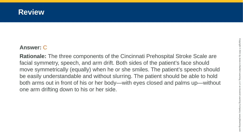
slide96. Review Answer: C
Rationale: The three components of the Cincinnati Prehospital Stroke Scale are facial symmetry, speech, and arm drift. Both sides of the patient’s face should move symmetrically (equally) when he or she smiles. The patient’s speech should be easily understandable and without slurring. The patient should be able to hold both arms out in front of his or her body—with eyes closed and palms up—without one arm drifting down to his or her side.<br>
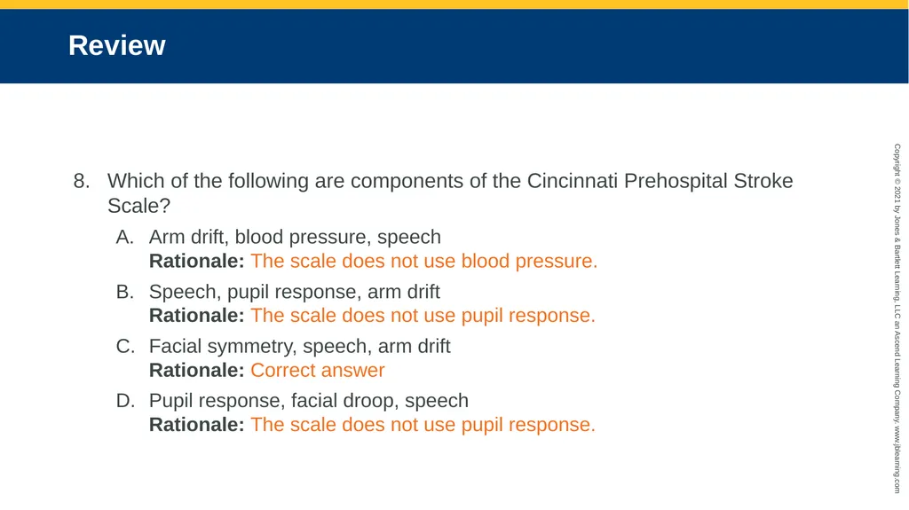
slide97. Review Which of the following are components of the Cincinnati Prehospital Stroke Scale?
Arm drift, blood pressure, speech Rationale: The scale does not use blood pressure.
Speech, pupil response, arm drift Rationale: The scale does not use pupil response.
Facial symmetry, speech, arm drift Rationale: Correct answer
Pupil response, facial droop, speech Rationale: The scale does not use pupil response.<br>
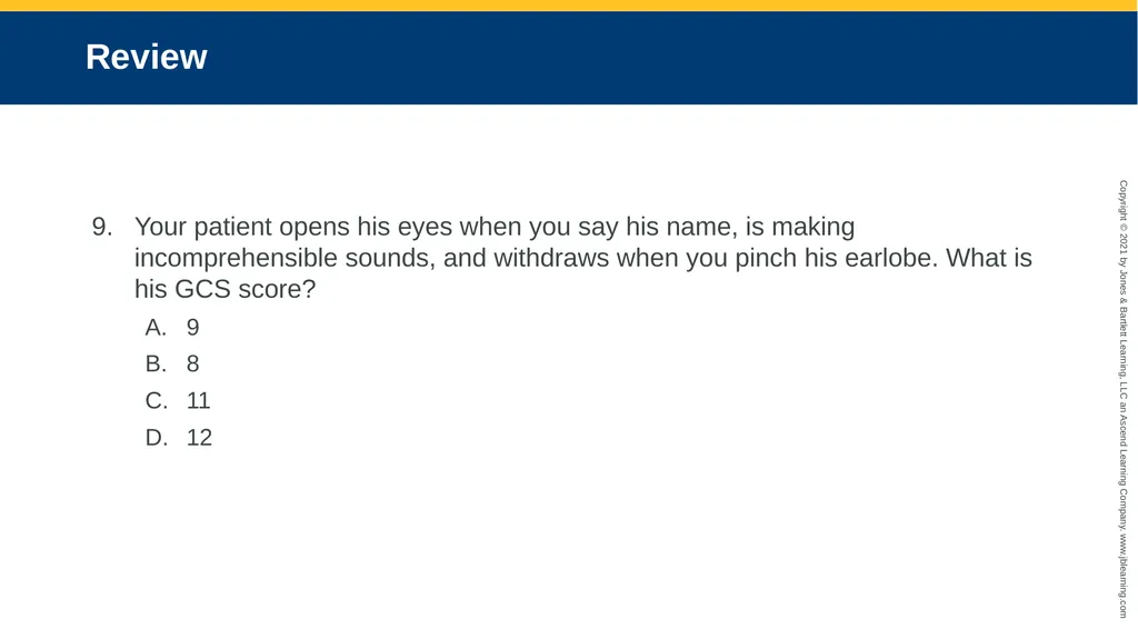
slide98. Review Your patient opens his eyes when you say his name, is making incomprehensible sounds, and withdraws when you pinch his earlobe. What is his GCS score?
9
8
11
12<br>
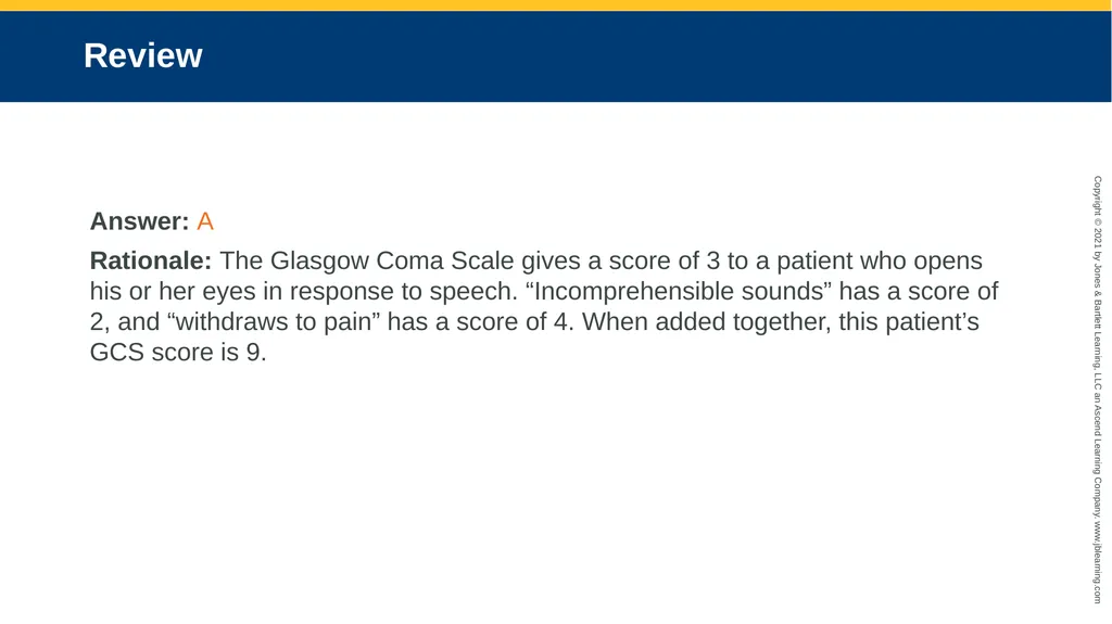
slide99. Review Answer: A
Rationale: The Glasgow Coma Scale gives a score of 3 to a patient who opens his or her eyes in response to speech. “Incomprehensible sounds” has a score of 2, and “withdraws to pain” has a score of 4. When added together, this patient’s GCS score is 9.<br>
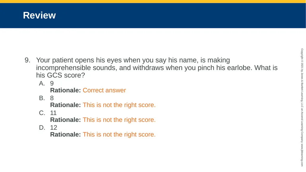
slide100. Review Your patient opens his eyes when you say his name, is making incomprehensible sounds, and withdraws when you pinch his earlobe. What is his GCS score?
9 Rationale: Correct answer
8 Rationale: This is not the right score.
11 Rationale: This is not the right score.
12 Rationale: This is not the right score.<br>
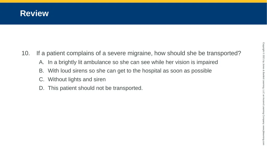
slide101. Review If a patient complains of a severe migraine, how should she be transported?
In a brightly lit ambulance so she can see while her vision is impaired
With loud sirens so she can get to the hospital as soon as possible
Without lights and siren
This patient should not be transported.<br>
slide102. Review Answer: C
Rationale: Treatment of a migraine headache is supportive; however, you should always assess the patient for other signs and symptoms that might indicate a more serious condition. Applying high-flow oxygen, if tolerated, may help ease the patient’s condition. When possible, provide a darkened and quiet environment because patients are sensitive to light and sound. Do not use lights and siren during transport.<br>
slide103. Review If a patient complains of a severe migraine, how should she be transported?
In a brightly lit ambulance so she can see while her vision is impaired Rationale: Migraine patients are sensitive to light.
With loud sirens so she can get to the hospital as soon as possible Rationale: Migraine patients are sensitive to loud noises.
Without lights and siren Rationale: Correct answer
This patient should not be transported. Rationale: A migraine could indicate a more serious condition.<br>
slide2. National EMS Education Standard Competencies (1 of 3) Medicine
Applies fundamental knowledge to provide basic emergency care and transportation based on assessment findings for an acutely ill patient.<br>
slide3. National EMS Education Standard Competencies (2 of 3) Neurology
Anatomy, presentations, and management of
Decreased level of responsiveness
Seizure
Stroke<br>
slide4. National EMS Education Standard Competencies (3 of 3) Neurology (cont’d)
Anatomy, physiology, pathophysiology, assessment, and management of
Stroke/transient ischemic attack
Seizure
Status epilepticus
Headache<br>
slide5. Introduction (1 of 3) Stroke is the fifth-leading cause of death and the leading cause of adult disability in the United States.
Common in geriatric patients
Contributing factors for stroke include family history and race.
New treatments are available.<br>
slide6. Introduction (2 of 3) Seizures and altered mental status may also occur.
Seizures may occur as a result of:
Recent or prior head injury
A brain tumor
Metabolic problems
Fever
A genetic disposition<br>
slide7. Introduction (3 of 3) Possible causes of altered mental status include:
Intoxication
Head injury
Hypoxia
Stroke
Metabolic disturbances
Treatment varies widely.<br>
slide8. Anatomy and Physiology (1 of 7) The brain is the body’s computer.
Controls breathing, speech, and all body functions
Three major parts: brainstem, cerebellum, and cerebrum
The cerebrum is the largest part.<br>
slide9. Anatomy and Physiology (2 of 7) FIGURE 18-1 The brain is well protected within the skull. The brain’s major parts are the cerebrum, the cerebellum, and the brainstem © Jones & Bartlett Learning.<br>
slide10. Anatomy and Physiology (3 of 7) The brainstem controls the most basic functions.
Breathing, blood pressure, swallowing, pupil constriction
The cerebellum controls muscle and body coordination.<br>
slide11. Anatomy and Physiology (4 of 7) The cerebrum is divided into right and left hemispheres.
Each controls activities on the opposite side of the body.
The front of the cerebrum controls emotion and thought.
The middle controls sensation and movement.
The back processes sight.<br>
slide12. Anatomy and Physiology (5 of 7) In most people, speech is controlled on the left side of the brain near the middle of the cerebrum.
Messages sent to and from the brain travel through nerves.
Twelve cranial nerves run directly from the brain to parts of the head.<br>
slide13. Anatomy and Physiology (6 of 7) The rest of the nerves join in the spinal cord and exit the brain through a large opening in the base of the skull called the foramen magnum.
At each vertebra in the neck and back, two nerves branch out (spinal nerves).
These carry signals to and from the body.<br>
slide14. Anatomy and Physiology (7 of 7) FIGURE 18-2 The spinal cord is the continuation of the
brainstem. It exits the skull at the foramen magnum and
extends down to the level of the second lumbar vertebra. © Jones & Bartlett Learning.<br>
slide15. Pathophysiology Many different disorders may cause brain dysfunction.
May affect the patient’s level of consciousness, speech, and voluntary muscle control
The brain is sensitive to changes in oxygen, glucose, and temperature.<br>
slide16. Headache (1 of 5) One of the most common complaints
Can be a symptom of another condition or a neurologic condition on its own
Only a small percentage of headaches are caused by a serious medical condition.
Tension headaches, migraines, and sinus headaches are the most common.<br>
slide17. Headache (2 of 5) Tension headaches
Caused by muscle contractions in the head and neck
Attributed to stress
Pain is usually described as squeezing, dull, or as an ache.
Usually do not require medical attention<br>
slide18. Headache (3 of 5) Migraine headaches
Thought to be caused by changes in blood vessel size in the base of the brain.
Pain is usually described as pounding, throbbing, and pulsating.
Often associated with nausea and vomiting, and may be preceded by visual changes
Can last for several hours or days<br>
slide19. Headache (4 of 5) Sinus headaches
Caused by pressure that is the result of fluid accumulation in the sinus cavities
Patients may also have cold-like symptoms of nasal congestion, cough, and fever.
Prehospital emergency care is not required.<br>
slide20. Headache (5 of 5) Serious conditions that include headache as a symptom are hemorrhagic stroke, brain tumor, and meningitis.<br>
slide21. Stroke Also called a cerebrovascular accident (CVA)
Interruption of blood flow to an area within the brain
Results in the loss of brain function
There are two main types of stroke: ischemic and hemorrhagic.<br>
slide22. Ischemic Stroke (1 of 2) Most common, accounting for 87% of strokes
Results from thrombosis or an embolus
Symptoms may range from nothing at all to complete paralysis.
Atherosclerosis in the blood vessels is often the cause.<br>
slide23. Ischemic Stroke (2 of 2) FIGURE 18-3 Atherosclerosis can damage the wall of a
cerebral artery, producing narrowing and/or a blood clot.
When a vessel is narrowed or completely blocked, blood
flow to part of the brain may be blocked, causing brain
cells to die because of the lack of adequate oxygenation. © Jones & Bartlett Learning.<br>
slide24. Hemorrhagic Stroke (1 of 2) Accounts for 13% of strokes
Results from bleeding inside the brain
Cerebral hemorrhages are often fatal.
People at high risk include those experiencing stress or exertion.
People at highest risk are those who have very high blood pressure.<br>
slide25. Hemorrhagic Stroke (2 of 2) Aneurysm
Swelling or enlargement of the wall of an artery resulting from a defect or weakening of the arterial wall FIGURE 18-5 An angiogram showing a cerebral aneurysm © Living Art Enterprises/Photo Researchers, Inc.<br>
slide26. Transient Ischemic Attack (TIA) Stroke-like symptoms go away on their own in less than 24 hours.
May be a warning sign of a larger stroke to come
About one-third of patients who have a TIA will experience a stroke.<br>
slide27. Signs and Symptoms of Stroke (1 of 4) Facial drooping
Sudden weakness or numbness in the face, arm, leg, or one side of body
Decreased or absent movement and sensation on one side of the body
Lack of muscle coordination (ataxia) or loss of balance<br>
slide28. Signs and Symptoms of Stroke (2 of 4) Sudden vision loss in one eye
Blurred and double vision
Difficulty swallowing
Decreased level of responsiveness
Speech disorders<br>
slide29. Signs and Symptoms of Stroke (3 of 4) Aphasia
Slurred speech (dysarthria)
Sudden and severe headache
Confusion
Dizziness<br>
slide30. Signs and Symptoms of Stroke (4 of 4) Weakness
Combativeness
Restlessness
Tongue deviation
Coma<br>
slide31. Left Hemisphere Stroke in the left cerebral hemisphere may cause aphasia.
Inability to produce or understand speech
Speech problems can vary widely.
May also cause paralysis of the right side of the body<br>
slide32. Right Hemisphere Stroke may cause paralysis of the left side of the body.
Usually, patients can understand language and are able to speak.
Patients may be oblivious to their problem (neglect).
Neglect and lack of pain cause many patients to delay seeking help.<br>
slide33. Bleeding in the Brain Patients may have high blood pressure.
May be the cause of the bleeding
May be caused by the bleeding, as a compensatory response
Increasing blood pressure is an important sign.
Significant drops in blood pressure may occur as the patient’s condition worsens.<br>
slide34. Hypoglycemia
Postictal state
Subdural or epidural bleeding Conditions That May Mimic Stroke (1 of 2)<br>
slide35. Conditions That May Mimic Stroke (2 of 2) FIGURE 18-6 Trauma to the head may result in intracranial bleeding. A. Bleeding outside the dura and under the skull is called epidural bleeding. B. Bleeding beneath the dura but outside the brain is called subdural bleeding. A, B: © Jones & Bartlett Learning.<br>
slide36. Seizures A neurologic episode caused by a surge of electrical activity in the brain
Can take the form of a convulsion and/or can be associated with a temporary alteration in consciousness.
Two basic groups: generalized and partial (focal)<br>
slide37. Results from abnormal electrical discharges from large areas of the brain
Typically characterized by unconsciousness and a generalized severe twitching of all muscles lasting several minutes or longer Generalized Seizure<br>
slide38. Does not involve any changes in motor activity
Characterized by a brief lapse of consciousness in which the patient seems to stare and not respond Absence Seizure<br>
slide39. Partial (Focal) Seizure (1 of 2) Focal-onset aware seizure
No change in the patient’s level of consciousness
May have numbness, weakness, dizziness, visual changes, or unusual smells/tastes
May have some twitching or brief paralysis<br>
slide40. Partial (Focal) Seizure (2 of 2) Focal-onset, impaired awareness seizure
Altered mental status
Results from abnormal discharges from the temporal lobe of the brain
Lip smacking, eye blinking, isolated jerking
Unpleasant smells, visual hallucinations, uncontrollable fear, repetitive physical behavior<br>
slide41. Aura Patients may experience an aura prior to a seizure.
Can include visual changes or hallucinations
People with a history of seizures recognize their auras and usually take steps to minimize injury.
Auras do not occur prior to every seizure, and not all patients with a seizure disorder experience an aura.<br>
slide42. Generalized Seizure Characterized by sudden loss of consciousness, chaotic muscle movement and tone, and apnea.
May exhibit bilateral muscle movement characterized by a cycle of muscle rigidity and relaxation
Typically lasts less than 5 minutes
Followed by a postictal state<br>
slide43. Absence Seizure Formerly called petit mal
May last for seconds
Patient fully recovers with a brief lapse of memory<br>
slide44. Status Epilepticus Seizures lasting more than 5 minutes are likely to progress to status epilepticus.
Seizures that continue every few minutes without the person regaining consciousness or last longer than 30 minutes<br>
slide45. Causes of Seizures (1 of 2)<br>
slide46. Causes of Seizures (2 of 2) Epileptic seizures usually can be controlled by medications.
Levetiracetam (Keppra)
Phenytoin (Dilantin)
Phenobarbital
Carbamazepine (Tegretol)
Valproate (Depakote)
Topiramate (Topamax)
Clonazepam (Klonopin)<br>
slide47. The Importance of Recognizing Seizures Recognize when a seizure is occurring and whether this episode differs from previous ones.
Recognize the postictal state and complications of seizures.
Identify other problems associated with seizures.<br>
slide48. The Postictal State After a seizure, the muscles relax and breathing becomes labored.
May be characterized by hemiparesis
Most commonly characterized by lethargy and confusion
If the patient does not improve, consider other possible underlying conditions.<br>
slide49. Syncope Seizures are often mistaken for syncope, or fainting.
Fainting typically occurs while the patient is standing.
Seizures may occur in any position.
Fainting is not associated with a postictal state.<br>
slide50. Altered Mental Status Aside from stroke and seizures, the most common neurologic emergency
Patient is not thinking clearly or is incapable of being aroused.
In some cases, the patient will be unconscious; in others, the patient may be alert but confused.<br>
slide51. Causes of AMS (1 of 2) Hypoglycemia
Hypoxemia
Intoxication
Delirium
Drug overdose
Unrecognized head injury<br>
slide52. Causes of AMS (2 of 2) Brain infection
Body temperature abnormality
Brain tumor
Overdose and/or poisoning<br>
slide53. Scene Size-up Make an early determination whether the cause is medical or trauma.
Look for threats to safety.
Follow standard precautions.
Consider the need for spinal motion restriction.
Call for additional resources early.<br>
slide54. Primary Assessment Look for and treat life-threatening conditions.
Perform a rapid exam.
Establish priorities of care based on assessment of the patient’s LOC and XABCs.<br>
slide55. History Taking Investigate the chief complaint.
For unresponsive patients, gather any history from family or bystanders.
If no one is around, quickly look for explanations for the AMS.
Try to determine the events leading up to the incident.
Obtain a SAMPLE history.<br>
slide56. Secondary Assessment (1 of 8) Vital signs
Significant intracranial bleeding leads to a great deal of pressure in the skull, compressing the brain.
Slow pulse and erratic respirations
High blood pressure
Changes in pupil size
If the patient has an AMS, check the blood glucose level.<br>
slide57. Secondary Assessment (2 of 8) Stroke assessment
Stroke scales evaluate the face, arms, and speech.
BE-FAST mnemonic
Cincinnati Prehospital Stroke Scale
Los Angeles Prehospital Stroke Screen
3-Item Stroke Severity Scale (LAG)
Glasgow Coma Scale (GCS) score<br>
slide58. Secondary Assessment (3 of 8)<br>
slide59. Secondary Assessment (4 of 8)<br>
slide60. Secondary Assessment (5 of 8)<br>
slide61. Secondary Assessment (6 of 8)<br>
slide62. Secondary Assessment (7 of 8) All patients with an altered mental status should also have a Glasgow Coma Scale (GCS) score calculated.<br>
slide63. Secondary Assessment (8 of 8)<br>
slide64. Reassessment Focus on reassessing the ABCs, vital signs, and interventions.
Compare baseline findings with updated information.
Watch carefully for changes in pulse, blood pressure, respirations, and GCS scores.
Notify the receiving facility of patient’s chief complaint and assessment findings.<br>
slide65. Emergency Medical Care (1 of 3) ED physicians determine if there is bleeding in the brain for patients with a suspected stroke with a CT scan of the head.
If no bleeding is present, the patient may be a candidate for blood clot dissolving medication.
Notify the hospital regarding the last time the patient was known to be without their current signs and symptoms of stroke.<br>
slide66. Emergency Medical Care (2 of 3) Patients who have had a seizure require definitive evaluation and treatment.
Supplemental oxygen is strongly advised.
For patients who are having a seizure:
Protect them from harm.
Maintain a clear airway by suctioning.
Provide oxygen as quickly as possible.
If head or neck trauma is suspected, provide spinal immobilization.<br>
slide67. Emergency Medical Care (3 of 3) For patients who continue to have a seizure, as in status epilepticus:
Suction the airway.
Provide positive pressure ventilations.
Transport quickly to the hospital.
Rendezvous with ALS, if possible.<br>
slide68. Emergency Medical Care: Headache You should be concerned if the patient complains of:
A sudden-onset, severe headache
A sudden headache with fever, seizures, altered mental status, or following trauma<br>
slide69. Emergency Medical Care: Migraine Always assess the patient for other signs and symptoms that might indicate a more serious condition.
Apply high-flow oxygen, if tolerated.
Provide a darkened, quiet environment.
Do not use lights and siren during transport.<br>
slide70. Emergency Medical Care: Stroke Support XABCs and provide rapid transport to a stroke center.
Maintain a SpO2 level of at least 94%.
Oxygen therapy not recommended unless the patient is in respiratory distress or is hypoxic.
If possible, transport to a designated stroke center.<br>
slide71. Emergency Medical Care: Seizure (1 of 2) The patient may be in a postictal state upon your arrival.
The patient may still be having a seizure:
Continue to assess and treat XABCs.
Protect the patient from harm.<br>
slide72. Emergency Medical Care: Seizure (2 of 2) If the patient refuses transport after a seizure:
Contact medical control.
Follow local protocols.<br>
slide73. Emergency Medical Care: Altered Mental Status Determine the cause.
Provide spinal motion restriction.
Provide airway and ventilation support.
Transport to the appropriate facility.<br>
slide74. Review A 41-year-old man presents with slow, irregular breathing; hypotension; and dilated pupils. These signs MOST likely indicate dysfunction of the:
brainstem.
hypothalamus.
cerebrum.
cerebellum.<br>
slide75. Review Answer: A
Rationale: The brainstem is responsible for functions such as breathing, blood pressure, and pupil constriction. Brainstem dysfunction would result in abnormal findings with these functions.<br>
slide76. Review A 41-year-old man presents with slow, irregular breathing; hypotension; and dilated pupils. These signs MOST likely indicate dysfunction of the:
brainstem. Rationale: Correct answer
hypothalamus. Rationale: The hypothalamus causes changes to occur in the heart rate, body temperature, and thirst.
cerebrum. Rationale: The cerebrum causes changes to occur in emotion, thought, touch, and movement.
cerebellum. Rationale: The cerebellum causes changes to occur in muscle control, body coordination, standing, walking, and writing.<br>
slide77. Review An acute ischemic stroke is caused by:
a ruptured cerebral artery.
increased intracranial pressure.
an acute rise in a person’s blood pressure.
a blocked cerebral artery.<br>
slide78. Review Answer: D
Rationale: There are two types of stroke—hemorrhagic and ischemic. A hemorrhagic stroke is caused by a ruptured cerebral artery (aneurysm), which causes bleeding within the brain and increased intracranial pressure. An ischemic stroke is caused by a blocked cerebral artery—either from a clot that grows locally (thrombus) or that travels to the brain from another part of the body (embolus).<br>
slide79. Review An acute ischemic stroke is caused by:
a ruptured cerebral artery. Rationale: This is known as a hemorrhagic stroke.
increased intracranial pressure. Rationale: This can be caused by swelling, bleeding, or tumors.
an acute rise in a person’s blood pressure. Rationale: This is known as a hypertensive crisis.
a blocked cerebral artery. Rationale: Correct answer<br>
slide80. Review A 56-year-old man experienced a sudden, severe headache and then became unresponsive. He has a history of high blood pressure. The MOST likely cause of his condition is a(n):
hemorrhagic stroke.
acute ischemic stroke.
severe migraine headache.
transient ischemic attack.<br>
slide81. Review Answer: A
Rationale: Hemorrhagic strokes are typically preceded by a sudden, severe headache (signals the rupture of a cerebral artery), after which the patient becomes unresponsive due to bleeding within the brain. Ischemic strokes and transient ischemic attacks generally do not cause a sudden, severe headache, and migraine headaches typically do not cause a loss of consciousness.<br>
slide82. Review A 56-year-old man experienced a sudden, severe headache and then became unresponsive. He has a history of high blood pressure. The MOST likely cause of his condition is a(n):
hemorrhagic stroke. Rationale: Correct answer
acute ischemic stroke. Rationale: This generally does not cause a sudden, severe headache.
severe migraine headache. Rationale: This generally does not cause a loss of consciousness.
transient ischemic attack. Rationale: This generally does not cause a sudden, severe headache.<br>
slide83. Review Unlike an ischemic stroke, a transient ischemic attack is characterized by all of the following, EXCEPT:
symptoms that resolve within 24 hours.
symptoms that persist for longer than 24 hours.
weakness or paralysis to one side of the body.
an acute onset of confusion and slurred speech.<br>
slide84. Review Answer: B
Rationale: Signs and symptoms of a transient ischemic attack (TIA) are usually identical to those of an acute ischemic stroke (eg, hemiparesis, slurred speech, confusion, facial droop). Unlike the ischemic stroke, however, the symptoms of a TIA usually resolve within 24 hours.<br>
slide85. Review Unlike an ischemic stroke, a transient ischemic attack is characterized by all of the following EXCEPT:
symptoms that resolve within 24 hours. Rationale: This is a sign of a transient ischemic attack.
symptoms that persist for longer than 24 hours. Rationale: Correct answer
weakness or paralysis to one side of the body. Rationale: This is a sign of a transient ischemic attack.
an acute onset of confusion and slurred speech. Rationale: This is a sign of a transient ischemic attack.<br>
slide86. Review A patient with a suspected stroke presents with slurred speech that is difficult for you to understand. This is referred to as:
aphasia.
dysphasia.
dysphagia.
dysarthria.<br>
slide87. Review Answer: D
Rationale: Dysarthria is defined as slurred, poorly articulated speech; it is common in stroke patients. Dysphasia is defined as difficulty speaking; the patient’s speech may or may not be slurred. Aphasia is the inability to speak. Dysphagia is defined as difficulty swallowing.<br>
slide88. Review A patient with a suspected stroke presents with slurred speech that is difficult for you to understand. This is referred to as:
aphasia. Rationale: Aphasia is the inability to produce or understand speech.
dysphasia. Rationale: Dysphasia is difficulty in speaking.
dysphagia. Rationale: Dysphagia is difficulty in swallowing.
dysarthria. Rationale: Correct answer<br>
slide89. Review A type of seizure that is characterized by severe twitching of all the body’s muscles and lasts for several minutes or longer is called a(n):
partial seizure.
absence seizure.
focal seizure.
generalized seizure.<br>
slide90. Review Answer: D
Rationale: Generalized seizures are characterized by generalized severe twitching of all of the body’s muscles; they often last for several minutes or longer. An absence seizure is characterized by a blank stare; generalized muscle twitching is absent.<br>
slide91. Review A type of seizure that is characterized by severe twitching of all the body’s muscles and lasts for several minutes or longer is called a(n):
partial seizure. Rationale: A partial seizure is broken down into simple (jerking of one part of the body) and complex (changes in behavior and emotion).
absence seizure. Rationale: An absence seizure does not involve any changes in motor activity.
focal seizure. Rationale: A focal seizure may cause twitching of the muscles, but it is not characterized by the dramatic severe twitching seen in a generalized seizure.
generalized seizure. Rationale: Correct answer<br>
slide92. Review The MOST important reason for promptly transporting a stroke patient to the hospital is:
a transient ischemic attack can be ruled out.
medications may be given to reverse the stroke.
the clot in the coronary artery may be dissolved.
he or she needs close blood pressure monitoring.<br>
slide93. Review Answer: B
Rationale: Fibrinolytic medications (clot busters) have been shown to reverse the symptoms of a stroke by dissolving the clot that is blocking the cerebral artery. However, for the patient to be eligible for this therapy, it must be initiated within 3 hours after the onset of symptoms. For this reason, prompt transport of the stroke patient is critical.<br>
slide94. Review The MOST important reason for promptly transporting a stroke patient to the hospital is because:
a transient ischemic attack can be ruled out. Rationale: In a TIA, stroke symptoms resolve (on their own), usually in less than 24 hours with no neurologic deficits.
medications may be given to reverse the stroke. Rationale: Correct answer
the clot in the coronary artery may be dissolved. Rationale: The coronary artery is in the heart and not the brain.
he or she needs close blood pressure monitoring. Rationale: Monitoring of a patient’s blood pressure is important, but dissolving the clot and stopping the progression of damage is more important.<br>
slide95. Review Which of the following are components of the Cincinnati Prehospital Stroke Scale?
Arm drift, blood pressure, speech
Speech, pupil response, arm drift
Facial symmetry, speech, arm drift
Pupil response, facial droop, speech<br>
slide96. Review Answer: C
Rationale: The three components of the Cincinnati Prehospital Stroke Scale are facial symmetry, speech, and arm drift. Both sides of the patient’s face should move symmetrically (equally) when he or she smiles. The patient’s speech should be easily understandable and without slurring. The patient should be able to hold both arms out in front of his or her body—with eyes closed and palms up—without one arm drifting down to his or her side.<br>
slide97. Review Which of the following are components of the Cincinnati Prehospital Stroke Scale?
Arm drift, blood pressure, speech Rationale: The scale does not use blood pressure.
Speech, pupil response, arm drift Rationale: The scale does not use pupil response.
Facial symmetry, speech, arm drift Rationale: Correct answer
Pupil response, facial droop, speech Rationale: The scale does not use pupil response.<br>
slide98. Review Your patient opens his eyes when you say his name, is making incomprehensible sounds, and withdraws when you pinch his earlobe. What is his GCS score?
9
8
11
12<br>
slide99. Review Answer: A
Rationale: The Glasgow Coma Scale gives a score of 3 to a patient who opens his or her eyes in response to speech. “Incomprehensible sounds” has a score of 2, and “withdraws to pain” has a score of 4. When added together, this patient’s GCS score is 9.<br>
slide100. Review Your patient opens his eyes when you say his name, is making incomprehensible sounds, and withdraws when you pinch his earlobe. What is his GCS score?
9 Rationale: Correct answer
8 Rationale: This is not the right score.
11 Rationale: This is not the right score.
12 Rationale: This is not the right score.<br>
slide101. Review If a patient complains of a severe migraine, how should she be transported?
In a brightly lit ambulance so she can see while her vision is impaired
With loud sirens so she can get to the hospital as soon as possible
Without lights and siren
This patient should not be transported.<br>
slide102. Review Answer: C
Rationale: Treatment of a migraine headache is supportive; however, you should always assess the patient for other signs and symptoms that might indicate a more serious condition. Applying high-flow oxygen, if tolerated, may help ease the patient’s condition. When possible, provide a darkened and quiet environment because patients are sensitive to light and sound. Do not use lights and siren during transport.<br>
slide103. Review If a patient complains of a severe migraine, how should she be transported?
In a brightly lit ambulance so she can see while her vision is impaired Rationale: Migraine patients are sensitive to light.
With loud sirens so she can get to the hospital as soon as possible Rationale: Migraine patients are sensitive to loud noises.
Without lights and siren Rationale: Correct answer
This patient should not be transported. Rationale: A migraine could indicate a more serious condition.<br>