
Chapter 20 Periodontal Examination Learning
Description: Chapter 20 Periodontal Examination Learning Objectives Describe components of a comprehensive periodontal examination List instruments used for a periodontal examination Explain technique for use of periodontal probe and explorers Explain
Related Topics
Download Presentation
"Chapter 20 Periodontal Examination Learning" is the property of its rightful owner. Permission is granted to download and print the materials on this website for personal, non-commercial use only, and to display it on your personal computer provided you do not modify the materials and that you retain all copyright notices contained in the materials. By downloading content from our website, you accept the terms of this agreement.
Presentation Transcript
slide1. Chapter 20
Periodontal Examination<br>
slide2. Learning Objectives Describe components of a comprehensive periodontal examination
List instruments used for a periodontal examination
Explain technique for use of periodontal probe and explorers
Explain how procedure will be described to patient 2<br>
slide3. Basic Instruments for Examination Mouth mirror
Probes:
Furcation probe
Plastic probe
Subgingival explorer 3<br>
slide4. The Mouth Mirror Purposes and Uses
Indirect Vision
Indirect Illumination
Transillumination
Retraction 4<br>
slide5. II. Procedure for Use Grasp and Rest
Retraction
Maintain Clear Vision 5<br>
slide6. Air–Water Syringe Purposes and Uses
Procedure for Use
To Improve and Facilitate Examination Procedures
To Improve Visibility of the Treatment Area during Instrumentation
To Prepare Teeth and/or Gingiva for Certain Procedures
Precautions 6<br>
slide7. Explorers General Purposes and Uses
Detect, by Tactile Sense, the Texture and Character of the Tooth Surfaces
Define the Extent of Instrumentation Needed and Guide Techniques
Evaluate the Completeness of Treatment 7<br>
slide8. II. Specific Explorers and Their Uses Subgingival Explorers
Use
Features for subgingival examination
Supragingival Explorers
Shepherd hook explorer
Pigtail or Cowhorn 8<br>
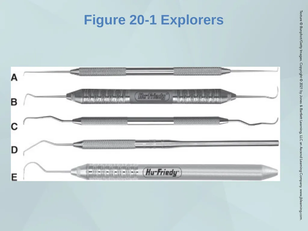
slide9. Figure 20-1 Explorers 9<br>
slide10. Basic Procedures for Use of Explorers Use of Sensory Stimuli
Tooth Surface Irregularities
Normal
Irregularities: Increases of elevations in tooth surface
Irregularities: Depressions, grooves 10<br>
slide11. III. Types of Stimuli Tactile
Auditory 11<br>
slide12. Explorers : Supragingival Procedures Use of Vision
Facial and Lingual Surfaces
Proximal Surfaces 12<br>
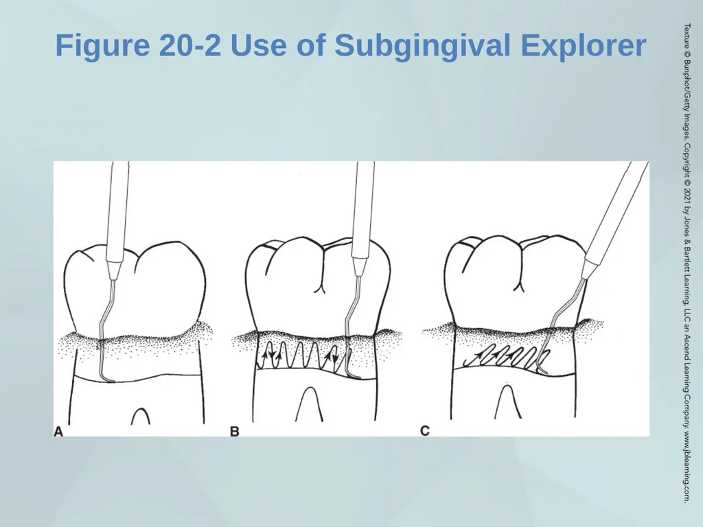
slide13. Explorers: Subgingival Procedures Essentials for Detection of Tooth Surface Irregularities 13<br>
slide14. Figure 20-2 Use of Subgingival Explorer 14<br>
slide15. Periodontal Probe Types of Probes
Standard or traditional probes
Automated probes 15<br>
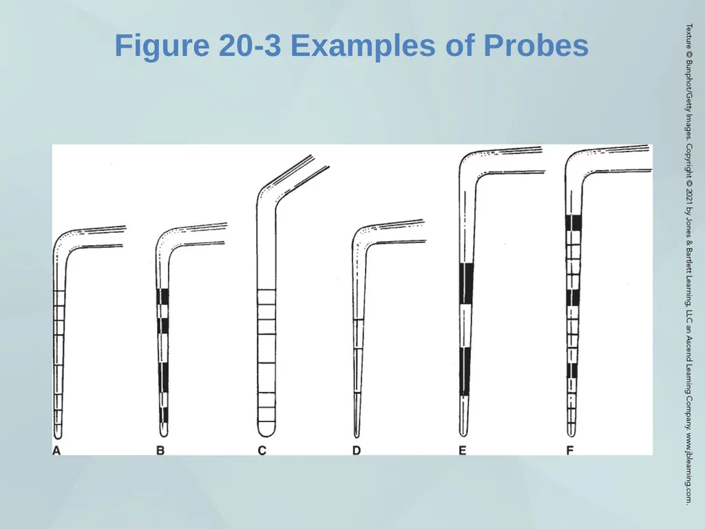
slide16. Figure 20-3 Examples of Probes 16<br>
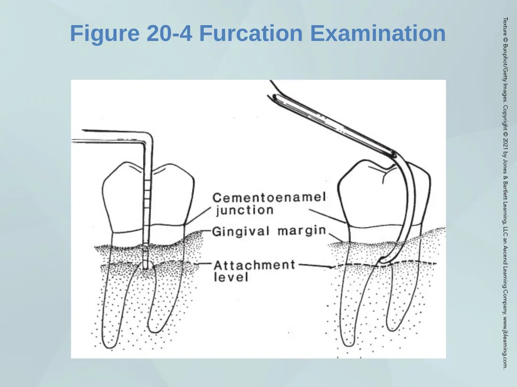
slide17. Figure 20-4 Furcation Examination 17<br>
slide18. II. Purposes and Uses Assess the Periodontal Status for Preparation of a Treatment Plan
Determine Clinical Attachment Level
Conduct Mucogingival Examination
Make Other Gingival Determinations
Guide Treatment
Evaluate Success and Completeness of Treatment
Evaluation at Continuing Care and Periodontal Maintenance Appointments 18<br>
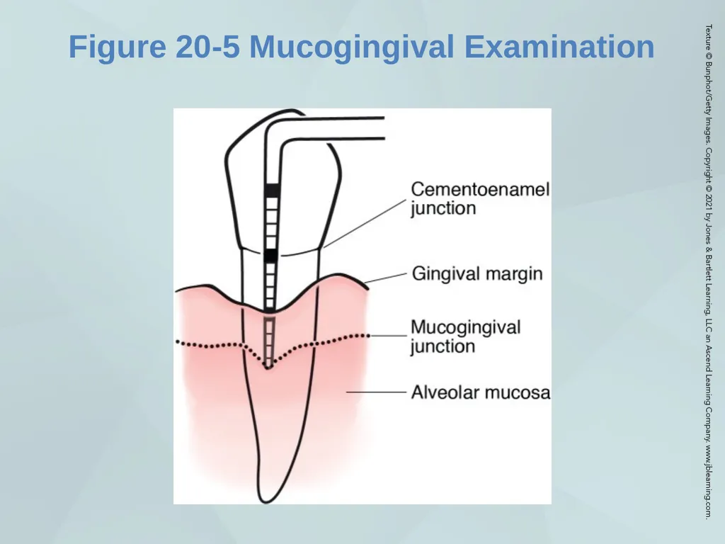
slide19. Figure 20-5 Mucogingival Examination 19<br>
slide20. III. Description of Manual Periodontal Probes Materials
Characteristics
Selection 20<br>
slide21. Guide to Periodontal Probing Pocket Characteristics
Evaluation of Tooth Surface
Factors that Affect Probe Determinations
Severity and Extent of Periodontal Disease
The Periodontal Probe
Placement Problems 21<br>
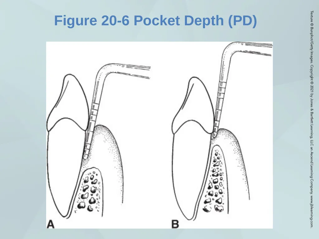
slide22. Figure 20-6 Pocket Depth (PD) 22<br>
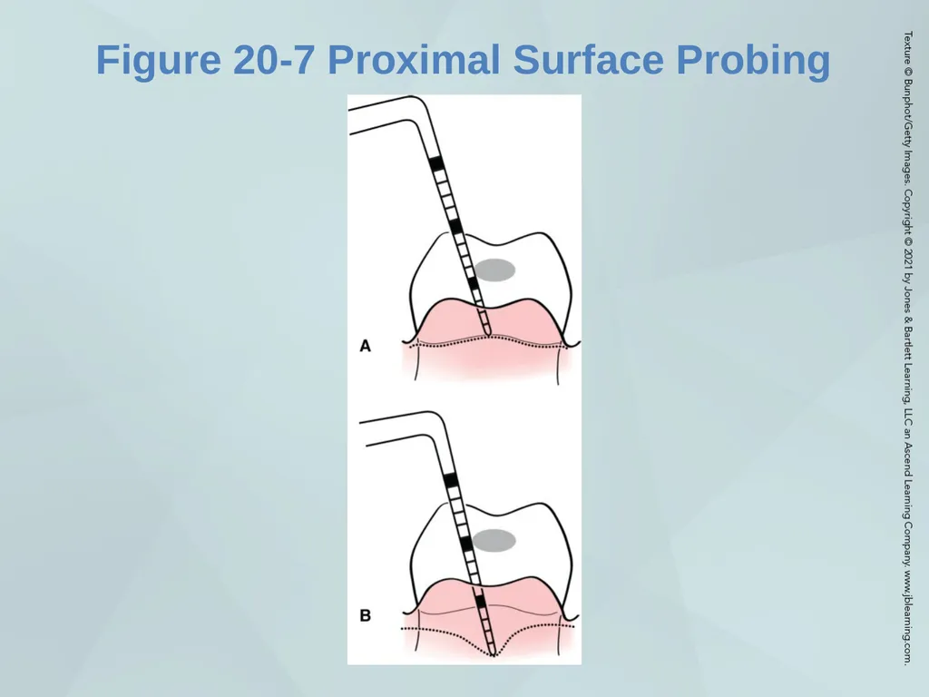
slide23. Figure 20-7 Proximal Surface Probing 23<br>
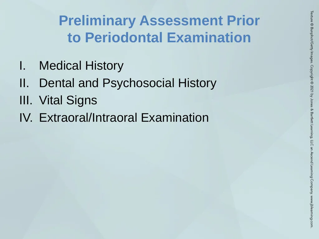
slide24. Preliminary Assessment Prior to Periodontal Examination Medical History
Dental and Psychosocial History
Vital Signs
Extraoral/Intraoral Examination 24<br>
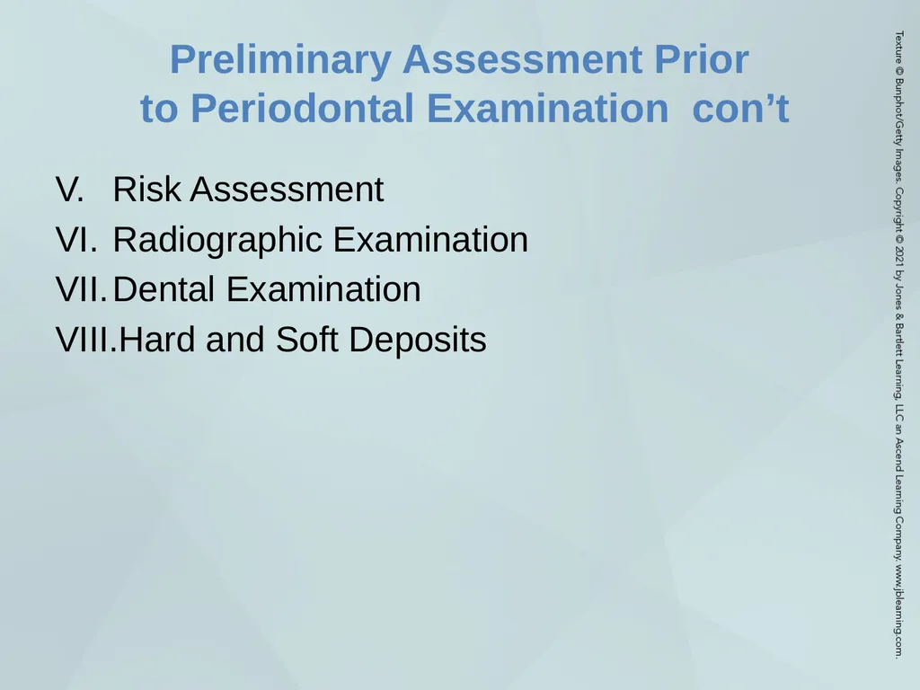
slide25. Preliminary Assessment Prior to Periodontal Examination con’t Risk Assessment
Radiographic Examination
Dental Examination
Hard and Soft Deposits 25<br>
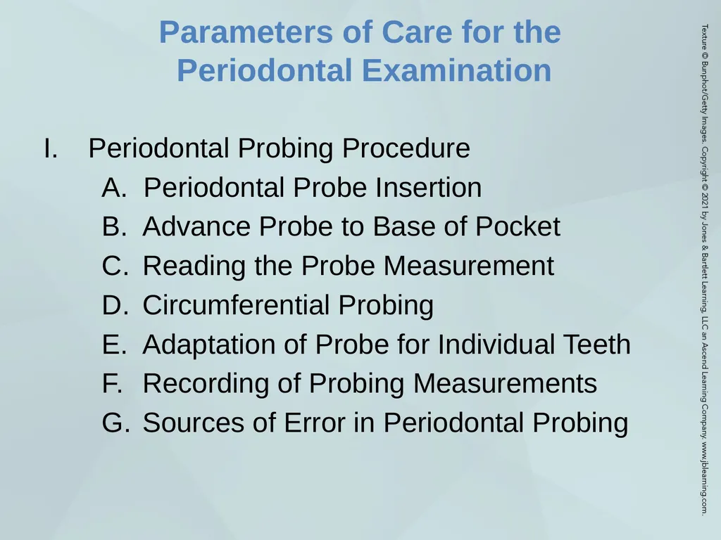
slide26. Parameters of Care for the Periodontal Examination Periodontal Probing Procedure
A. Periodontal Probe Insertion
Advance Probe to Base of Pocket
Reading the Probe Measurement
Circumferential Probing
Adaptation of Probe for Individual Teeth
Recording of Probing Measurements
Sources of Error in Periodontal Probing 26<br>
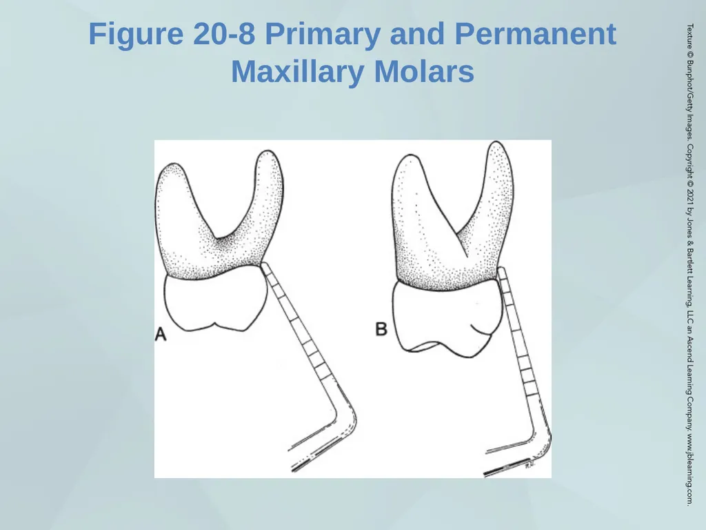
slide27. Figure 20-8 Primary and Permanent Maxillary Molars 27<br>
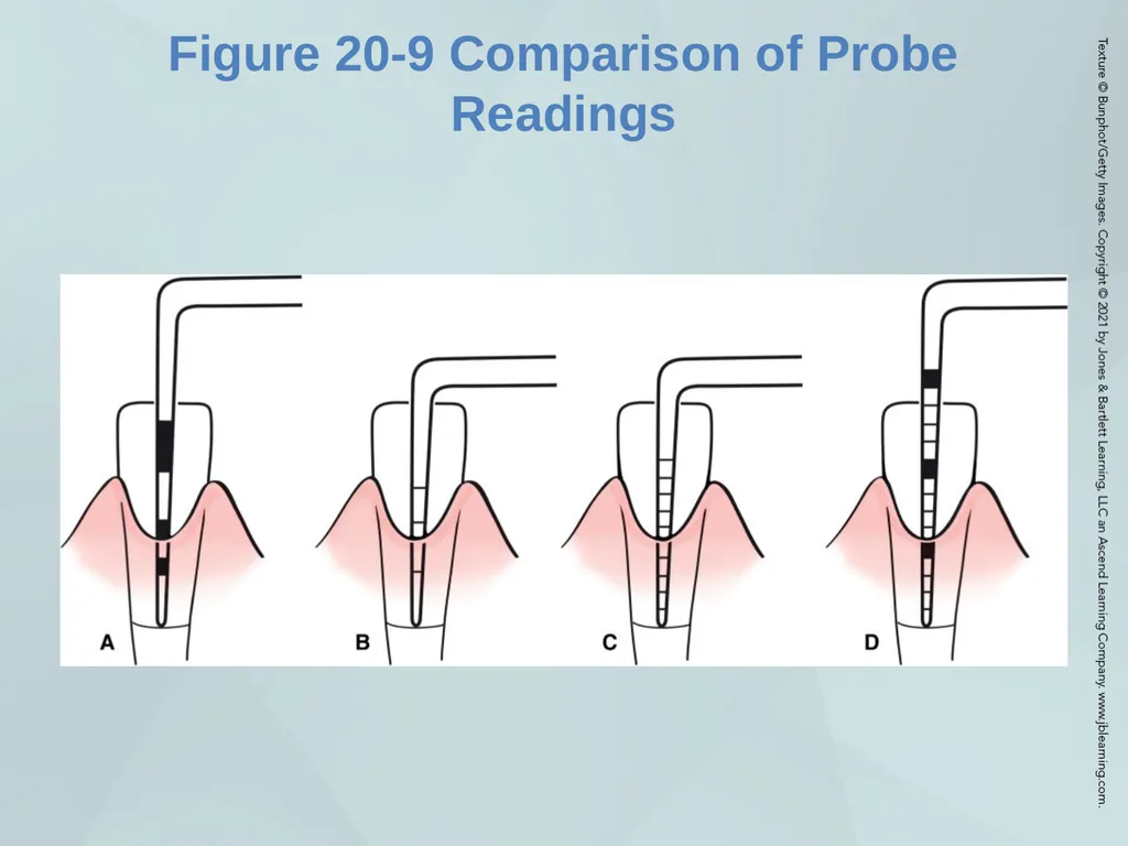
slide28. Figure 20-9 Comparison of Probe Readings 28<br>
slide29. Parameters of Care for the Periodontal Examination con’t Circumferential Probing
Adaptation of Probe for Individual Teeth
Recording of Probing Measurements
Sources of Error in Periodontal Probing 29<br>
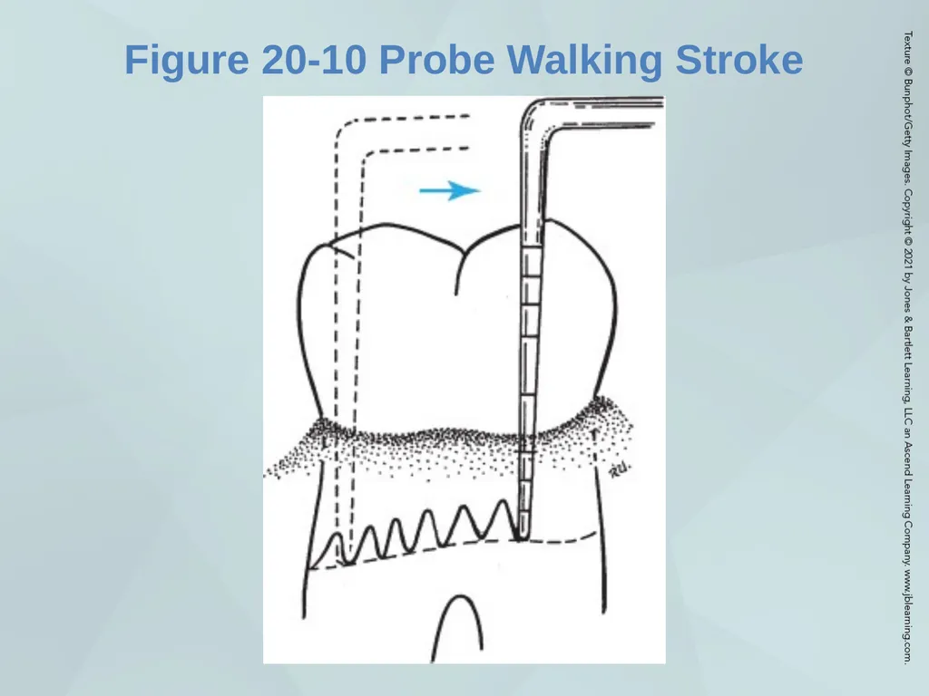
slide30. Figure 20-10 Probe Walking Stroke 30<br>
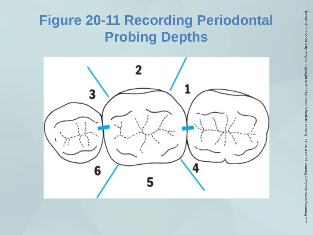
slide31. Figure 20-11 Recording Periodontal Probing Depths 31<br>
slide32. II. Clinical Attachment Level Rationale
Procedure 32<br>
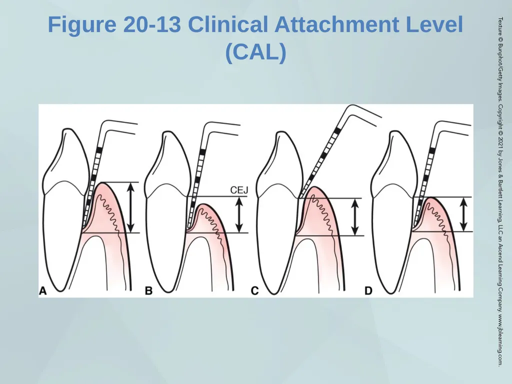
slide33. Figure 20-13 Clinical Attachment Level (CAL) 33<br>
slide34. III. Mucogingival Examination Methods for Identifying MGJ
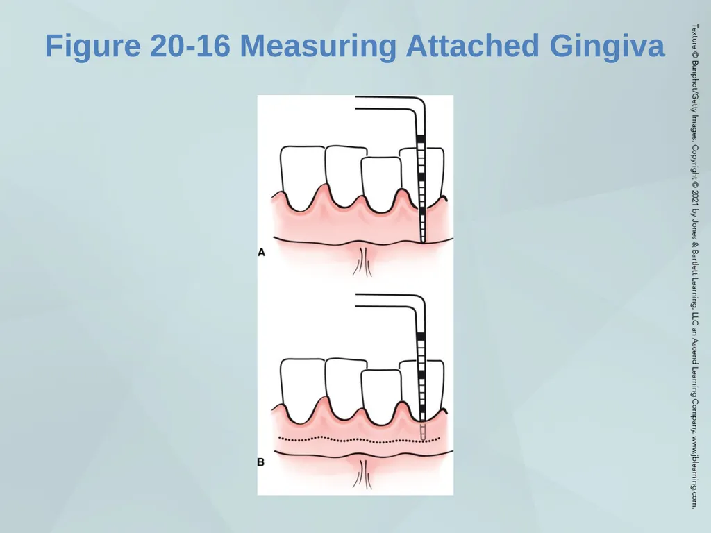
Measurement of Width of Attached Gingiva
Areas at Risk for Mucogingival Conditions 34<br>
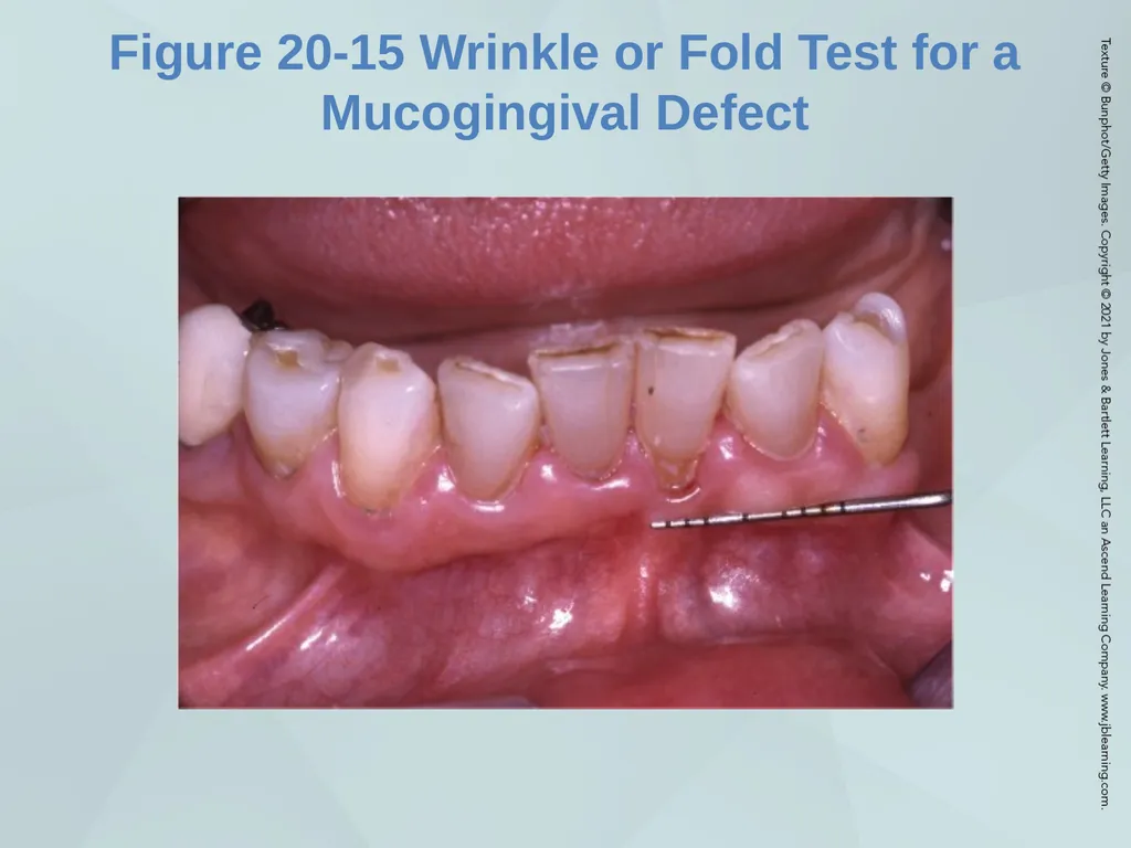
slide35. Figure 20-15 Wrinkle or Fold Test for a Mucogingival Defect 35<br>
slide36. Figure 20-16 Measuring Attached Gingiva 36<br>
slide37. IV. Mobility Examination Procedure for Determination of Mobility
Record Degree of Movement
Scale
Recording
Letter N means normal mobility
Chart form 37<br>
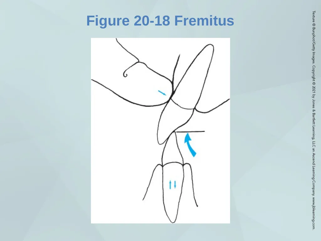
slide38. V. Fremitus Definition
Procedure for Determination of Fremitus 38<br>
slide39. Figure 20-18 Fremitus 39<br>
slide40. VI. Furcation Examination Anatomic Features
Bifurcation (teeth with two roots)
Examination Methods
Furcation measurement procedure
Furcation grades
Difficulties encountered in measurement of the furcation 40<br>
slide41. Radiographic Changes in Periodontal Disease Bone Level
Normal Bone Level
Bone Level in Periodontal Disease
Shape of Remaining Bone
Horizontal Bone Loss
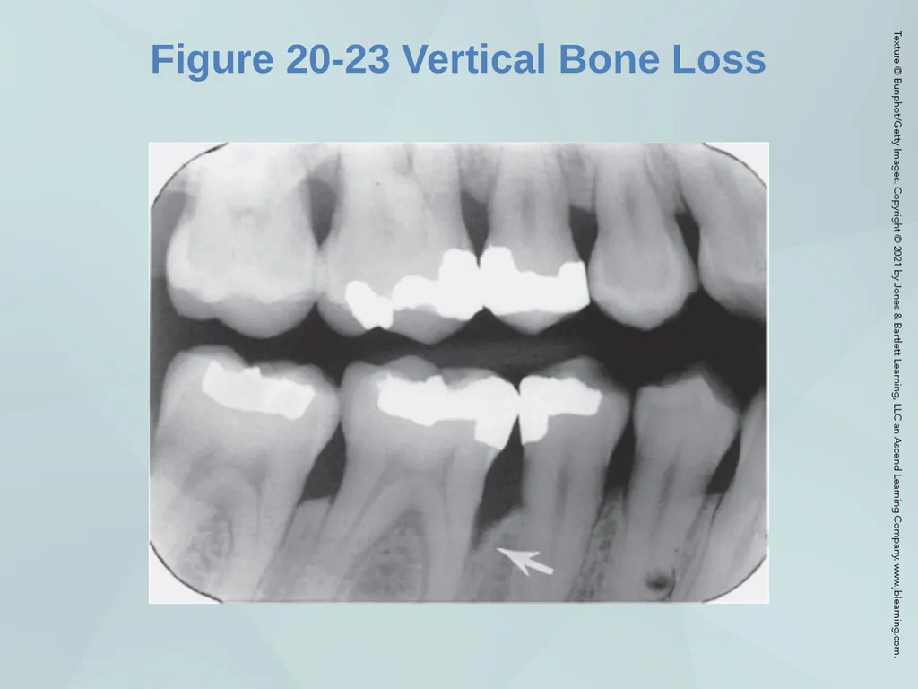
Angular or Vertical Bone Loss 41<br>
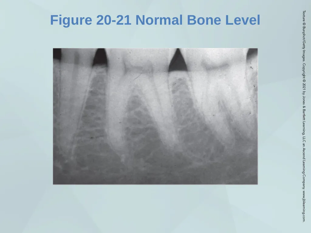
slide42. Figure 20-21 Normal Bone Level 42<br>
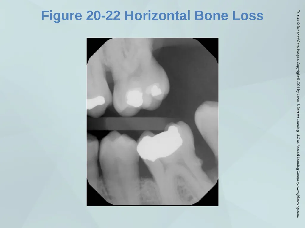
slide43. Figure 20-22 Horizontal Bone Loss 43<br>
slide44. Figure 20-23 Vertical Bone Loss 44<br>
slide45. Radiographic Changes in Periodontal Disease con’t Crestal Lamina Dura
Normal
Evidence of Disease
Furcation Involvement
Normal
Evidence of Disease 45<br>
slide46. Radiographic Changes in Periodontal Disease con’t Periodontal Ligament Space
Normal
Evidence of Disease 46<br>
slide47. Other Radiographic Findings Calculus
Overhanging Restorations
Dental Caries 47<br>
slide48. Documentation Findings by clinical observation and periodontal examination
Mobility, fremitus, occlusal problems, and other findings that will tie the complete oral plan for dental hygiene care into the overall major treatment plan 48<br>
slide49. Factors to Teach the Patient Need for comprehensive examination
Value of various components of the comprehensive examination
Why bleeding can occur when probing
Relation of PD measurements to normal sulci
Significance of the periodontal findings 49<br>
slide2. Learning Objectives Describe components of a comprehensive periodontal examination
List instruments used for a periodontal examination
Explain technique for use of periodontal probe and explorers
Explain how procedure will be described to patient 2<br>
slide3. Basic Instruments for Examination Mouth mirror
Probes:
Furcation probe
Plastic probe
Subgingival explorer 3<br>
slide4. The Mouth Mirror Purposes and Uses
Indirect Vision
Indirect Illumination
Transillumination
Retraction 4<br>
slide5. II. Procedure for Use Grasp and Rest
Retraction
Maintain Clear Vision 5<br>
slide6. Air–Water Syringe Purposes and Uses
Procedure for Use
To Improve and Facilitate Examination Procedures
To Improve Visibility of the Treatment Area during Instrumentation
To Prepare Teeth and/or Gingiva for Certain Procedures
Precautions 6<br>
slide7. Explorers General Purposes and Uses
Detect, by Tactile Sense, the Texture and Character of the Tooth Surfaces
Define the Extent of Instrumentation Needed and Guide Techniques
Evaluate the Completeness of Treatment 7<br>
slide8. II. Specific Explorers and Their Uses Subgingival Explorers
Use
Features for subgingival examination
Supragingival Explorers
Shepherd hook explorer
Pigtail or Cowhorn 8<br>
slide9. Figure 20-1 Explorers 9<br>
slide10. Basic Procedures for Use of Explorers Use of Sensory Stimuli
Tooth Surface Irregularities
Normal
Irregularities: Increases of elevations in tooth surface
Irregularities: Depressions, grooves 10<br>
slide11. III. Types of Stimuli Tactile
Auditory 11<br>
slide12. Explorers : Supragingival Procedures Use of Vision
Facial and Lingual Surfaces
Proximal Surfaces 12<br>
slide13. Explorers: Subgingival Procedures Essentials for Detection of Tooth Surface Irregularities 13<br>
slide14. Figure 20-2 Use of Subgingival Explorer 14<br>
slide15. Periodontal Probe Types of Probes
Standard or traditional probes
Automated probes 15<br>
slide16. Figure 20-3 Examples of Probes 16<br>
slide17. Figure 20-4 Furcation Examination 17<br>
slide18. II. Purposes and Uses Assess the Periodontal Status for Preparation of a Treatment Plan
Determine Clinical Attachment Level
Conduct Mucogingival Examination
Make Other Gingival Determinations
Guide Treatment
Evaluate Success and Completeness of Treatment
Evaluation at Continuing Care and Periodontal Maintenance Appointments 18<br>
slide19. Figure 20-5 Mucogingival Examination 19<br>
slide20. III. Description of Manual Periodontal Probes Materials
Characteristics
Selection 20<br>
slide21. Guide to Periodontal Probing Pocket Characteristics
Evaluation of Tooth Surface
Factors that Affect Probe Determinations
Severity and Extent of Periodontal Disease
The Periodontal Probe
Placement Problems 21<br>
slide22. Figure 20-6 Pocket Depth (PD) 22<br>
slide23. Figure 20-7 Proximal Surface Probing 23<br>
slide24. Preliminary Assessment Prior to Periodontal Examination Medical History
Dental and Psychosocial History
Vital Signs
Extraoral/Intraoral Examination 24<br>
slide25. Preliminary Assessment Prior to Periodontal Examination con’t Risk Assessment
Radiographic Examination
Dental Examination
Hard and Soft Deposits 25<br>
slide26. Parameters of Care for the Periodontal Examination Periodontal Probing Procedure
A. Periodontal Probe Insertion
Advance Probe to Base of Pocket
Reading the Probe Measurement
Circumferential Probing
Adaptation of Probe for Individual Teeth
Recording of Probing Measurements
Sources of Error in Periodontal Probing 26<br>
slide27. Figure 20-8 Primary and Permanent Maxillary Molars 27<br>
slide28. Figure 20-9 Comparison of Probe Readings 28<br>
slide29. Parameters of Care for the Periodontal Examination con’t Circumferential Probing
Adaptation of Probe for Individual Teeth
Recording of Probing Measurements
Sources of Error in Periodontal Probing 29<br>
slide30. Figure 20-10 Probe Walking Stroke 30<br>
slide31. Figure 20-11 Recording Periodontal Probing Depths 31<br>
slide32. II. Clinical Attachment Level Rationale
Procedure 32<br>
slide33. Figure 20-13 Clinical Attachment Level (CAL) 33<br>
slide34. III. Mucogingival Examination Methods for Identifying MGJ
Measurement of Width of Attached Gingiva
Areas at Risk for Mucogingival Conditions 34<br>
slide35. Figure 20-15 Wrinkle or Fold Test for a Mucogingival Defect 35<br>
slide36. Figure 20-16 Measuring Attached Gingiva 36<br>
slide37. IV. Mobility Examination Procedure for Determination of Mobility
Record Degree of Movement
Scale
Recording
Letter N means normal mobility
Chart form 37<br>
slide38. V. Fremitus Definition
Procedure for Determination of Fremitus 38<br>
slide39. Figure 20-18 Fremitus 39<br>
slide40. VI. Furcation Examination Anatomic Features
Bifurcation (teeth with two roots)
Examination Methods
Furcation measurement procedure
Furcation grades
Difficulties encountered in measurement of the furcation 40<br>
slide41. Radiographic Changes in Periodontal Disease Bone Level
Normal Bone Level
Bone Level in Periodontal Disease
Shape of Remaining Bone
Horizontal Bone Loss
Angular or Vertical Bone Loss 41<br>
slide42. Figure 20-21 Normal Bone Level 42<br>
slide43. Figure 20-22 Horizontal Bone Loss 43<br>
slide44. Figure 20-23 Vertical Bone Loss 44<br>
slide45. Radiographic Changes in Periodontal Disease con’t Crestal Lamina Dura
Normal
Evidence of Disease
Furcation Involvement
Normal
Evidence of Disease 45<br>
slide46. Radiographic Changes in Periodontal Disease con’t Periodontal Ligament Space
Normal
Evidence of Disease 46<br>
slide47. Other Radiographic Findings Calculus
Overhanging Restorations
Dental Caries 47<br>
slide48. Documentation Findings by clinical observation and periodontal examination
Mobility, fremitus, occlusal problems, and other findings that will tie the complete oral plan for dental hygiene care into the overall major treatment plan 48<br>
slide49. Factors to Teach the Patient Need for comprehensive examination
Value of various components of the comprehensive examination
Why bleeding can occur when probing
Relation of PD measurements to normal sulci
Significance of the periodontal findings 49<br>