
Chapter 46 Urinary Elimination Copyright © 2017,
DP
Published · 39 slides · 0 views
1 / 1













Description
Chapter 46 Urinary Elimination Copyright 2017, Elsevier Inc. All Rights Reserved. Scientific Knowledge Base Kidneys Ureters Bladder Urethra Copyright 2017, Elsevier Inc. All Rights Reserved. 2 1. A patient with a long-standing history
Related Topics
Share
Embed code
Download this presentation From Below
"Chapter 46 Urinary Elimination Copyright © 2017," is the property of its rightful owner. Permission is granted to download and print the materials on this website for personal, non-commercial use only, and to display it on your personal computer provided you do not modify the materials and that you retain all copyright notices contained in the materials. By downloading content from our website, you accept the terms of this agreement.
Presentation Transcript
01
Chapter 46 Urinary Elimination Copyright © 2017, Elsevier Inc. All Rights Reserved.<br>
02
Scientific Knowledge Base Kidneys
Ureters
Bladder
Urethra Copyright © 2017, Elsevier Inc. All Rights Reserved. 2<br>
Ureters
Bladder
Urethra Copyright © 2017, Elsevier Inc. All Rights Reserved. 2<br>
03
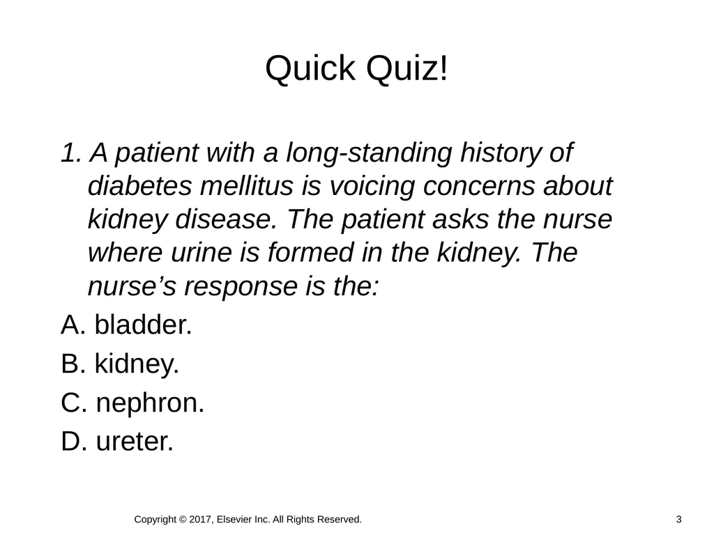
1. A patient with a long-standing history of diabetes mellitus is voicing concerns about kidney disease. The patient asks the nurse where urine is formed in the kidney. The nurse’s response is the:
A. bladder.
B. kidney.
C. nephron.
D. ureter. Quick Quiz! Copyright © 2017, Elsevier Inc. All Rights Reserved. 3<br>
A. bladder.
B. kidney.
C. nephron.
D. ureter. Quick Quiz! Copyright © 2017, Elsevier Inc. All Rights Reserved. 3<br>
04
Mrs. Vallero is a 65-year-old woman who has been in the hospital for 4 days with problems related to heart failure, fluid retention, and diabetes. She has a history of urinary retention secondary to neuropathy caused by her diabetes.
Mrs. Vallero’s indwelling urinary catheter was removed 2 days ago and subsequently was replaced yesterday at 6 a.m. because of her inability to urinate more than 100 mL at a time, being incontinent of small amounts of urine, complaints of urinary urgency, and lower abdominal pain. Case Study Copyright © 2017, Elsevier Inc. All Rights Reserved. 4<br>
Mrs. Vallero’s indwelling urinary catheter was removed 2 days ago and subsequently was replaced yesterday at 6 a.m. because of her inability to urinate more than 100 mL at a time, being incontinent of small amounts of urine, complaints of urinary urgency, and lower abdominal pain. Case Study Copyright © 2017, Elsevier Inc. All Rights Reserved. 4<br>
05
Brain structures influence bladder function.
Voiding: Bladder contraction + urethral sphincter and pelvic floor muscle relaxation
1. Bladder wall stretching signals micturition center.
2. Impulses from the micturition center in the brain respond to or ignore this urge, thus making urination under voluntary control.
3. When a person is ready to void, the central nervous system sends a message to the micturition centers, the external sphincter relaxes and the bladder empties. Act of Urination Copyright © 2017, Elsevier Inc. All Rights Reserved. 5<br>
Voiding: Bladder contraction + urethral sphincter and pelvic floor muscle relaxation
1. Bladder wall stretching signals micturition center.
2. Impulses from the micturition center in the brain respond to or ignore this urge, thus making urination under voluntary control.
3. When a person is ready to void, the central nervous system sends a message to the micturition centers, the external sphincter relaxes and the bladder empties. Act of Urination Copyright © 2017, Elsevier Inc. All Rights Reserved. 5<br>
06
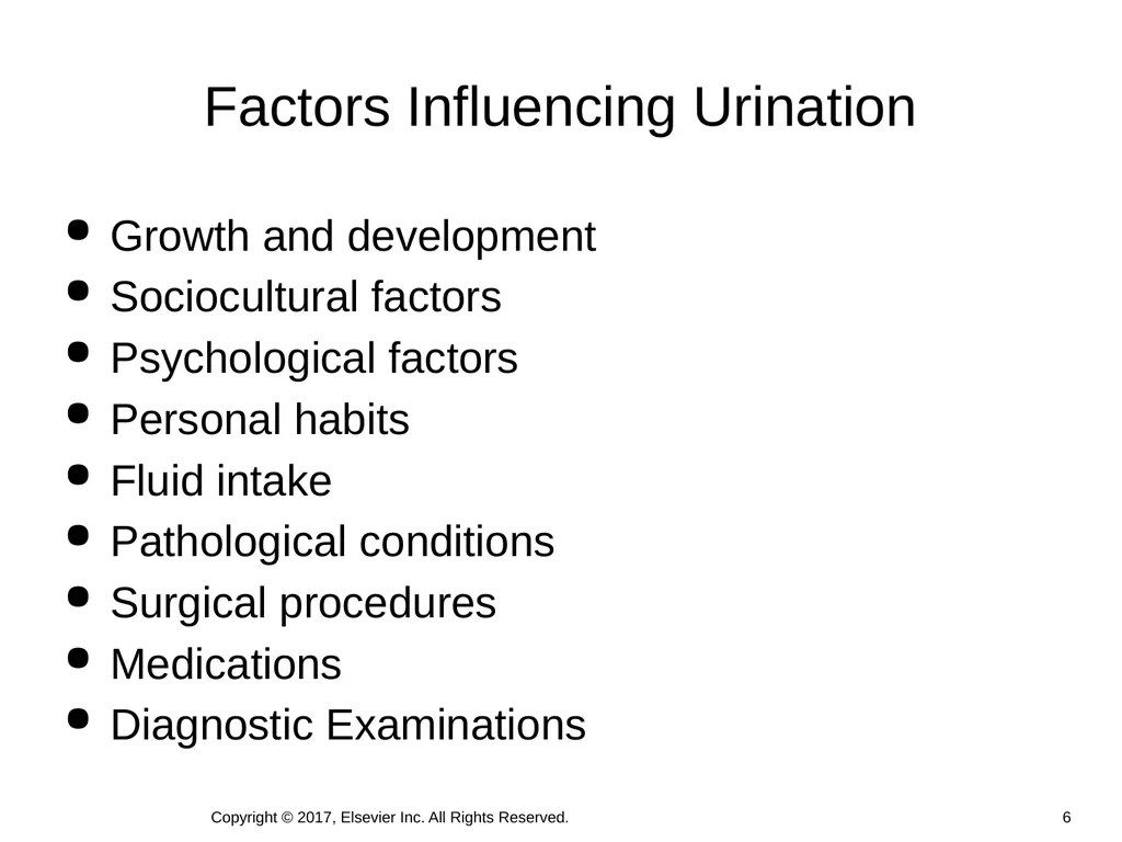
Growth and development
Sociocultural factors
Psychological factors
Personal habits
Fluid intake
Pathological conditions
Surgical procedures
Medications
Diagnostic Examinations Factors Influencing Urination Copyright © 2017, Elsevier Inc. All Rights Reserved. 6<br>
Sociocultural factors
Psychological factors
Personal habits
Fluid intake
Pathological conditions
Surgical procedures
Medications
Diagnostic Examinations Factors Influencing Urination Copyright © 2017, Elsevier Inc. All Rights Reserved. 6<br>
07
Sandy notes that the urinary catheter was removed at 7 a.m. this morning, and the patient has no recorded urine output for the day. Mrs. Vallero verifies that she has only “dribbled” urine. While making rounds, Sandy talks with Mrs. Vallero, who says she is worried because “I thought this was all under control.”
The health care provider is notified, and an order is obtained for an intermittent catheterization. The registered nurse on the day shift catheterizes Mrs. Vallero at 3 p.m. with a return of 600 mL of pale, clear yellow urine. Case Study (Cont.) Copyright © 2017, Elsevier Inc. All Rights Reserved. 7<br>
The health care provider is notified, and an order is obtained for an intermittent catheterization. The registered nurse on the day shift catheterizes Mrs. Vallero at 3 p.m. with a return of 600 mL of pale, clear yellow urine. Case Study (Cont.) Copyright © 2017, Elsevier Inc. All Rights Reserved. 7<br>
08
As Sandy prepares to assess Mrs. Vallero again, she remembers that urinary problems are common in patients who have diabetes and in older adults. Age alone does not cause incontinence. She recalls that patients with urinary retention sometimes leak or “dribble” urine and are then misdiagnosed as incontinent.
She knows that patients generally void at least every 6 to 8 hours, and that Mrs. Vallero’s recent catheterization, her decreased mobility, and her history of diabetes make her more prone to urinary retention, incontinence of small amounts of urine, and urinary tract infection (UTI). Case Study (Cont.) Copyright © 2017, Elsevier Inc. All Rights Reserved. 8<br>
She knows that patients generally void at least every 6 to 8 hours, and that Mrs. Vallero’s recent catheterization, her decreased mobility, and her history of diabetes make her more prone to urinary retention, incontinence of small amounts of urine, and urinary tract infection (UTI). Case Study (Cont.) Copyright © 2017, Elsevier Inc. All Rights Reserved. 8<br>
09
Common Urinary Elimination Problems Urinary retention
An accumulation of urine due to the inability of the bladder to empty
Urinary tract infection
Results from catheterization or procedure
Urinary incontinence
Involuntary leakage of urine Copyright © 2017, Elsevier Inc. All Rights Reserved. 9<br>
An accumulation of urine due to the inability of the bladder to empty
Urinary tract infection
Results from catheterization or procedure
Urinary incontinence
Involuntary leakage of urine Copyright © 2017, Elsevier Inc. All Rights Reserved. 9<br>
10
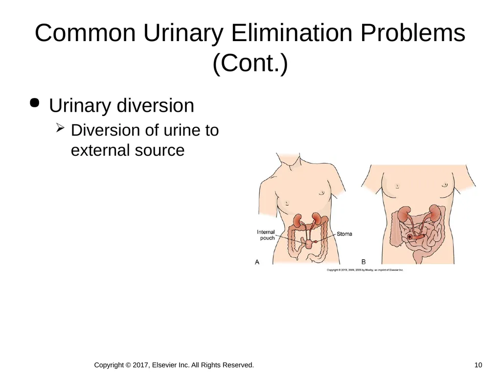
Common Urinary Elimination Problems (Cont.) Urinary diversion
Diversion of urine to external source 10 Copyright © 2017, Elsevier Inc. All Rights Reserved.<br>
Diversion of urine to external source 10 Copyright © 2017, Elsevier Inc. All Rights Reserved.<br>
11
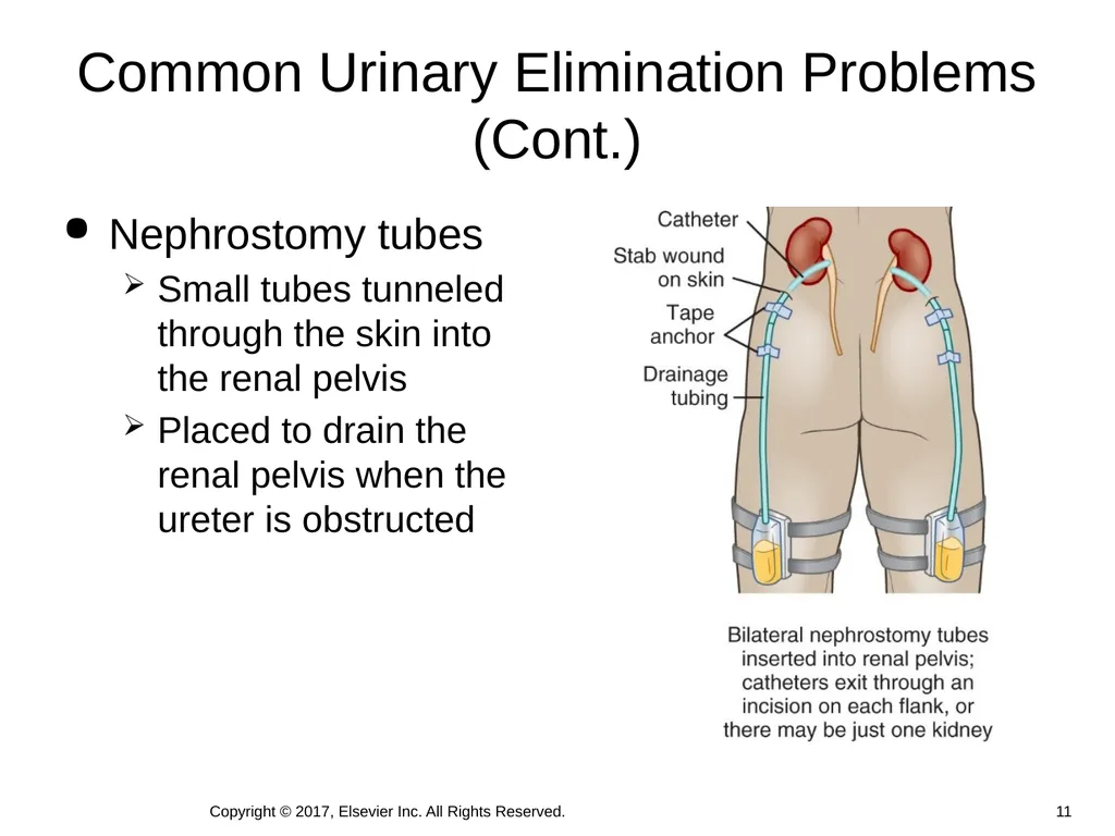
Common Urinary Elimination Problems (Cont.) Nephrostomy tubes
Small tubes tunneled through the skin into the renal pelvis
Placed to drain the renal pelvis when the ureter is obstructed Copyright © 2017, Elsevier Inc. All Rights Reserved. 11<br>
Small tubes tunneled through the skin into the renal pelvis
Placed to drain the renal pelvis when the ureter is obstructed Copyright © 2017, Elsevier Inc. All Rights Reserved. 11<br>
12
2. A health care provider may suspect that a patient is experiencing urinary retention when the patient has:
A. large amounts of voided cloudy urine.
B. pain in the suprapubic region.
C. spasms and difficulty during urination.
D. small amounts of urine voided two to three times per hour. Quick Quiz! Copyright © 2017, Elsevier Inc. All Rights Reserved. 12<br>
A. large amounts of voided cloudy urine.
B. pain in the suprapubic region.
C. spasms and difficulty during urination.
D. small amounts of urine voided two to three times per hour. Quick Quiz! Copyright © 2017, Elsevier Inc. All Rights Reserved. 12<br>
13
Sandy knows that she will need to assess whether Mrs. Vallero feels the urge to urinate. She determines that no one has taken Mrs. Vallero to the bathroom recently. Sandy also needs to find out more about her patient’s urination patterns at home because Mrs. Vallero has verbalized anxiety about her present voiding patterns.
Previous clinical experience has taught Sandy that palpation of the abdomen over a distended bladder causes some discomfort, and that the patient often experiences an urge to urinate. Mrs. Vallero grimaces when her abdomen is palpated and says she has a little dolor (pain). Case Study (Cont.) Copyright © 2017, Elsevier Inc. All Rights Reserved. 13<br>
Previous clinical experience has taught Sandy that palpation of the abdomen over a distended bladder causes some discomfort, and that the patient often experiences an urge to urinate. Mrs. Vallero grimaces when her abdomen is palpated and says she has a little dolor (pain). Case Study (Cont.) Copyright © 2017, Elsevier Inc. All Rights Reserved. 13<br>
14
Nursing Knowledge Base Infection control and hygiene
Growth and development
Psychosocial considerations Copyright © 2017, Elsevier Inc. All Rights Reserved. 14<br>
Growth and development
Psychosocial considerations Copyright © 2017, Elsevier Inc. All Rights Reserved. 14<br>
15
Critical Thinking Successful critical thinking requires synthesis of
Knowledge
Experience
Information gathered from patients
Critical thinking attitudes
Intellectual and professional standards 15 Copyright © 2017, Elsevier Inc. All Rights Reserved.<br>
Knowledge
Experience
Information gathered from patients
Critical thinking attitudes
Intellectual and professional standards 15 Copyright © 2017, Elsevier Inc. All Rights Reserved.<br>
16
Through the patient’s eyes
Self-care ability
Cultural considerations
Health literacy
Nursing history
Pattern of urination
Symptoms of urinary alterations Nursing Process: Assessment Copyright © 2017, Elsevier Inc. All Rights Reserved. 16<br>
Self-care ability
Cultural considerations
Health literacy
Nursing history
Pattern of urination
Symptoms of urinary alterations Nursing Process: Assessment Copyright © 2017, Elsevier Inc. All Rights Reserved. 16<br>
17
Physical assessment
Kidneys
Bladder
External genitalia and urethral meatus
Perineal skin Assessment (Cont.) 17 Copyright © 2017, Elsevier Inc. All Rights Reserved.<br>
Kidneys
Bladder
External genitalia and urethral meatus
Perineal skin Assessment (Cont.) 17 Copyright © 2017, Elsevier Inc. All Rights Reserved.<br>
18
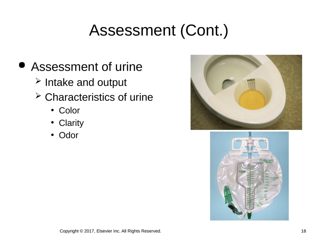
Assessment (Cont.) Assessment of urine
Intake and output
Characteristics of urine
Color
Clarity
Odor 18 Copyright © 2017, Elsevier Inc. All Rights Reserved.<br>
Intake and output
Characteristics of urine
Color
Clarity
Odor 18 Copyright © 2017, Elsevier Inc. All Rights Reserved.<br>
19
Findings:
Patient is able to palpate bladder, indicating bladder distention. During palpation, patient states she has the sensation of bladder fullness.
Patient complains of dribbling frequently and being unable to urinate. Case Study (Cont.) Copyright © 2017, Elsevier Inc. All Rights Reserved. 19<br>
Patient is able to palpate bladder, indicating bladder distention. During palpation, patient states she has the sensation of bladder fullness.
Patient complains of dribbling frequently and being unable to urinate. Case Study (Cont.) Copyright © 2017, Elsevier Inc. All Rights Reserved. 19<br>
20
3. A young girl is having problems urinating postoperatively. You remember that children may have trouble voiding:
A. in bathrooms other than their own.
B. in a urinal.
C. while lying in bed.
D. in the presence of a person other than one of their parents. Quick Quiz! Copyright © 2017, Elsevier Inc. All Rights Reserved. 20<br>
A. in bathrooms other than their own.
B. in a urinal.
C. while lying in bed.
D. in the presence of a person other than one of their parents. Quick Quiz! Copyright © 2017, Elsevier Inc. All Rights Reserved. 20<br>
21
Laboratory and Diagnostic Testing Nursing responsibilities before testing:
Ensure a signed consent is completed.
Assess the patient for any allergies.
Administer bowel-cleansing agents as ordered.
Ensure that the patient adheres to the appropriate pretest diet or nothing by mouth (NPO).
Responsibilities after testing include:
Assessing I&O
Assessing voiding and urine
Encouraging fluid intake Copyright © 2017, Elsevier Inc. All Rights Reserved. 21<br>
Ensure a signed consent is completed.
Assess the patient for any allergies.
Administer bowel-cleansing agents as ordered.
Ensure that the patient adheres to the appropriate pretest diet or nothing by mouth (NPO).
Responsibilities after testing include:
Assessing I&O
Assessing voiding and urine
Encouraging fluid intake Copyright © 2017, Elsevier Inc. All Rights Reserved. 21<br>
22
Nursing Diagnosis Nursing diagnoses common to patients with urinary elimination problems:
Functional urinary incontinence
Stress urinary incontinence
Urge urinary incontinence
Risk for infection
Toileting self-care deficit
Impaired skin integrity
Impaired urinary elimination
Urinary retention 22 Copyright © 2017, Elsevier Inc. All Rights Reserved.<br>
Functional urinary incontinence
Stress urinary incontinence
Urge urinary incontinence
Risk for infection
Toileting self-care deficit
Impaired skin integrity
Impaired urinary elimination
Urinary retention 22 Copyright © 2017, Elsevier Inc. All Rights Reserved.<br>
23
Planning Goals and outcomes
Set realistic and individualized goals along with relevant outcomes
Collaborate with the patient
Setting priorities
Patient’s immediate physical and safety needs
Patient expectations and readiness to perform some self-care activities
Teamwork and collaboration 23 Copyright © 2017, Elsevier Inc. All Rights Reserved.<br>
Set realistic and individualized goals along with relevant outcomes
Collaborate with the patient
Setting priorities
Patient’s immediate physical and safety needs
Patient expectations and readiness to perform some self-care activities
Teamwork and collaboration 23 Copyright © 2017, Elsevier Inc. All Rights Reserved.<br>
24
Nursing diagnosis: Urinary retention related to weakened detrusor muscle and recent removal of indwelling urinary catheter.
Goal: Mrs. Vallero will have normal micturition within 1 month. Case Study (Cont.) Copyright © 2017, Elsevier Inc. All Rights Reserved. 24<br>
Goal: Mrs. Vallero will have normal micturition within 1 month. Case Study (Cont.) Copyright © 2017, Elsevier Inc. All Rights Reserved. 24<br>
25
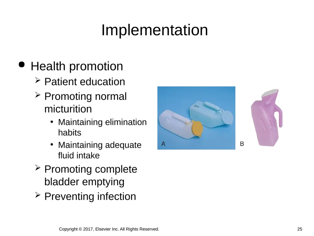
Implementation Health promotion
Patient education
Promoting normal micturition
Maintaining elimination habits
Maintaining adequate fluid intake
Promoting complete bladder emptying
Preventing infection Copyright © 2017, Elsevier Inc. All Rights Reserved. 25<br>
Patient education
Promoting normal micturition
Maintaining elimination habits
Maintaining adequate fluid intake
Promoting complete bladder emptying
Preventing infection Copyright © 2017, Elsevier Inc. All Rights Reserved. 25<br>
26
Interventions for urinary retention care include:
Assist with toileting every 2 to 3 hours while awake.
Instruct the patient/family to record urinary output as appropriate.
Have Mrs. Vallero take a warm bath if unable to urinate.
Use Credé’s method with each attempted void. Case Study (Cont.) Copyright © 2017, Elsevier Inc. All Rights Reserved. 26<br>
Assist with toileting every 2 to 3 hours while awake.
Instruct the patient/family to record urinary output as appropriate.
Have Mrs. Vallero take a warm bath if unable to urinate.
Use Credé’s method with each attempted void. Case Study (Cont.) Copyright © 2017, Elsevier Inc. All Rights Reserved. 26<br>
27
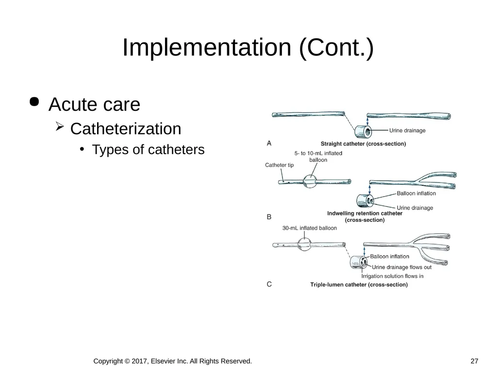
Implementation (Cont.) Acute care
Catheterization
Types of catheters Copyright © 2017, Elsevier Inc. All Rights Reserved. 27<br>
Catheterization
Types of catheters Copyright © 2017, Elsevier Inc. All Rights Reserved. 27<br>
28
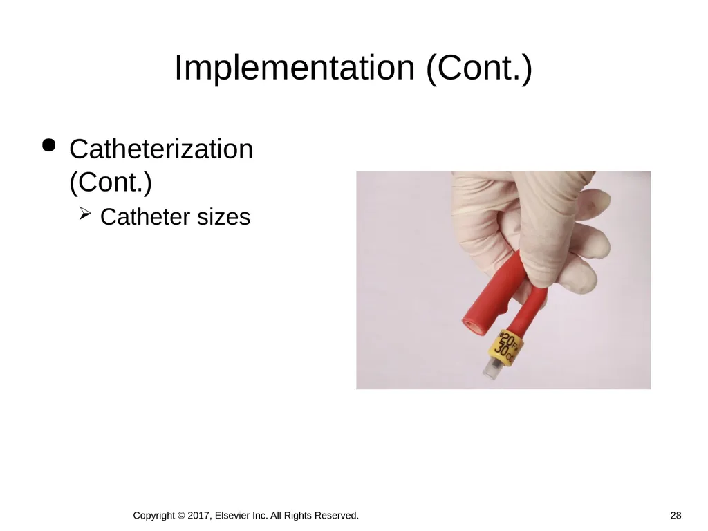
Implementation (Cont.) Catheterization (Cont.)
Catheter sizes Copyright © 2017, Elsevier Inc. All Rights Reserved. 28<br>
Catheter sizes Copyright © 2017, Elsevier Inc. All Rights Reserved. 28<br>
29
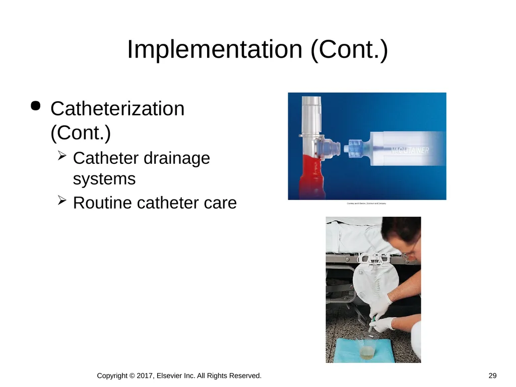
Implementation (Cont.) Catheterization (Cont.)
Catheter drainage systems
Routine catheter care Copyright © 2017, Elsevier Inc. All Rights Reserved. 29<br>
Catheter drainage systems
Routine catheter care Copyright © 2017, Elsevier Inc. All Rights Reserved. 29<br>
30
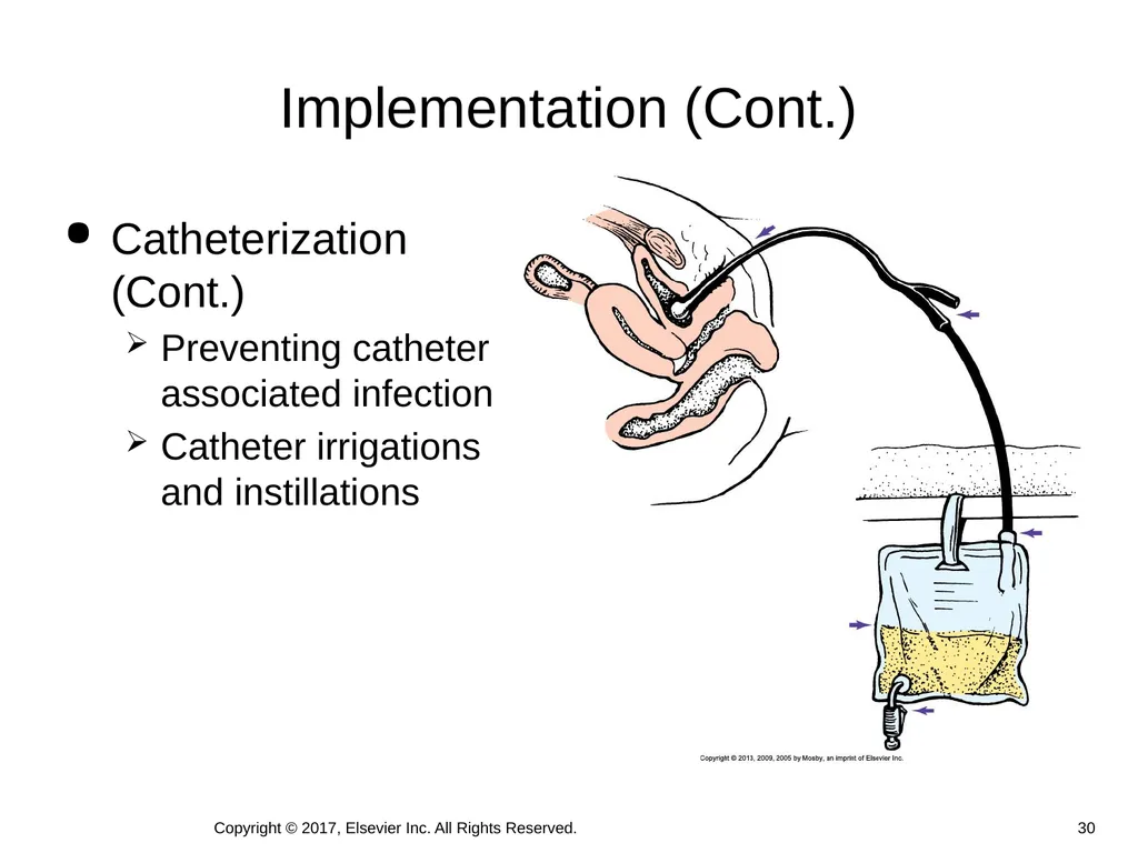
Implementation (Cont.) Catheterization (Cont.)
Preventing catheter associated infection
Catheter irrigations and instillations Copyright © 2017, Elsevier Inc. All Rights Reserved. 30<br>
Preventing catheter associated infection
Catheter irrigations and instillations Copyright © 2017, Elsevier Inc. All Rights Reserved. 30<br>
31
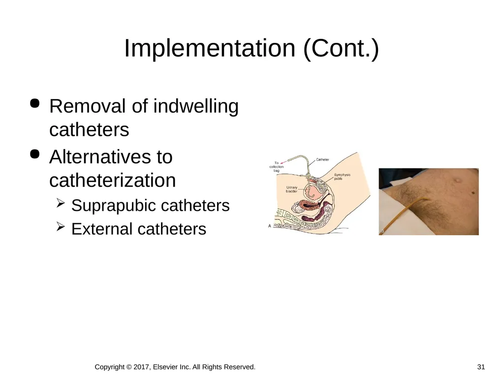
Implementation (Cont.) Removal of indwelling catheters
Alternatives to catheterization
Suprapubic catheters
External catheters 31 Copyright © 2017, Elsevier Inc. All Rights Reserved.<br>
Alternatives to catheterization
Suprapubic catheters
External catheters 31 Copyright © 2017, Elsevier Inc. All Rights Reserved.<br>
32
Urinary diversions
Incontinent diversions
Changing a pouch
Gently cleanse the skin surrounding the stoma
Measure the stoma and cut the opening in the pouch
Remove the adhesive backing and apply the pouch
Press firmly into place over the stoma.
Observe the appearance of the stoma and surrounding skin.
Continent diversions
Orthopic neobladder Implementation (Cont.) 32 Copyright © 2017, Elsevier Inc. All Rights Reserved.<br>
Incontinent diversions
Changing a pouch
Gently cleanse the skin surrounding the stoma
Measure the stoma and cut the opening in the pouch
Remove the adhesive backing and apply the pouch
Press firmly into place over the stoma.
Observe the appearance of the stoma and surrounding skin.
Continent diversions
Orthopic neobladder Implementation (Cont.) 32 Copyright © 2017, Elsevier Inc. All Rights Reserved.<br>
33
Medications
Antimuscarinics: treat urgency, frequency, nocturia and urgency UI
Bethanechol: treat urinary retention
Tamsulosin and silodosin: relax smooth muscle
Finasteride and dutasteride: shrink the prostate
Antibiotics: treat urinary tract infections
Be familiar with the medications and indications for all medications your patient is taking. Implementation (Cont.) 33 Copyright © 2017, Elsevier Inc. All Rights Reserved.<br>
Antimuscarinics: treat urgency, frequency, nocturia and urgency UI
Bethanechol: treat urinary retention
Tamsulosin and silodosin: relax smooth muscle
Finasteride and dutasteride: shrink the prostate
Antibiotics: treat urinary tract infections
Be familiar with the medications and indications for all medications your patient is taking. Implementation (Cont.) 33 Copyright © 2017, Elsevier Inc. All Rights Reserved.<br>
34
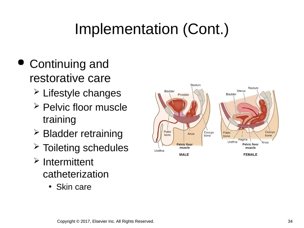
Implementation (Cont.) Continuing and restorative care
Lifestyle changes
Pelvic floor muscle training
Bladder retraining
Toileting schedules
Intermittent catheterization
Skin care 34 Copyright © 2017, Elsevier Inc. All Rights Reserved.<br>
Lifestyle changes
Pelvic floor muscle training
Bladder retraining
Toileting schedules
Intermittent catheterization
Skin care 34 Copyright © 2017, Elsevier Inc. All Rights Reserved.<br>
35
Through the patient’s eyes
Assess the patient’s self-image, social interactions, sexuality, and emotional status
Patient outcomes
Use the expected outcomes developed during planning to determine whether interventions were effective
Evaluate for changes in the patient’s voiding pattern and/or presence of symptoms
Evaluate patient/caregiver compliance with the plan Evaluation Copyright © 2017, Elsevier Inc. All Rights Reserved. 35<br>
Assess the patient’s self-image, social interactions, sexuality, and emotional status
Patient outcomes
Use the expected outcomes developed during planning to determine whether interventions were effective
Evaluate for changes in the patient’s voiding pattern and/or presence of symptoms
Evaluate patient/caregiver compliance with the plan Evaluation Copyright © 2017, Elsevier Inc. All Rights Reserved. 35<br>
36
Sandy talks with Mrs. Vallero the next evening. The patient’s care plan incorporates scheduled voiding, oral fluids, and use of Credé’s method of manual compression during voiding. She palpates Mrs. Vallero’s bladder and assists her to the toilet.
After making sure she is comfortable and leaving the call light in place, Sandy instructs her to use Credé’s method of manual compression. She returns to measure Mrs. Vallero’s urinary output and evaluates for bladder residual using an ultrasound bladder scan. Case Study (Cont.) Copyright © 2017, Elsevier Inc. All Rights Reserved. 36<br>
After making sure she is comfortable and leaving the call light in place, Sandy instructs her to use Credé’s method of manual compression. She returns to measure Mrs. Vallero’s urinary output and evaluates for bladder residual using an ultrasound bladder scan. Case Study (Cont.) Copyright © 2017, Elsevier Inc. All Rights Reserved. 36<br>
37
Ask Mrs. Vallero about her urge to void, sensation of bladder fullness, and dribbling episodes.
Have Mrs. Vallero keep a log of her pattern of elimination, including urine output volumes with each voiding, during the 1-month period.
Ask Mrs. Vallero if she continues to have lower abdominal pain. Case Study (Cont.) Copyright © 2017, Elsevier Inc. All Rights Reserved. 37<br>
Have Mrs. Vallero keep a log of her pattern of elimination, including urine output volumes with each voiding, during the 1-month period.
Ask Mrs. Vallero if she continues to have lower abdominal pain. Case Study (Cont.) Copyright © 2017, Elsevier Inc. All Rights Reserved. 37<br>
38
Mrs. Vallero is concerned about regaining her urinary function. Sandy develops the following outcome for her: At the end of the teaching session, Mrs. Vallero will be able to describe approaches to promote normal urinary elimination habits. Case Study (Cont.) Copyright © 2017, Elsevier Inc. All Rights Reserved. 38<br>
39
Follow principles of surgical and medical asepsis as indicated
Identify patients at risk for latex allergies
Identify patients with allergies to povidone-iodine (Betadine). Provide alternatives such as chlorhexidine. Safety Guidelines for Nursing Skills 39 Copyright © 2017, Elsevier Inc. All Rights Reserved.<br>
Identify patients at risk for latex allergies
Identify patients with allergies to povidone-iodine (Betadine). Provide alternatives such as chlorhexidine. Safety Guidelines for Nursing Skills 39 Copyright © 2017, Elsevier Inc. All Rights Reserved.<br>