
Chapter 7 THE CELLULAR BASIS OF INHERITANCE
BR
Published · 31 slides · 0 views
1 / 1





Description
Chapter 7 THE CELLULAR BASIS OF INHERITANCE PowerPoint Image Slideshow Concepts of Biology This work is licensed under cc by 4.0 license. Credit: OpenStax: modified by M. F. Sega J. Wedincamp for ALG 18 grant through addition of ppt
Related Topics
Share
Embed code
Download this presentation From Below
"Chapter 7 THE CELLULAR BASIS OF INHERITANCE" is the property of its rightful owner. Permission is granted to download and print the materials on this website for personal, non-commercial use only, and to display it on your personal computer provided you do not modify the materials and that you retain all copyright notices contained in the materials. By downloading content from our website, you accept the terms of this agreement.
Presentation Transcript
01
Chapter 7 THE CELLULAR BASIS OF INHERITANCE
PowerPoint Image Slideshow Concepts of Biology This work is licensed under cc by 4.0 license. Credit: OpenStax: modified by M. F. Sega & J. Wedincamp for ALG 18 grant through addition of ppt slides with texts made using Openstax textbook information.<br>
PowerPoint Image Slideshow Concepts of Biology This work is licensed under cc by 4.0 license. Credit: OpenStax: modified by M. F. Sega & J. Wedincamp for ALG 18 grant through addition of ppt slides with texts made using Openstax textbook information.<br>
02
Cellular basis of inheritance Inheritance refers to the transmission of the genetic material from the parents to the offspring
It involves two types of reproduction:
Asexual
Sexual
At the cellular level the inheritance of the genetic material is different between asexual versus sexual reproduction<br>
It involves two types of reproduction:
Asexual
Sexual
At the cellular level the inheritance of the genetic material is different between asexual versus sexual reproduction<br>
03
asexual reproduction Asexual reproduction
– only requires one individual (prefix “a-” means without)
- implies cell divisions by MITOSIS or similar
- making identical individuals or clones
- not successful during evolution
- in case the environment changes might result in killing the whole population or species since all are identical.
- Examples – bacteria, mold<br>
– only requires one individual (prefix “a-” means without)
- implies cell divisions by MITOSIS or similar
- making identical individuals or clones
- not successful during evolution
- in case the environment changes might result in killing the whole population or species since all are identical.
- Examples – bacteria, mold<br>
04
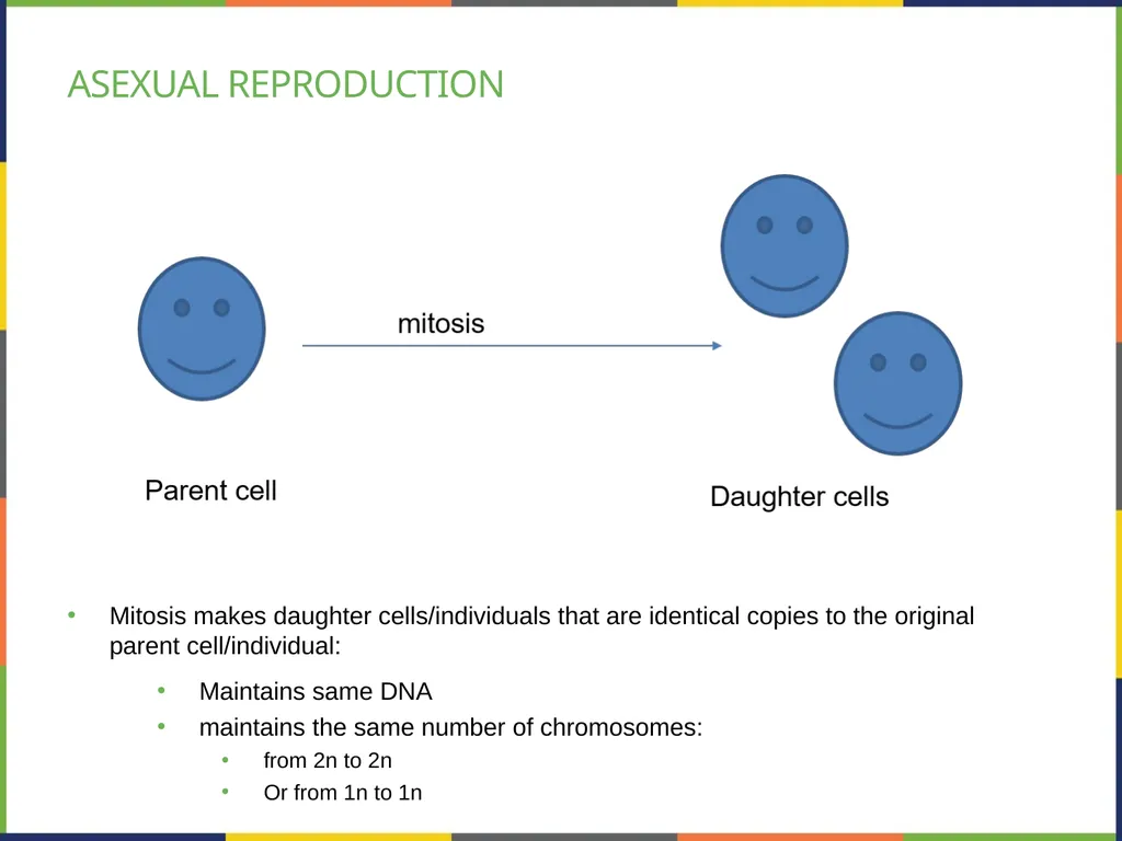
Asexual reproduction Mitosis makes daughter cells/individuals that are identical copies to the original parent cell/individual:
Maintains same DNA
maintains the same number of chromosomes:
from 2n to 2n
Or from 1n to 1n<br>
Maintains same DNA
maintains the same number of chromosomes:
from 2n to 2n
Or from 1n to 1n<br>
05
Sexual reproduction Sexual reproduction
– requires two sexes
- using gametes (sex cells) made by MEIOSIS
- results in making different individuals
- successful during evolution
- in case the environment changes, the fittest survive and save the species from extinction.
What makes the offspring/individuals different is the making of the gametes through MEIOSIS.<br>
– requires two sexes
- using gametes (sex cells) made by MEIOSIS
- results in making different individuals
- successful during evolution
- in case the environment changes, the fittest survive and save the species from extinction.
What makes the offspring/individuals different is the making of the gametes through MEIOSIS.<br>
06
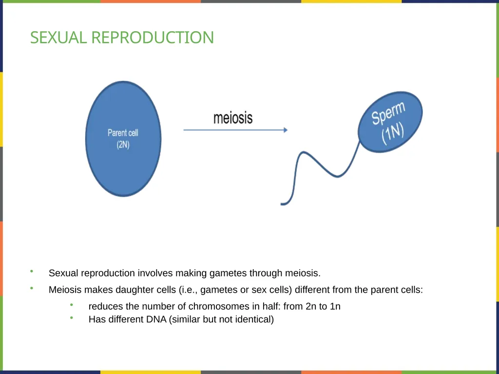
Sexual reproduction Sexual reproduction involves making gametes through meiosis.
Meiosis makes daughter cells (i.e., gametes or sex cells) different from the parent cells:
reduces the number of chromosomes in half: from 2n to 1n
Has different DNA (similar but not identical)<br>
Meiosis makes daughter cells (i.e., gametes or sex cells) different from the parent cells:
reduces the number of chromosomes in half: from 2n to 1n
Has different DNA (similar but not identical)<br>
07
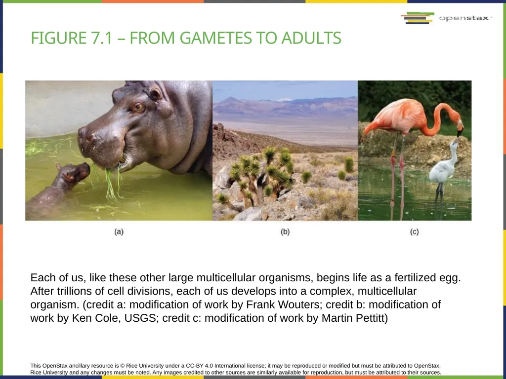
This OpenStax ancillary resource is © Rice University under a CC-BY 4.0 International license; it may be reproduced or modified but must be attributed to OpenStax, Rice University and any changes must be noted. Any images credited to other sources are similarly available for reproduction, but must be attributed to their sources. Each of us, like these other large multicellular organisms, begins life as a fertilized egg. After trillions of cell divisions, each of us develops into a complex, multicellular organism. (credit a: modification of work by Frank Wouters; credit b: modification of work by Ken Cole, USGS; credit c: modification of work by Martin Pettitt) Figure 7.1 – from gametes to adults<br>
08
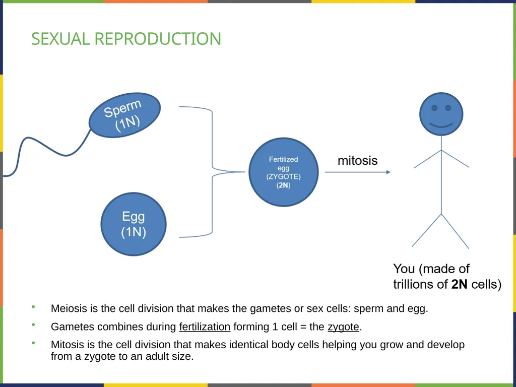
Sexual reproduction Meiosis is the cell division that makes the gametes or sex cells: sperm and egg.
Gametes combines during fertilization forming 1 cell = the zygote.
Mitosis is the cell division that makes identical body cells helping you grow and develop from a zygote to an adult size.<br>
Gametes combines during fertilization forming 1 cell = the zygote.
Mitosis is the cell division that makes identical body cells helping you grow and develop from a zygote to an adult size.<br>
09
Meiosis & sex cells Meiosis is referring to the nucleus division (see previous chapter)
It is preceded by INTERPHASE (see previous chapter)
It involves 2 successive divisions without s-phase in between:
Meiosis I: starts with 1 cell that is 2n and makes 2 cells that are 1n
Meiosis II: the cells resulted from Meiosis I divide again forming 2 cells each, so a total of 4 haploid cells (these are the sex cells)<br>
It is preceded by INTERPHASE (see previous chapter)
It involves 2 successive divisions without s-phase in between:
Meiosis I: starts with 1 cell that is 2n and makes 2 cells that are 1n
Meiosis II: the cells resulted from Meiosis I divide again forming 2 cells each, so a total of 4 haploid cells (these are the sex cells)<br>
10
meiosis Parent cell goes through S-phase once during the interphase;
Then, it goes through 2 successive divisions: meiosis I & II<br>
Then, it goes through 2 successive divisions: meiosis I & II<br>
11
Meiosis I & II Preceded by Interphase (review S-phase previous chapter):
Meiosis I:
Prophase I, Prometaphase I, Metaphase I, Anaphase I, Telophase I
Meiosis II:
Prophase II, Prometaphase II, Metaphase II, Anaphase II, Telophase II<br>
Meiosis I:
Prophase I, Prometaphase I, Metaphase I, Anaphase I, Telophase I
Meiosis II:
Prophase II, Prometaphase II, Metaphase II, Anaphase II, Telophase II<br>
12
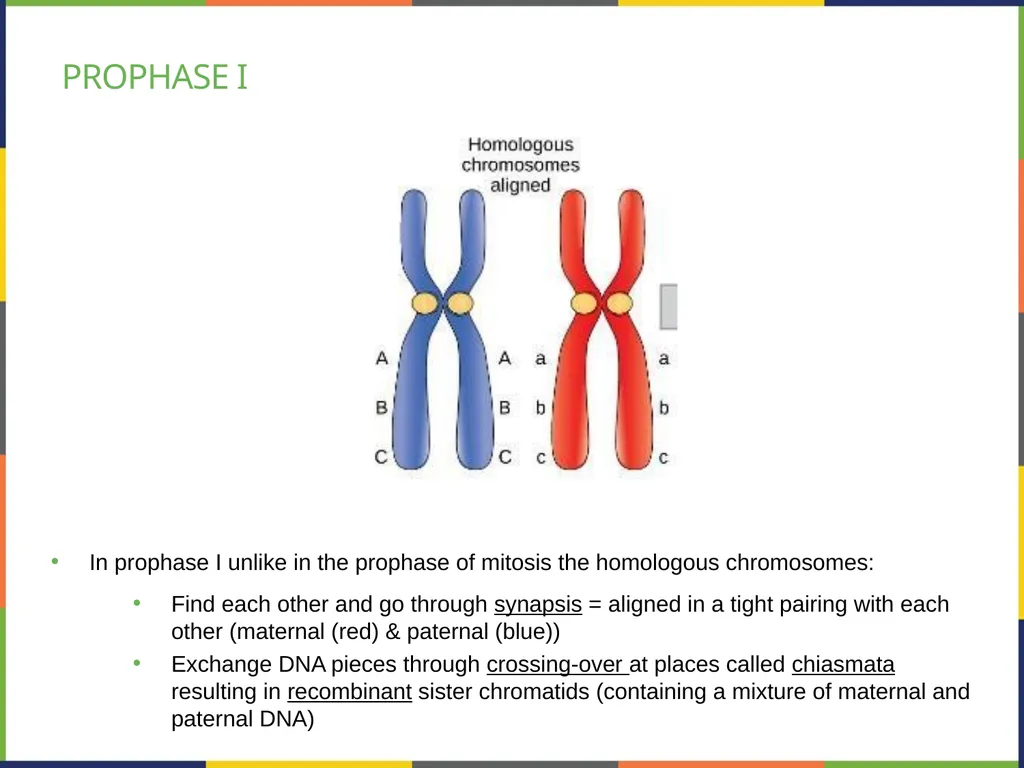
Prophase I In prophase I unlike in the prophase of mitosis the homologous chromosomes:
Find each other and go through synapsis = aligned in a tight pairing with each other (maternal (red) & paternal (blue))
Exchange DNA pieces through crossing-over at places called chiasmata resulting in recombinant sister chromatids (containing a mixture of maternal and paternal DNA)<br>
Find each other and go through synapsis = aligned in a tight pairing with each other (maternal (red) & paternal (blue))
Exchange DNA pieces through crossing-over at places called chiasmata resulting in recombinant sister chromatids (containing a mixture of maternal and paternal DNA)<br>
13
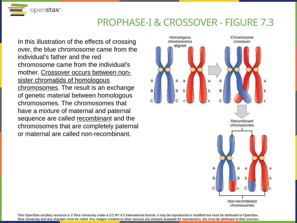
This OpenStax ancillary resource is © Rice University under a CC-BY 4.0 International license; it may be reproduced or modified but must be attributed to OpenStax, Rice University and any changes must be noted. Any images credited to other sources are similarly available for reproduction, but must be attributed to their sources. In this illustration of the effects of crossing over, the blue chromosome came from the individual’s father and the red chromosome came from the individual’s mother. Crossover occurs between non-sister chromatids of homologous chromosomes. The result is an exchange of genetic material between homologous chromosomes. The chromosomes that have a mixture of maternal and paternal sequence are called recombinant and the chromosomes that are completely paternal or maternal are called non-recombinant. Prophase-I & Crossover - Figure 7.3<br>
14
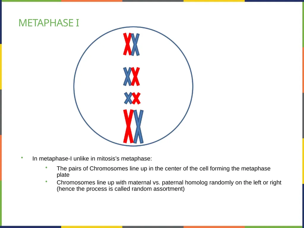
Metaphase I In metaphase-I unlike in mitosis’s metaphase:
The pairs of Chromosomes line up in the center of the cell forming the metaphase plate
Chromosomes line up with maternal vs. paternal homolog randomly on the left or right (hence the process is called random assortment)<br>
The pairs of Chromosomes line up in the center of the cell forming the metaphase plate
Chromosomes line up with maternal vs. paternal homolog randomly on the left or right (hence the process is called random assortment)<br>
15
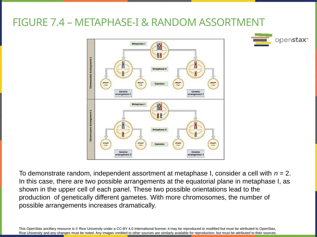
This OpenStax ancillary resource is © Rice University under a CC-BY 4.0 International license; it may be reproduced or modified but must be attributed to OpenStax, Rice University and any changes must be noted. Any images credited to other sources are similarly available for reproduction, but must be attributed to their sources. To demonstrate random, independent assortment at metaphase I, consider a cell with n = 2. In this case, there are two possible arrangements at the equatorial plane in metaphase I, as shown in the upper cell of each panel. These two possible orientations lead to the production of genetically different gametes. With more chromosomes, the number of possible arrangements increases dramatically. Figure 7.4 – metaphase-I & random assortment<br>
16
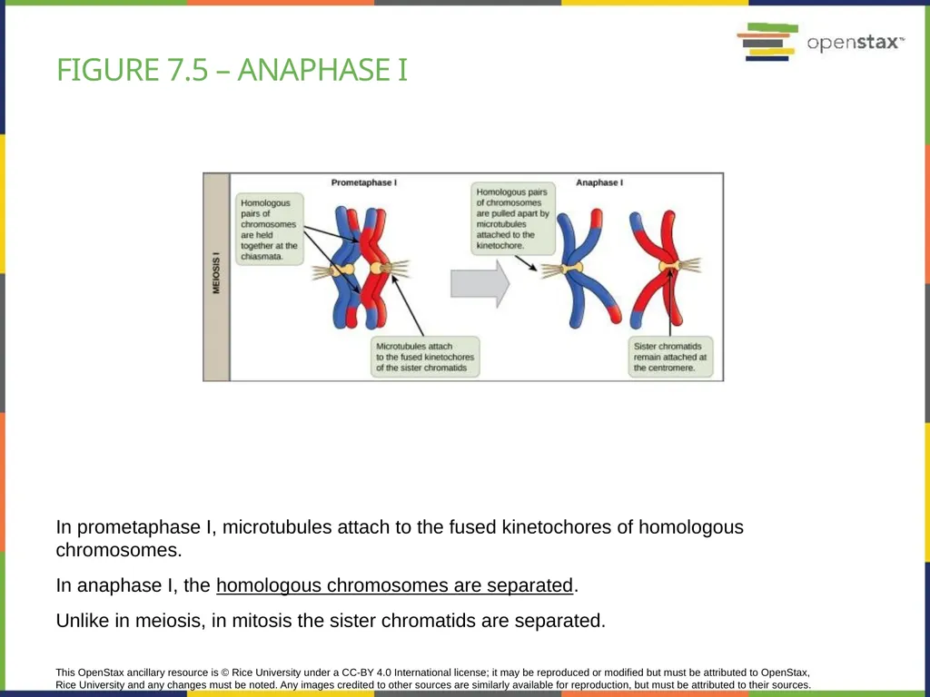
This OpenStax ancillary resource is © Rice University under a CC-BY 4.0 International license; it may be reproduced or modified but must be attributed to OpenStax, Rice University and any changes must be noted. Any images credited to other sources are similarly available for reproduction, but must be attributed to their sources. In prometaphase I, microtubules attach to the fused kinetochores of homologous chromosomes.
In anaphase I, the homologous chromosomes are separated.
Unlike in meiosis, in mitosis the sister chromatids are separated. Figure 7.5 – Anaphase I<br>
In anaphase I, the homologous chromosomes are separated.
Unlike in meiosis, in mitosis the sister chromatids are separated. Figure 7.5 – Anaphase I<br>
17
Telophase-I & cytokinesis Telophase I – chromosomes arrive at the poles of the cell
Cytokinesis – follows telophase and ends the meiosis I process.
The result is:
Two daughter cells
Each daughter cell is haploid
Interkinesis (i.e. a period of rest) follows shortly and then Meiosis II begins<br>
Cytokinesis – follows telophase and ends the meiosis I process.
The result is:
Two daughter cells
Each daughter cell is haploid
Interkinesis (i.e. a period of rest) follows shortly and then Meiosis II begins<br>
18
Meiosis – II The events of Meiosis II are identical with mitosis; briefly:
Prophase II – chromosomes condense
Prometaphase II – mitotic spindles attaches to each chromatid of each chromosome; nuclear membrane breaks down
Metaphase II – the chromosomes individually line up forming metaphase plate
Anaphase II – spindles pull the sister chromatids apart
Telophase II – nuclei of the future daughter cells form
Followed by cytokinesis.<br>
Prophase II – chromosomes condense
Prometaphase II – mitotic spindles attaches to each chromatid of each chromosome; nuclear membrane breaks down
Metaphase II – the chromosomes individually line up forming metaphase plate
Anaphase II – spindles pull the sister chromatids apart
Telophase II – nuclei of the future daughter cells form
Followed by cytokinesis.<br>
19
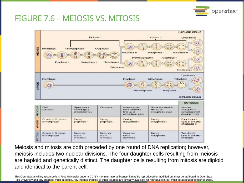
This OpenStax ancillary resource is © Rice University under a CC-BY 4.0 International license; it may be reproduced or modified but must be attributed to OpenStax, Rice University and any changes must be noted. Any images credited to other sources are similarly available for reproduction, but must be attributed to their sources. Meiosis and mitosis are both preceded by one round of DNA replication; however, meiosis includes two nuclear divisions. The four daughter cells resulting from meiosis are haploid and genetically distinct. The daughter cells resulting from mitosis are diploid and identical to the parent cell. Figure 7.6 – meiosis vs. mitosis<br>
20
Meiosis vs. mitosis<br>
21
Meiosis and evolution Meiosis results in sex cells that have different DNA compared to the parent cells; this is the result of:
Crossing-over
Random assortment
Additional variability comes from the randomness of fertilization.
All these 3 process will result in completely different offspring with new features.
New features could allow offspring to survive an eventual change in the environment; which will prevent species extinction.<br>
Crossing-over
Random assortment
Additional variability comes from the randomness of fertilization.
All these 3 process will result in completely different offspring with new features.
New features could allow offspring to survive an eventual change in the environment; which will prevent species extinction.<br>
22
Errors in Meiosis Disorders
due to abnormal number of chromosomes
Due to abnormal structure of chromosomes
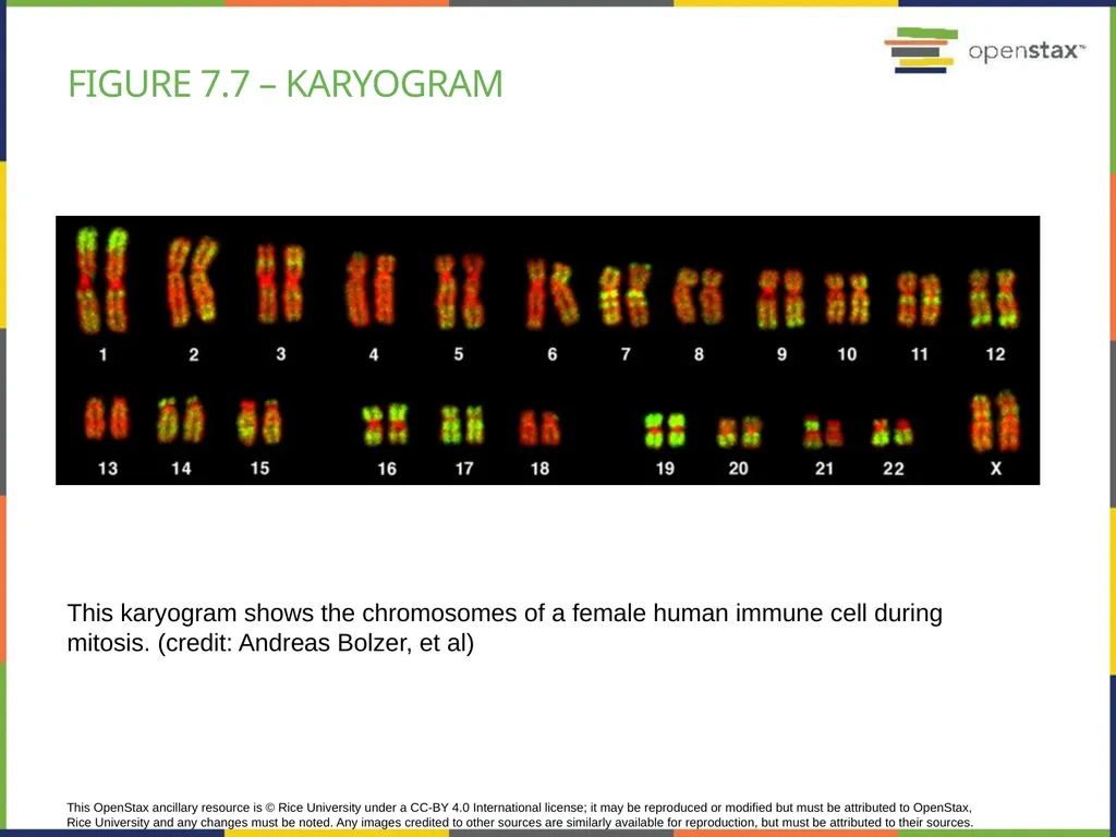
Abnormalities could be seen on the karyogram (a picture of the stained chromosomes organized in pairs of homologs)
Karyotyping is a method to stain the chromosomes and organizing them in pairs
This allows to compare the chromosomes' structure (patterns of staining), length, number.<br>
due to abnormal number of chromosomes
Due to abnormal structure of chromosomes
Abnormalities could be seen on the karyogram (a picture of the stained chromosomes organized in pairs of homologs)
Karyotyping is a method to stain the chromosomes and organizing them in pairs
This allows to compare the chromosomes' structure (patterns of staining), length, number.<br>
23
This OpenStax ancillary resource is © Rice University under a CC-BY 4.0 International license; it may be reproduced or modified but must be attributed to OpenStax, Rice University and any changes must be noted. Any images credited to other sources are similarly available for reproduction, but must be attributed to their sources. This karyogram shows the chromosomes of a female human immune cell during mitosis. (credit: Andreas Bolzer, et al) Figure 7.7 – karyogram<br>
24
Chromosome number Correct number is called Euploidy
Abnormal number is known as Aneuploidy
different number of sets of chromosomes is called polyploidy
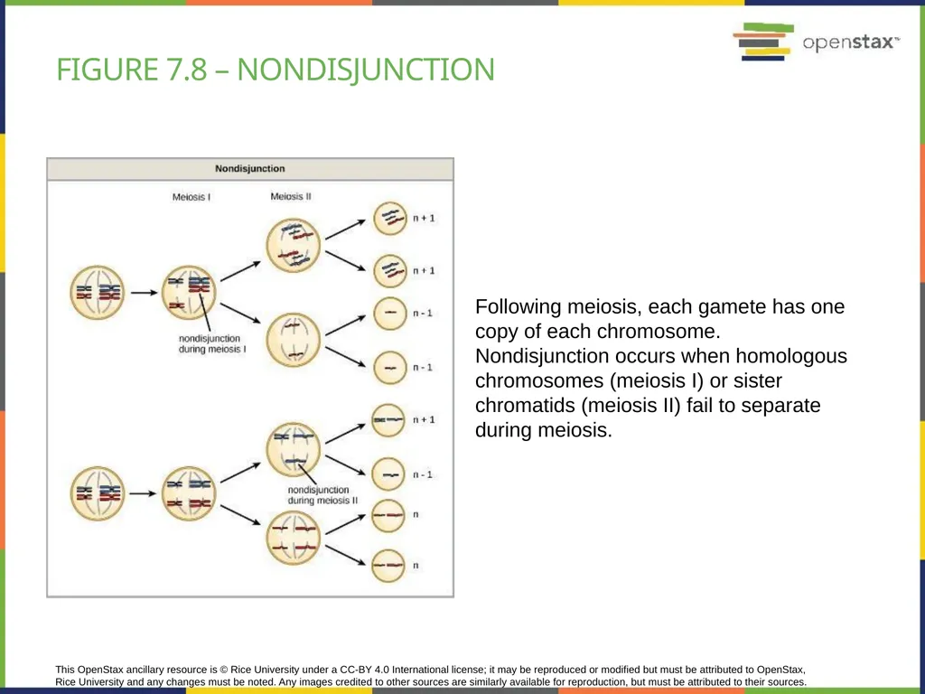
Aneuploidy results from a process called nondisjunction of the chromosomes
Nondisjunction occurs when homologous chromosomes (meiosis I) or sister chromatids (meiosis II) fail to separate during meiosis
This could result from incorrect spindle attachment to each of the chromosomes (meiosis I) or sister chromatids (meisosi II)<br>
Abnormal number is known as Aneuploidy
different number of sets of chromosomes is called polyploidy
Aneuploidy results from a process called nondisjunction of the chromosomes
Nondisjunction occurs when homologous chromosomes (meiosis I) or sister chromatids (meiosis II) fail to separate during meiosis
This could result from incorrect spindle attachment to each of the chromosomes (meiosis I) or sister chromatids (meisosi II)<br>
25
This OpenStax ancillary resource is © Rice University under a CC-BY 4.0 International license; it may be reproduced or modified but must be attributed to OpenStax, Rice University and any changes must be noted. Any images credited to other sources are similarly available for reproduction, but must be attributed to their sources. Following meiosis, each gamete has one copy of each chromosome. Nondisjunction occurs when homologous chromosomes (meiosis I) or sister chromatids (meiosis II) fail to separate during meiosis. Figure 7.8 – nondisjunction<br>
26
Aneuploidy Monosomy – loss 1 chromosome
Turner syndrome – XO
Trisomy – gain of 1 chromosome
Autosomes = body chromosomes
Down syndrome – chromosome 21
Sex chromosomes
Any more than 1 X chromosomes result is a Barr-body formation due to X-inactivation
Triplo-X – XXX
Klinefelter syndrome - XXY<br>
Turner syndrome – XO
Trisomy – gain of 1 chromosome
Autosomes = body chromosomes
Down syndrome – chromosome 21
Sex chromosomes
Any more than 1 X chromosomes result is a Barr-body formation due to X-inactivation
Triplo-X – XXX
Klinefelter syndrome - XXY<br>
27
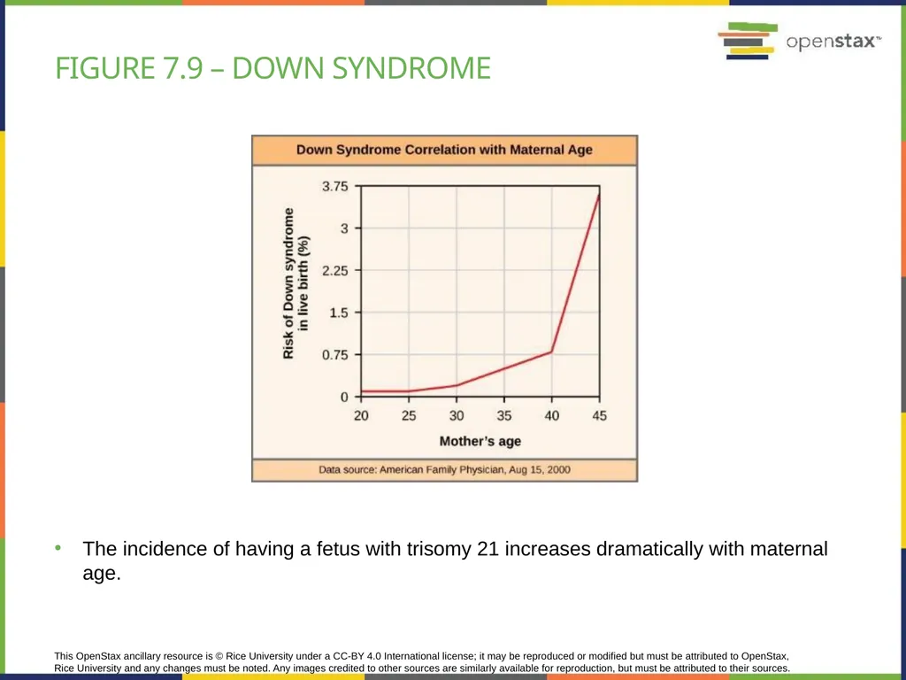
This OpenStax ancillary resource is © Rice University under a CC-BY 4.0 International license; it may be reproduced or modified but must be attributed to OpenStax, Rice University and any changes must be noted. Any images credited to other sources are similarly available for reproduction, but must be attributed to their sources. The incidence of having a fetus with trisomy 21 increases dramatically with maternal age. Figure 7.9 – down syndrome<br>
28
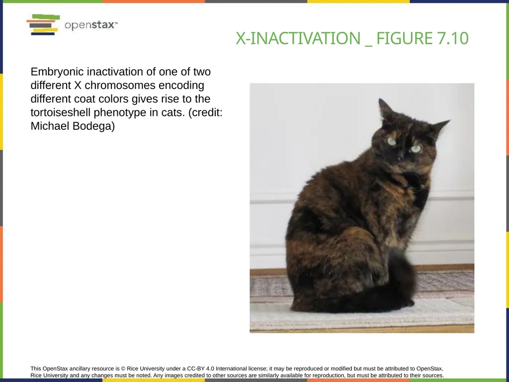
This OpenStax ancillary resource is © Rice University under a CC-BY 4.0 International license; it may be reproduced or modified but must be attributed to OpenStax, Rice University and any changes must be noted. Any images credited to other sources are similarly available for reproduction, but must be attributed to their sources. Embryonic inactivation of one of two different X chromosomes encoding different coat colors gives rise to the tortoiseshell phenotype in cats. (credit: Michael Bodega) X-inactivation _ Figure 7.10<br>
29
abnormal structure of chromosomes Abnormal structural chromosomes result from:
Deletion
Insertion
Duplication
Translocation
Could result in cancer or other diseases<br>
Deletion
Insertion
Duplication
Translocation
Could result in cancer or other diseases<br>
30
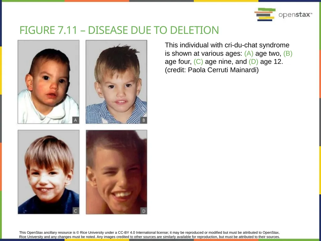
This OpenStax ancillary resource is © Rice University under a CC-BY 4.0 International license; it may be reproduced or modified but must be attributed to OpenStax, Rice University and any changes must be noted. Any images credited to other sources are similarly available for reproduction, but must be attributed to their sources. This individual with cri-du-chat syndrome is shown at various ages: (A) age two, (B) age four, (C) age nine, and (D) age 12. (credit: Paola Cerruti Mainardi) Figure 7.11 – disease due to deletion<br>
31
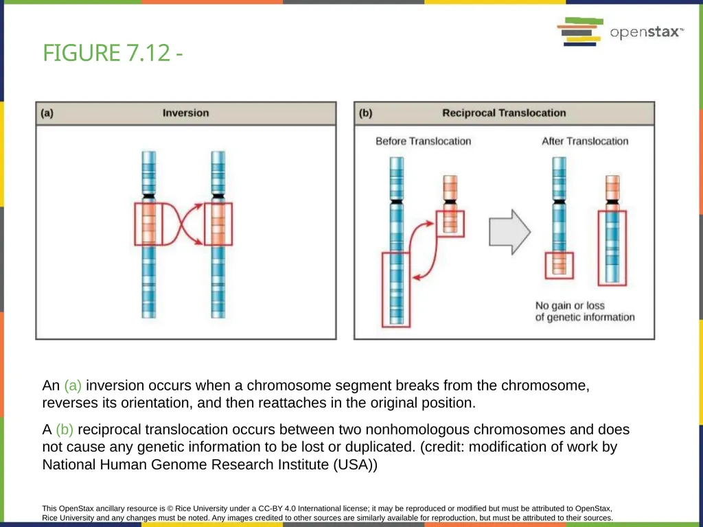
This OpenStax ancillary resource is © Rice University under a CC-BY 4.0 International license; it may be reproduced or modified but must be attributed to OpenStax, Rice University and any changes must be noted. Any images credited to other sources are similarly available for reproduction, but must be attributed to their sources. An (a) inversion occurs when a chromosome segment breaks from the chromosome, reverses its orientation, and then reattaches in the original position.
A (b) reciprocal translocation occurs between two nonhomologous chromosomes and does not cause any genetic information to be lost or duplicated. (credit: modification of work by National Human Genome Research Institute (USA)) Figure 7.12 -<br>
A (b) reciprocal translocation occurs between two nonhomologous chromosomes and does not cause any genetic information to be lost or duplicated. (credit: modification of work by National Human Genome Research Institute (USA)) Figure 7.12 -<br>