/
Chapter 13 Meiosis and Sexual Chapter 13 Meiosis and Sexual

Chapter 13 Meiosis and Sexual - PowerPoint Presentation

celsa-spraggs
celsa-spraggs . @celsa-spraggs
Follow
377 views
Uploaded On 2018-11-10

Chapter 13 Meiosis and Sexual - PPT Presentation

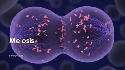
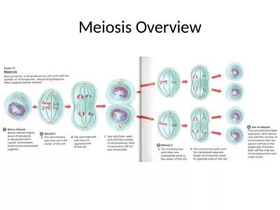
Life Cycles Overview Variations on a Theme Living organisms are distinguished by their ability to reproduce their own kind Genetics is the scientific study of heredity and variation Heredity ID: 726936

chromosomes pearson cummings meiosis pearson chromosomes meiosis cummings benjamin publishing education 2008 copyright chromatids haploid metaphase fig cells homologous

Share:

Link:

Embed:

Download Presentation from below link

Download Presentation The PPT/PDF document "Chapter 13 Meiosis and Sexual" is the property of its rightful owner. Permission is granted to download and print the materials on this web site for personal, non-commercial use only, and to display it on your personal computer provided you do not modify the materials and that you retain all copyright notices contained in the materials. By downloading content from our website, you accept the terms of this agreement.


Presentation Transcript

Slide1

Chapter 13

Meiosis and Sexual

Life CyclesSlide2

Overview: Variations on a Theme

Living organisms are distinguished by their ability to reproduce their own kind

Genetics

is the scientific study of heredity and variationHeredity is the transmission of traits from one generation to the nextVariation is demonstrated by the differences in appearance that offspring show from parents and siblings

Copyright © 2008 Pearson Education Inc., publishing as Pearson Benjamin CummingsSlide3

Fig. 13-1Slide4

Concept 13.1: Offspring acquire genes from parents by inheriting chromosomes

In a literal sense, children do not inherit particular physical traits from their parents

It is genes that are actually inherited

Copyright © 2008 Pearson Education Inc., publishing as Pearson Benjamin CummingsSlide5

Inheritance of Genes

Genes

are the units of heredity, and are made up of segments of DNA

Genes are passed to the next generation through reproductive cells called gametes (sperm and eggs) Each gene has a specific location called a locus

on a certain chromosomeMost DNA is packaged into chromosomesOne set of chromosomes is inherited from each parent

Copyright © 2008 Pearson Education Inc., publishing as Pearson Benjamin CummingsSlide6

Comparison of Asexual and Sexual Reproduction

In

asexual reproduction

, one parent produces genetically identical offspring by mitosisA clone is a group of genetically identical individuals from the same parentIn sexual reproduction, two parents give rise to offspring that have unique combinations of genes inherited from the two parents

Video: Hydra Budding

Copyright © 2008 Pearson Education Inc., publishing as Pearson Benjamin CummingsSlide7

Fig. 13-2

(a) Hydra

(b) Redwoods

Parent

Bud

0.5 mmSlide8

Fig. 13-2a

(a) Hydra

0.5 mm

Bud

ParentSlide9

Fig. 13-2b

(b) RedwoodsSlide10

Concept 13.2: Fertilization and meiosis alternate in sexual life cycles

A

life cycle

is the generation-to-generation sequence of stages in the reproductive history of an organism

Copyright © 2008 Pearson Education Inc., publishing as Pearson Benjamin CummingsSlide11

Sets of Chromosomes in Human Cells

Human

somatic cells

(any cell other than a gamete) have 23 pairs of chromosomesA karyotype is an ordered display of the pairs of chromosomes from a cell The two chromosomes in each pair are called homologous chromosomes, or homologs

Chromosomes in a homologous pair are the same length and carry genes controlling the same inherited characters

Copyright © 2008 Pearson Education Inc., publishing as Pearson Benjamin CummingsSlide12

Fig. 13-3

APPLICATION

TECHNIQUE

Pair of homologous

replicated chromosomes

5 µm

Centromere

Sister

chromatids

Metaphase

chromosomeSlide13

Fig. 13-3a

APPLICATIONSlide14

Fig. 13-3b

TECHNIQUE

Pair of homologous

replicated chromosomes

Centromere

Sister

chromatids

Metaphase

chromosome

5 µmSlide15

The

sex chromosomes

are called X and Y

Human females have a homologous pair of X chromosomes (XX)Human males have one X and one Y chromosomeThe 22 pairs of chromosomes that do not determine sex are called autosomes

Copyright © 2008 Pearson Education Inc., publishing as Pearson Benjamin CummingsSlide16

Each pair of homologous chromosomes includes one chromosome from each parent

The 46 chromosomes in a human somatic cell are two sets of 23: one from the mother and one from the father

A

diploid cell (2n) has two sets of chromosomesFor humans, the diploid number is 46 (2n = 46)

Copyright © 2008 Pearson Education Inc., publishing as Pearson Benjamin CummingsSlide17

In a cell in which DNA synthesis has occurred, each chromosome is replicated

Each replicated chromosome consists of two identical sister chromatids

Copyright © 2008 Pearson Education Inc., publishing as Pearson Benjamin CummingsSlide18

Fig. 13-4

Key

Maternal set of

chromosomes (

n

= 3)

Paternal set of

chromosomes (

n

= 3)

2

n

= 6

Centromere

Two sister chromatids

of one replicated

chromosome

Two nonsister

chromatids in

a homologous pair

Pair of homologous

chromosomes

(one from each set)Slide19

A gamete (sperm or egg) contains a single set of chromosomes, and is

haploid

(

n)For humans, the haploid number is 23 (n = 23)Each set of 23 consists of 22 autosomes and a single sex chromosomeIn an unfertilized egg (ovum), the sex chromosome is XIn a sperm cell, the sex chromosome may be either X or Y

Copyright © 2008 Pearson Education Inc., publishing as Pearson Benjamin CummingsSlide20

Fertilization

is the union of gametes (the sperm and the egg)

The fertilized egg is called a

zygote and has one set of chromosomes from each parent The zygote produces somatic cells by mitosis and develops into an adult

Behavior of Chromosome Sets in the Human Life Cycle

Copyright © 2008 Pearson Education Inc., publishing as Pearson Benjamin CummingsSlide21

At sexual maturity, the ovaries and testes produce haploid gametes

Gametes are the only types of human cells produced by

meiosis

, rather than mitosisMeiosis results in one set of chromosomes in each gameteFertilization and meiosis alternate in sexual life cycles to maintain chromosome number

Copyright © 2008 Pearson Education Inc., publishing as Pearson Benjamin CummingsSlide22

Fig. 13-5

Key

Haploid (

n

)

Diploid (2

n

)

Haploid gametes (

n = 23

)

Egg (

n

)

Sperm (

n

)

MEIOSIS

FERTILIZATION

Ovary

Testis

Diploid

zygote

(2

n

= 46)

Mitosis and

development

Multicellular diploid

adults (2

n

= 46)Slide23

The Variety of Sexual Life Cycles

The alternation of meiosis and fertilization is common to all organisms that reproduce sexually

The three main types of sexual life cycles differ in the timing of meiosis and fertilization

Copyright © 2008 Pearson Education Inc., publishing as Pearson Benjamin CummingsSlide24

In animals, meiosis produces gametes, which undergo no further cell division before fertilization

Gametes are the only haploid cells in animals

Gametes fuse to form a diploid zygote that divides by mitosis to develop into a multicellular organism

Copyright © 2008 Pearson Education Inc., publishing as Pearson Benjamin CummingsSlide25

Fig. 13-6

Key

Haploid (

n

)

Diploid (2

n

)

n

n

Gametes

n

n

n

Mitosis

MEIOSIS

FERTILIZATION

MEIOSIS

2

n

2

n

Zygote

2

n

Mitosis

Diploid

multicellular

organism

(a) Animals

Spores

Diploid

multicellular

organism

(sporophyte)

(b) Plants and some algae

2

n

Mitosis

Gametes

Mitosis

n

n

n

Zygote

FERTILIZATION

n

n

n

Mitosis

Zygote

(c) Most fungi and some protists

MEIOSIS

FERTILIZATION

2

n

Gametes

n

n

Mitosis

Haploid multi-

cellular organism

(gametophyte)

Haploid unicellular or

multicellular organismSlide26

Fig. 13-6a

Key

Haploid (

n

)

Diploid (2

n

)

Gametes

n

n

n

2

n

2

n

Zygote

MEIOSIS

FERTILIZATION

Mitosis

Diploid

multicellular

organism

(a) AnimalsSlide27

Plants and some algae exhibit an

alternation of generations

This life cycle includes both a diploid and haploid multicellular stage

The diploid organism, called the sporophyte, makes haploid spores by meiosis

Copyright © 2008 Pearson Education Inc., publishing as Pearson Benjamin CummingsSlide28

Each spore grows by mitosis into a haploid organism called a

gametophyte

A gametophyte makes haploid gametes by mitosis

Fertilization of gametes results in a diploid sporophyte

Copyright © 2008 Pearson Education Inc., publishing as Pearson Benjamin CummingsSlide29

Fig. 13-6b

Key

Haploid (

n

)

Diploid (2

n

)

n

n

n

n

n

2

n

2

n

Mitosis

Mitosis

Mitosis

Zygote

Spores

Gametes

MEIOSIS

FERTILIZATION

Diploid

multicellular

organism

(sporophyte)

Haploid multi-

cellular organism

(gametophyte)

(b) Plants and some algaeSlide30

In most fungi and some protists, the only diploid stage is the single-celled zygote; there is no multicellular diploid stage

The zygote produces haploid cells by meiosis

Each haploid cell grows by mitosis into a haploid multicellular organism

The haploid adult produces gametes by mitosis

Copyright © 2008 Pearson Education Inc., publishing as Pearson Benjamin CummingsSlide31

Fig. 13-6c

Key

Haploid (

n

)

Diploid (2

n

)

Mitosis

Mitosis

Gametes

Zygote

Haploid unicellular or

multicellular organism

MEIOSIS

FERTILIZATION

n

n

n

n

n

2

n

(c) Most fungi and some protistsSlide32

Depending on the type of life cycle, either haploid or diploid cells can divide by mitosis

However, only diploid cells can undergo meiosis

In all three life cycles, the halving and doubling of chromosomes contributes to genetic variation in offspring

Copyright © 2008 Pearson Education Inc., publishing as Pearson Benjamin CummingsSlide33

Concept 13.3: Meiosis reduces the number of chromosome sets from diploid to haploid

Like mitosis, meiosis is preceded by the replication of chromosomes

Meiosis takes place in two sets of cell divisions, called

meiosis I and meiosis IIThe two cell divisions result in four daughter cells, rather than the two daughter cells in mitosisEach daughter cell has only half as many chromosomes as the parent cell

Copyright © 2008 Pearson Education Inc., publishing as Pearson Benjamin CummingsSlide34

The Stages of Meiosis

In the first cell division (meiosis I), homologous chromosomes separate

Meiosis I results in two haploid daughter cells with replicated chromosomes; it is called the reductional division

In the second cell division (meiosis II), sister chromatids separateMeiosis II results in four haploid daughter cells with unreplicated chromosomes; it is called the equational division

Copyright © 2008 Pearson Education Inc., publishing as Pearson Benjamin CummingsSlide35

Fig. 13-7-1

Interphase

Homologous pair of chromosomes

in diploid parent cell

Chromosomes

replicate

Homologous pair of replicated chromosomes

Sister

chromatids

Diploid cell with

replicated

chromosomesSlide36

Fig. 13-7-2

Interphase

Homologous pair of chromosomes

in diploid parent cell

Chromosomes

replicate

Homologous pair of replicated chromosomes

Sister

chromatids

Diploid cell with

replicated

chromosomes

Meiosis

I

Homologous

chromosomes

separate

1

Haploid cells with

replicated chromosomes Slide37

Fig. 13-7-3

Interphase

Homologous pair of chromosomes

in diploid parent cell

Chromosomes

replicate

Homologous pair of replicated chromosomes

Sister

chromatids

Diploid cell with

replicated

chromosomes

Meiosis

I

Homologous

chromosomes

separate

1

Haploid cells with

replicated chromosomes

Meiosis

II

2

Sister chromatids

separate

Haploid cells with unreplicated chromosomes Slide38

Meiosis I is preceded by interphase, in which chromosomes are replicated to form sister chromatids

The sister chromatids are genetically identical and joined at the centromere

The single centrosome replicates, forming two centrosomes

BioFlix: Meiosis

Copyright © 2008 Pearson Education Inc., publishing as Pearson Benjamin CummingsSlide39

Fig. 13-8

Prophase I

Metaphase I

Anaphase I

Telophase I and

Cytokinesis

Prophase II

Metaphase II

Anaphase II

Telophase II and

Cytokinesis

Centrosome

(with centriole pair)

Sister

chromatids

Chiasmata

Spindle

Homologous

chromosomes

Fragments

of nuclear

envelope

Centromere

(with kinetochore)

Metaphase

plate

Microtubule

attached to

kinetochore

Sister chromatids

remain attached

Homologous

chromosomes

separate

Cleavage

furrow

Sister chromatids

separate

Haploid daughter cells

formingSlide40

Division in meiosis I occurs in four phases:

Prophase I

– Metaphase I– Anaphase I– Telophase I and cytokinesis

Copyright © 2008 Pearson Education Inc., publishing as Pearson Benjamin CummingsSlide41

Metaphase I

Fig. 13-8a

Prophase I

Anaphase I

Telophase I and

Cytokinesis

Centrosome

(with centriole pair)

Sister

chromatids

Chiasmata

Spindle

Homologous

chromosomes

Fragments

of nuclear

envelope

Centromere

(with kinetochore)

Metaphase

plate

Microtubule

attached to

kinetochore

Sister chromatids

remain attached

Homologous

chromosomes

separate

Cleavage

furrowSlide42

Prophase I

Prophase I typically occupies more than 90% of the time required for meiosis

Chromosomes begin to condense

In synapsis, homologous chromosomes loosely pair up, aligned gene by gene

Copyright © 2008 Pearson Education Inc., publishing as Pearson Benjamin CummingsSlide43

In

crossing over

, nonsister chromatids exchange DNA segments

Each pair of chromosomes forms a tetrad, a group of four chromatidsEach tetrad usually has one or more chiasmata, X-shaped regions where crossing over occurred

Copyright © 2008 Pearson Education Inc., publishing as Pearson Benjamin CummingsSlide44

Metaphase I

In metaphase I, tetrads line up at the metaphase plate, with one chromosome facing each pole

Microtubules from one pole are attached to the kinetochore of one chromosome of each tetrad

Microtubules from the other pole are attached to the kinetochore of the other chromosome

Copyright © 2008 Pearson Education Inc., publishing as Pearson Benjamin CummingsSlide45

Fig. 13-8b

Prophase I

Metaphase I

Centrosome

(with centriole pair)

Sister

chromatids

Chiasmata

Spindle

Centromere

(with kinetochore)

Metaphase

plate

Homologous

chromosomes

Fragments

of nuclear

envelope

Microtubule

attached to

kinetochoreSlide46

Anaphase I

In anaphase I, pairs of homologous chromosomes separate

One chromosome moves toward each pole, guided by the spindle apparatus

Sister chromatids remain attached at the centromere and move as one unit toward the pole

Copyright © 2008 Pearson Education Inc., publishing as Pearson Benjamin CummingsSlide47

Telophase I and Cytokinesis

In the beginning of telophase I, each half of the cell has a haploid set of chromosomes; each chromosome still consists of two sister chromatids

Cytokinesis usually occurs simultaneously, forming two haploid daughter cells

Copyright © 2008 Pearson Education Inc., publishing as Pearson Benjamin CummingsSlide48

In animal cells, a cleavage furrow forms; in plant cells, a cell plate forms

No chromosome replication occurs between the end of meiosis I and the beginning of meiosis II because the chromosomes are already replicated

Copyright © 2008 Pearson Education Inc., publishing as Pearson Benjamin CummingsSlide49

Fig. 13-8c

Anaphase I

Telophase I and

Cytokinesis

Sister chromatids

remain attached

Homologous

chromosomes

separate

Cleavage

furrowSlide50

Division in meiosis II also occurs in four phases:

Prophase II

– Metaphase II– Anaphase II– Telophase II and cytokinesisMeiosis II is very similar to mitosis

Copyright © 2008 Pearson Education Inc., publishing as Pearson Benjamin CummingsSlide51

Fig. 13-8d

Prophase II

Metaphase II

Anaphase II

Telophase II and

Cytokinesis

Sister chromatids

separate

Haploid daughter cells

formingSlide52

Prophase II

In prophase II, a spindle apparatus forms

In late prophase II, chromosomes (each still composed of two chromatids) move toward the metaphase plate

Copyright © 2008 Pearson Education Inc., publishing as Pearson Benjamin CummingsSlide53

Metaphase II

In metaphase II, the sister chromatids are arranged at the metaphase plate

Because of crossing over in meiosis I, the two sister chromatids of each chromosome are no longer genetically identical

The kinetochores of sister chromatids attach to microtubules extending from opposite poles

Copyright © 2008 Pearson Education Inc., publishing as Pearson Benjamin CummingsSlide54

Fig. 13-8e

Prophase II

Metaphase IISlide55

Anaphase II

In anaphase II, the sister chromatids separate

The sister chromatids of each chromosome now move as two newly individual chromosomes toward opposite poles

Copyright © 2008 Pearson Education Inc., publishing as Pearson Benjamin CummingsSlide56

Telophase II and Cytokinesis

In telophase II, the chromosomes arrive at opposite poles

Nuclei form, and the chromosomes begin decondensing

Copyright © 2008 Pearson Education Inc., publishing as Pearson Benjamin CummingsSlide57

Cytokinesis separates the cytoplasm

At the end of meiosis, there are four daughter cells, each with a haploid set of unreplicated chromosomes

Each daughter cell is genetically distinct from the others and from the parent cell

Copyright © 2008 Pearson Education Inc., publishing as Pearson Benjamin CummingsSlide58

Fig. 13-8f

Anaphase II

Telephase II and

Cytokinesis

Sister chromatids

separate

Haploid daughter cells

formingSlide59

A Comparison of Mitosis and Meiosis

Mitosis conserves the number of chromosome sets, producing cells that are genetically identical to the parent cell

Meiosis reduces the number of chromosomes sets from two (diploid) to one (haploid), producing cells that differ genetically from each other and from the parent cell

The mechanism for separating sister chromatids is virtually identical in meiosis II and mitosis

Copyright © 2008 Pearson Education Inc., publishing as Pearson Benjamin CummingsSlide60

Fig. 13-9

MITOSIS

MEIOSIS

MEIOSIS

I

Prophase

I

Chiasma

Homologous

chromosome

pair

Chromosome

replication

Parent cell

2

n

= 6

Chromosome

replication

Replicated chromosome

Prophase

Metaphase

Metaphase

I

Anaphase

I

Telophase

I

Haploid

n

= 3

Daughter

cells of

meiosis

I

Anaphase

Telophase

2

n

2

n

Daughter cells

of mitosis

n

n

n

n

MEIOSIS

II

Daughter cells of meiosis

II

SUMMARY

Meiosis

Occurs during interphase before meiosis

I

begins

Two, each including prophase, metaphase, anaphase, and

telophase

Occurs during prophase

I

along with crossing over

between nonsister chromatids; resulting chiasmata

hold pairs together due to sister chromatid cohesion

Four, each haploid (

n

), containing half as many chromosomes

as the parent cell; genetically different from the parent

cell and from each other

Produces gametes; reduces number of chromosomes by half

and introduces genetic variability amoung the gametes

Mitosis

Occurs during interphase before

mitosis begins

One, including prophase, metaphase,

anahase, and telophase

Does not occur

Two, each diploid (2

n

) and genetically

identical to the parent cell

Enables multicellular adult to arise from

zygote; produces cells for growth, repair,

and, in some species, asexual reproduction

Property

DNA

replication

Number of

divisions

Synapsis of

homologous

chromosomes

Number of

daughter cells

and genetic

composition

Role in the

animal bodySlide61

Fig. 13-9a

MITOSIS

MEIOSIS

MEIOSIS

I

Prophase

I

Chiasma

Chromosome

replication

Homologous

chromosome

pair

Chromosome

replication

2

n

= 6

Parent cell

Prophase

Replicated chromosome

Metaphase

Metaphase

I

Anaphase

I

Telophase

I

Haploid

n

= 3

Daughter

cells of

meiosis

I

MEIOSIS

II

Daughter cells of meiosis

II

n

n

n

n

2

n

2

n

Daughter cells

of mitosis

Anaphase

TelophaseSlide62

Fig. 13-9b

SUMMARY

Meiosis

Mitosis

Property

DNA

replication

Number of

divisions

Occurs during interphase before

mitosis begins

One, including prophase, metaphase,

anaphase, and telophase

Synapsis of

homologous

chromosomes

Does not occur

Number of

daughter cells

and genetic

composition

Two, each diploid (2

n

) and genetically

identical to the parent cell

Role in the

animal body

Enables multicellular adult to arise from

zygote; produces cells for growth, repair,

and, in some species, asexual reproduction

Occurs during interphase before meiosis

I

begins

Two, each including prophase, metaphase, anaphase, and

telophase

Occurs during prophase

I

along with crossing over

between nonsister chromatids; resulting chiasmata

hold pairs together due to sister chromatid cohesion

Four, each haploid (

n

), containing half as many chromosomes

as the parent cell; genetically different from the parent

cell and from each other

Produces gametes; reduces number of chromosomes by half

and introduces genetic variability among the gametesSlide63

Three events are unique to meiosis, and all three occur in meiosis l:

Synapsis and crossing over in prophase I: Homologous chromosomes physically connect and exchange genetic information

– At the metaphase plate, there are paired homologous chromosomes (tetrads), instead of individual replicated chromosomes– At anaphase I, it is homologous chromosomes, instead of sister chromatids, that separate

Copyright © 2008 Pearson Education Inc., publishing as Pearson Benjamin CummingsSlide64

Sister chromatid cohesion allows sister chromatids of a single chromosome to stay together through meiosis I

Protein complexes called cohesins are responsible for this cohesion

In mitosis, cohesins are cleaved at the end of metaphase

In meiosis, cohesins are cleaved along the chromosome arms in anaphase I (separation of homologs) and at the centromeres in anaphase II (separation of sister chromatids)

Copyright © 2008 Pearson Education Inc., publishing as Pearson Benjamin CummingsSlide65

Fig. 13-10

EXPERIMENT

RESULTS

Shugoshin

+

(normal)

+

Spore case

Fluorescent label

Metaphase

I

Shugoshin

Anaphase

I

Metaphase

II

Anaphase

II

Mature

spores

OR

Spore

Two of three possible arrange-

ments of labeled chromosomes

Shugoshin

+

Shugoshin

Spore cases (%)

100

80

60

40

20

0

?

?

?

?

?

?

?

?Slide66

Fig. 13-10a

EXPERIMENT

Shugoshin

+

(normal)

Spore case

Fluorescent label

Metaphase

I

Anaphase

I

Metaphase

II

Anaphase

II

Mature

spores

Spore

OR

Two of three possible arrange-

ments of labeled chromosomes

Shugoshin

?

?

?

?

?

?

?

?Slide67

Fig. 13-10b

RESULTS

Shugoshin

+

Shugoshin

Spore cases (%)

100

80

60

40

20

0Slide68

Concept 13.4: Genetic variation produced in sexual life cycles contributes to evolution

Mutations (changes in an organism’s DNA) are the original source of genetic diversity

Mutations create different versions of genes called alleles

Reshuffling of alleles during sexual reproduction produces genetic variation

Copyright © 2008 Pearson Education Inc., publishing as Pearson Benjamin CummingsSlide69

Origins of Genetic Variation Among Offspring

The behavior of chromosomes during meiosis and fertilization is responsible for most of the variation that arises in each generation

Three mechanisms contribute to genetic variation:

Independent assortment of chromosomesCrossing overRandom fertilization

Copyright © 2008 Pearson Education Inc., publishing as Pearson Benjamin CummingsSlide70

Independent Assortment of Chromosomes

Homologous pairs of chromosomes orient randomly at metaphase I of meiosis

In independent assortment, each pair of chromosomes sorts maternal and paternal homologues into daughter cells independently of the other pairs

Copyright © 2008 Pearson Education Inc., publishing as Pearson Benjamin CummingsSlide71

The number of combinations possible when chromosomes assort independently into gametes is 2

n

, where

n is the haploid numberFor humans (n = 23), there are more than 8 million (223) possible combinations of chromosomes

Copyright © 2008 Pearson Education Inc., publishing as Pearson Benjamin CummingsSlide72

Fig. 13-11-1

Possibility 1

Possibility 2

Two equally probable

arrangements of

chromosomes at

metaphase

ISlide73

Fig. 13-11-2

Possibility 1

Possibility 2

Two equally probable

arrangements of

chromosomes at

metaphase

I

Metaphase

IISlide74

Fig. 13-11-3

Possibility 1

Possibility 2

Two equally probable

arrangements of

chromosomes at

metaphase

I

Metaphase

II

Daughter

cells

Combination 1

Combination 2

Combination 3

Combination 4Slide75

Crossing Over

Crossing over produces

recombinant chromosomes

, which combine genes inherited from each parentCrossing over begins very early in prophase I, as homologous chromosomes pair up gene by gene

Copyright © 2008 Pearson Education Inc., publishing as Pearson Benjamin CummingsSlide76

In crossing over, homologous portions of two nonsister chromatids trade places

Crossing over contributes to genetic variation by combining DNA from two parents into a single chromosome

Copyright © 2008 Pearson Education Inc., publishing as Pearson Benjamin CummingsSlide77

Fig. 13-12-1

Prophase

I

of meiosis

Pair of

homologs

Nonsister

chromatids

held together

during synapsisSlide78

Fig. 13-12-2

Prophase

I

of meiosis

Pair of

homologs

Nonsister

chromatids

held together

during synapsis

Chiasma

Centromere

TEMSlide79

Fig. 13-12-3

Prophase

I

of meiosis

Pair of

homologs

Nonsister

chromatids

held together

during synapsis

Chiasma

Centromere

Anaphase

I

TEMSlide80

Fig. 13-12-4

Prophase

I

of meiosis

Pair of

homologs

Nonsister

chromatids

held together

during synapsis

Chiasma

Centromere

Anaphase

I

Anaphase

II

TEMSlide81

Fig. 13-12-5

Prophase

I

of meiosis

Pair of

homologs

Nonsister

chromatids

held together

during synapsis

Chiasma

Centromere

Anaphase

I

Anaphase

II

Daughter

cells

Recombinant chromosomes

TEMSlide82

Random Fertilization

Random fertilization adds to genetic variation because any sperm can fuse with any ovum (unfertilized egg)

The fusion of two gametes (each with 8.4 million possible chromosome combinations from independent assortment) produces a zygote with any of about 70 trillion diploid combinations

Copyright © 2008 Pearson Education Inc., publishing as Pearson Benjamin CummingsSlide83

Crossing over adds even more variation

Each zygote has a unique genetic identity

Animation: Genetic Variation

Copyright © 2008 Pearson Education Inc., publishing as Pearson Benjamin CummingsSlide84

The Evolutionary Significance of Genetic Variation Within Populations

Natural selection results in the accumulation of genetic variations favored by the environment

Sexual reproduction contributes to the genetic variation in a population, which originates from mutations

Copyright © 2008 Pearson Education Inc., publishing as Pearson Benjamin CummingsSlide85

Fig. 13-UN1

Prophase

I

: Each homologous pair undergoes

synapsis and crossing over between nonsisterchromatids.

Metaphase

I:

Chromosomes line up as homolo-

gous pairs on the metaphase plate.

Anaphase

I:

Homologs separate from each other;

sister chromatids remain joined at the centromere.Slide86

Fig. 13-UN2

F

HSlide87

Fig. 13-UN3Slide88

Fig. 13-UN4Slide89

You should now be able to:

Distinguish between the following terms: somatic cell and gamete; autosome and sex chromosomes; haploid and diploid

Describe the events that characterize each phase of meiosis

Describe three events that occur during meiosis I but not mitosisName and explain the three events that contribute to genetic variation in sexually reproducing organisms

Copyright © 2008 Pearson Education Inc., publishing as Pearson Benjamin Cummings