Common presentation of a relatively Uncommon disease- An unusual case of Fever. Dr. Shubham Malani. Department of Medicine Dr. D. Y. Patil Medical College. A 24 year old male, student, came with co Fever moderate to high grade since 1
"Common presentation of a relatively Uncommon" is the property of its rightful owner. Permission is granted to
download and print the materials on this website for personal, non-commercial use only, and to display it
on your personal computer provided you do not modify the materials and that you retain all copyright
notices contained in the materials. By downloading content from our website, you accept the terms of this
agreement.
Presentation Transcript
01
Common presentation of a relatively Uncommon disease- An unusual case of Fever. Dr. Shubham Malani.
Department of Medicine
Dr. D. Y. Patil Medical College.<br>
02
A 24 year old male, student, came with c/o
Fever moderate to high grade since 1 month
Progressive breathlessness since 1 month which has worsened since 3 days
Generalized weakness and fatigability
There was no history of cough, chest pain, vomiting, bleeding manifestations or any other complaints .<br>
03
PAST HISTORY :
Patient had history of similar episode of Anemia with jaundice and fever admitted twice in last year, details of which were unavailable.
PERSONAL HISTORY :
Appetite was reduced.
Sleep was reduced.
Vegetarian by diet.
Bladder & bowel were unaltered.
No addictions.<br>
04
On Examination: The patient was drowsy but oriented.
Severe pallor and icterus was present.
Febrile, Temperature – 103oF
PR : 108/min
RR : 40 /min
BP : 110 / 70 mmhg
SPO2 was low. 76% on oxygen enriched room air.
JVP was raised upto the angle of jaw .
Clubbing grade II present.
Bilateral Pitting Pedal edema was present .<br>
05
CNS : Drowsy but oriented. No focal neurological deficit. Plantars- Flexor.
RS : Tachypnoea with B/l basal crepitations were present.
P/A : Soft , Mild hepatosplenomegaly.
CVS : Tachycardia was present. No S3/S4/murmur.
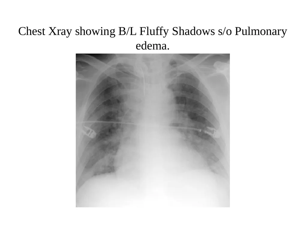
Chest Xray showed bilateral fluffy shadows.
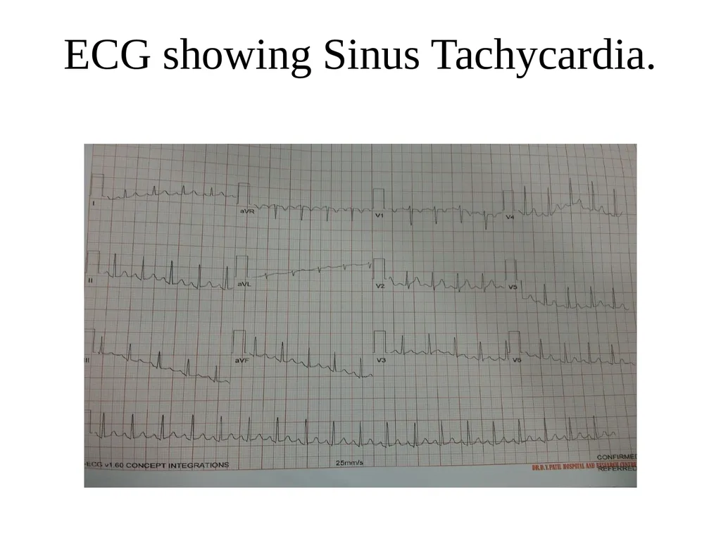
ECG suggestive of sinus tachycardia.
In view of above findings & as patient was unable to maintain saturation on oxygen ,he was intubated.<br>
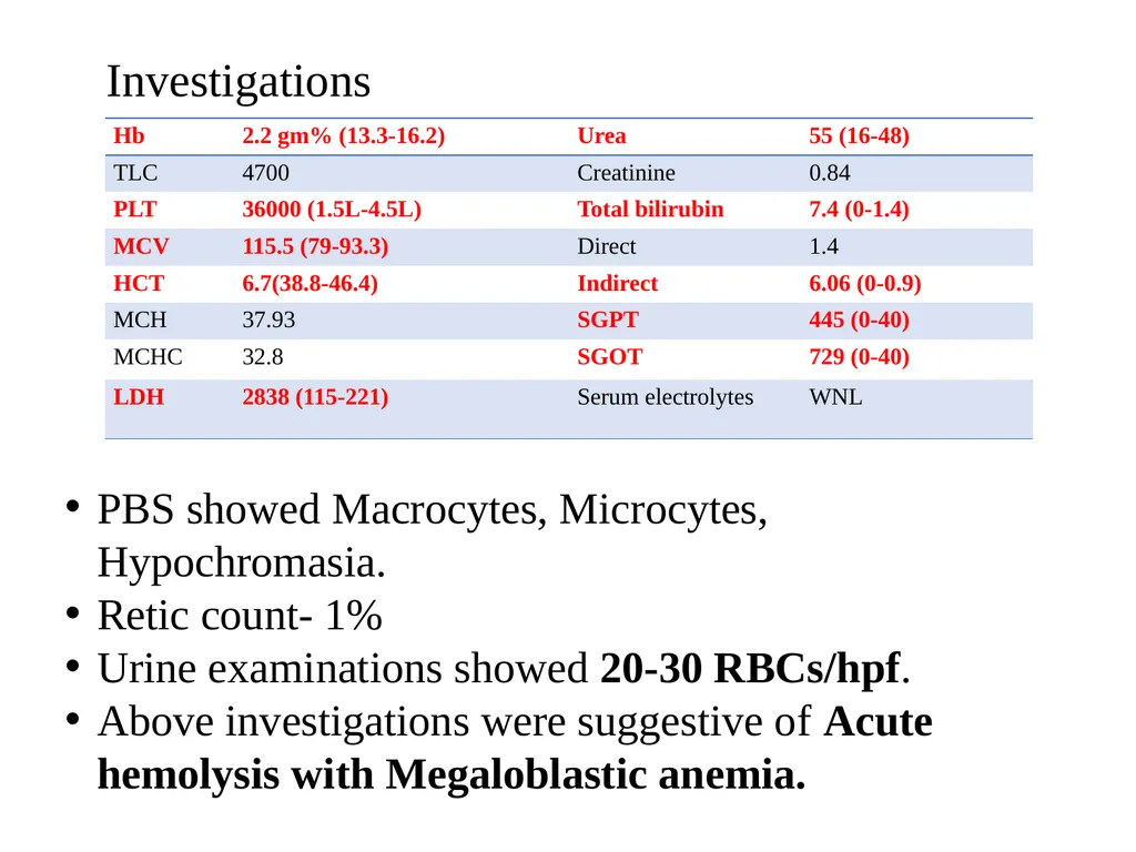
Investigations PBS showed Macrocytes, Microcytes, Hypochromasia.
Retic count- 1%
Urine examinations showed 20-30 RBCs/hpf.
Above investigations were suggestive of Acute hemolysis with Megaloblastic anemia.<br>
09
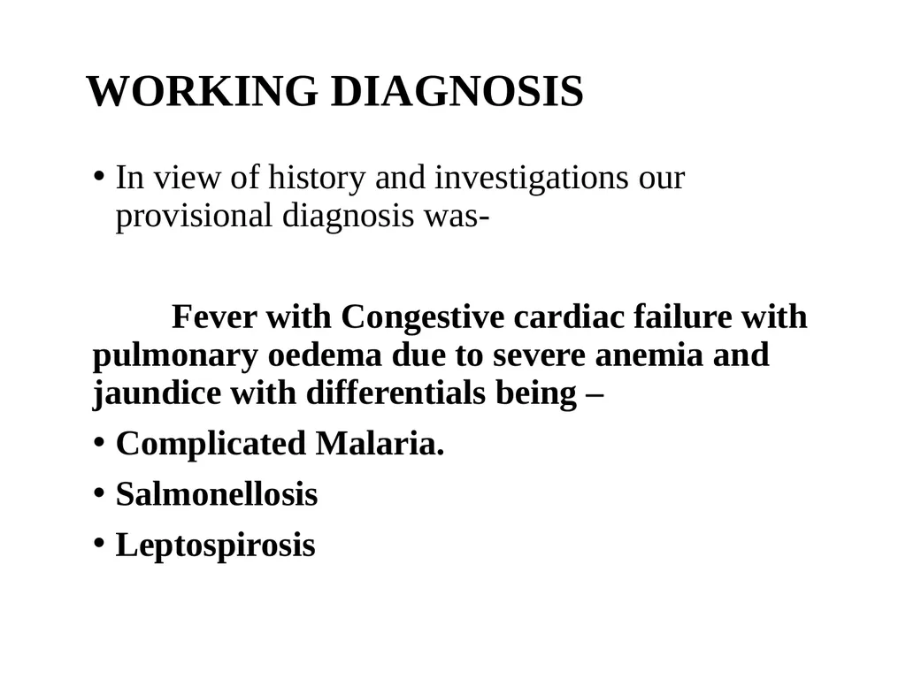
WORKING DIAGNOSIS In view of history and investigations our provisional diagnosis was-
Fever with Congestive cardiac failure with pulmonary oedema due to severe anemia and jaundice with differentials being –
Complicated Malaria.
Salmonellosis
Leptospirosis<br>
10
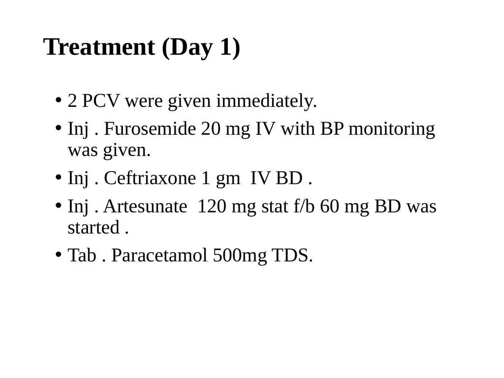
Treatment (Day 1) 2 PCV were given immediately.
Inj . Furosemide 20 mg IV with BP monitoring was given.
Inj . Ceftriaxone 1 gm IV BD .
Inj . Artesunate 120 mg stat f/b 60 mg BD was started .
Tab . Paracetamol 500mg TDS.<br>
11
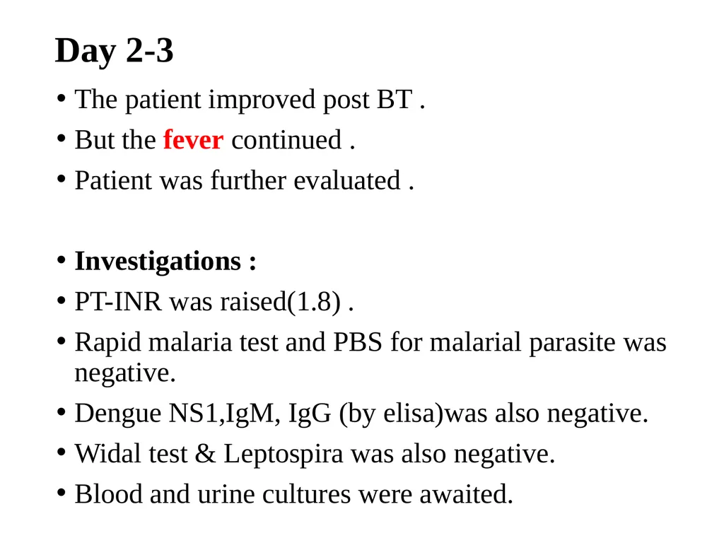
Day 2-3 The patient improved post BT .
But the fever continued .
Patient was further evaluated .
Investigations :
PT-INR was raised(1.8) .
Rapid malaria test and PBS for malarial parasite was negative.
Dengue NS1,IgM, IgG (by elisa)was also negative.
Widal test & Leptospira was also negative.
Blood and urine cultures were awaited.<br>
12
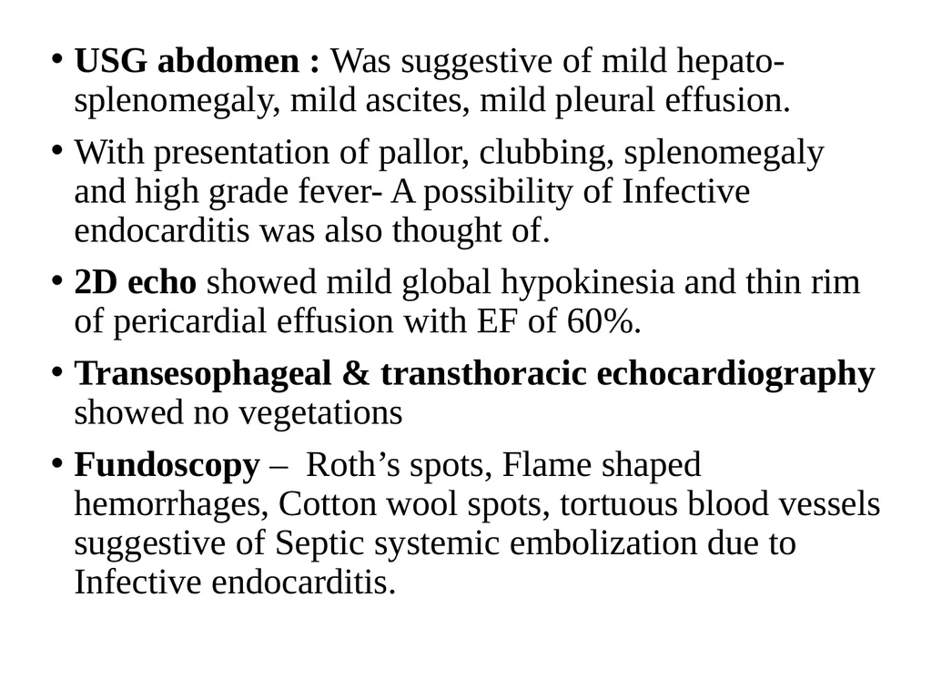
USG abdomen : Was suggestive of mild hepato-splenomegaly, mild ascites, mild pleural effusion.
With presentation of pallor, clubbing, splenomegaly and high grade fever- A possibility of Infective endocarditis was also thought of.
2D echo showed mild global hypokinesia and thin rim of pericardial effusion with EF of 60%.
Transesophageal & transthoracic echocardiography showed no vegetations
Fundoscopy – Roth’s spots, Flame shaped hemorrhages, Cotton wool spots, tortuous blood vessels suggestive of Septic systemic embolization due to Infective endocarditis.<br>
13
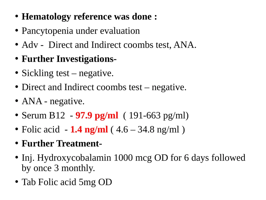
Hematology reference was done :
Pancytopenia under evaluation
Adv - Direct and Indirect coombs test, ANA.
Further Investigations-
Sickling test – negative.
Direct and Indirect coombs test – negative.
ANA - negative.
Serum B12 - 97.9 pg/ml ( 191-663 pg/ml)
Folic acid - 1.4 ng/ml ( 4.6 – 34.8 ng/ml )
Further Treatment-
Inj. Hydroxycobalamin 1000 mcg OD for 6 days followed by once 3 monthly.
Tab Folic acid 5mg OD<br>
14
The following day (Day 4-5) Congestive cardiac failure and hematocrit was improving.
The consciousness and general condition of the patient was getting better.
Patient was weaned off the ventilator.
Further PCV and FFP was given .
Fever however continued ,thus evaluating further ….<br>
15
OGDoscopy showed antral gastritis.
Bone marrow aspiration - Erythroid hyperplasia with dual maturation (Megaloblastic and micronormoblastic)
Urine culture showed no growth
Blood culture showed no growth
Bone marrow culture also was negative.<br>
16
Despite of all the measures fever still continued and no cause was found.
Then on recording further history It was found that patient had history of consumption of unpasteurized raw milk and contact with cattle for more than 1 year.
Following which Brucella antibody was sent and IgM antibody was found to be strongly positive (1.35).
Brucellosis induced Acute Hemolysis with Vit. B12 and Folic acid deficiency with Infective Endocarditis.<br>
17
Treatment Tab. Rifampicin 600 mg OD for 6 weeks
Tab. Doxycycline 100mg BD for 6 weeks
Following this treatment Patient recovered and is now doing well and came for follow-up after 1 month with repeat antibody titre positive. He was advised to continue the same treatment . After 3 months his repeat Antibody titre is Negative.<br>
18
Discussion. Human brucellosis is a zoonosis usually associated with occupational or domestic exposure to infected animals or their products.
The incubation period varies from 1 week to 3 months.
According to a study 14% of patients with brucellosis can present with Pancytopenia.
Brucellosis induced acute hemolysis due to Vit. B12 and folic acid deficiency presents as Pancytopenia and is a rare presentation of Brucellosis.<br>
19
Take home message. As brucellosis is common in our country, and pancytopenia is a common presentation, it should be included in the differential diagnosis of all those clinical conditions presenting as Fever with Pancytopenia.<br>
20
References Brucellosis induced acute hemolytic anemia in a severe vitamin b12 deficient individual presenting as pancytopenia: a case report ;2018 ; Mathi Manoj Kumar R and Subramony H ; Department of General Medicine,Apollo Hospital, Chennai.
Hematological Findings in adult with brucellosis ; Aypak A, et al. Pediatr Int. 2015.<br>