Cortical Visual Impairment Case History Six month
Description: Cortical Visual Impairment Case History Six month old ex 24-week premature infant has been recently discharged from the NICU with tracheostomy, feeding tube SP NEC and has increased tone and back arching. Mother of infant is concerned that
Related Topics
Download Presentation
"Cortical Visual Impairment Case History Six month" is the property of its rightful owner. Permission is granted to download and print the materials on this website for personal, non-commercial use only, and to display it on your personal computer provided you do not modify the materials and that you retain all copyright notices contained in the materials. By downloading content from our website, you accept the terms of this agreement.
Presentation Transcript
slide1. Cortical Visual Impairment<br>
slide2. Case History Six month old ex 24-week premature infant has been recently discharged from the NICU with tracheostomy, feeding tube S/P NEC and has increased tone and back arching.
Mother of infant is concerned that child looks past her, stares at lights and ceiling fans and looks away when reaching for an object.<br>
slide3. Definition Bilateral visual impairment due to retrogeniculate brain damage
Eye exam typically normal
Displays atypical behaviors
May see optic atrophy and nystagmus<br>
slide4. Causes Structural: brain malformations, tumors
Vascular: hypoxic/ischemic event, perinatal stroke
Infectious: meningitis, encephalitis
Inflammatory: vasculitis
Trauma: TBI, shaken baby
Metabolic: neonatal hypoglycemia, mitochondrial disease, lysosomal disorders
Neurologic disease: seizure, hydrocephalus<br>
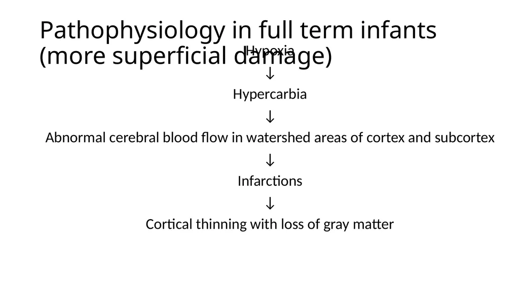
slide5. Pathophysiology in full term infants (more superficial damage) Hypoxia
↓
Hypercarbia
↓
Abnormal cerebral blood flow in watershed areas of cortex and subcortex
↓
Infarctions
↓
Cortical thinning with loss of gray matter<br>
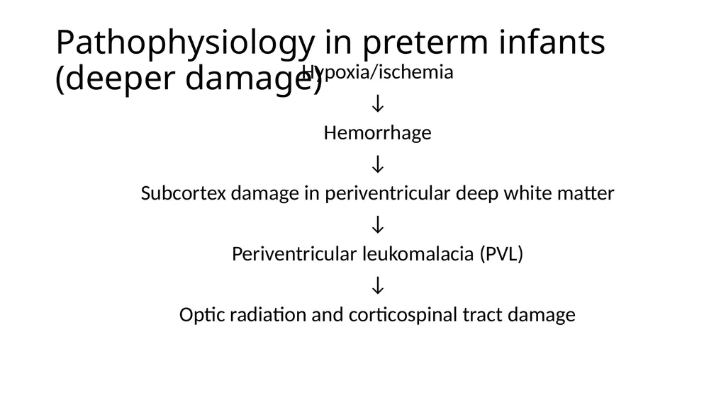
slide6. Pathophysiology in preterm infants (deeper damage) Hypoxia/ischemia
↓
Hemorrhage
↓
Subcortex damage in periventricular deep white matter
↓
Periventricular leukomalacia (PVL)
↓
Optic radiation and corticospinal tract damage<br>
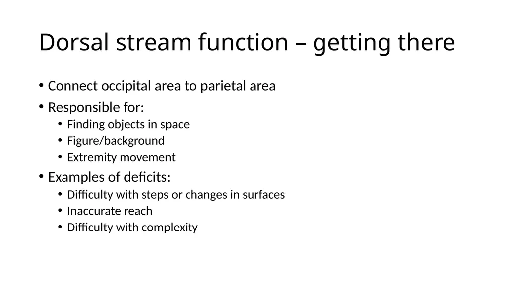
slide7. Dorsal stream function – getting there Connect occipital area to parietal area
Responsible for:
Finding objects in space
Figure/background
Extremity movement
Examples of deficits:
Difficulty with steps or changes in surfaces
Inaccurate reach
Difficulty with complexity<br>
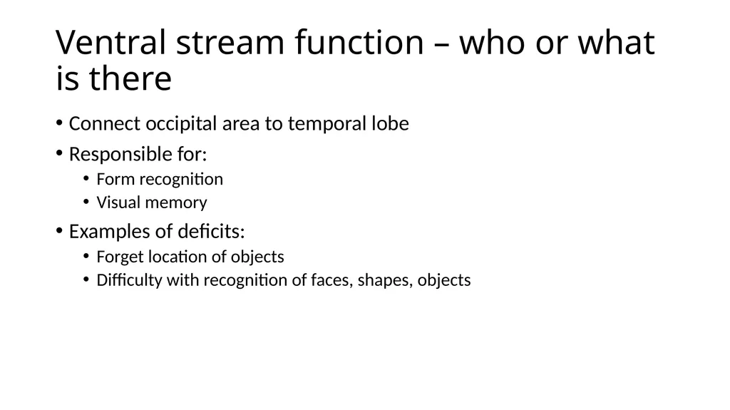
slide8. Ventral stream function – who or what is there Connect occipital area to temporal lobe
Responsible for:
Form recognition
Visual memory
Examples of deficits:
Forget location of objects
Difficulty with recognition of faces, shapes, objects<br>
slide9. Visual acuity Variable
Acuity may be normal
Deficit may be higher level
Processing or interpretation of visual information
May be called cognitive visual dysfunction<br>
slide10. Visual field Various types may be seen:
Swiss cheese or islands
Hemianopsia
Inferior field loss<br>
slide11. Characteristics in infants/delayed children Variable, poor or atypical response
Eccentric viewing
Latency
Preference for familiarity
Light gazing
Color preference
Difficulty with complexity
Preference for certain visual fields
Better visual performance with movement<br>
slide12. Characteristics in older children with higher function Slow and inefficient visual performance
Deficiency of visual perception and integration
Contrast sensitivity impairment
Difficulty with complexity
Short visual attention
Poor visual memory
Difficulty with object recognition/face recognition
Expressive delay<br>
slide13. CVI Treatment Systemic therapy
disease process may cause or prevent improvement
treat underlying medical conditions (eg: seizures, hydrocephalus)<br>
slide14. CVI Treatment Correct refractive error
Treat accommodative insufficiency
Treat amblyopia
Refer for vision services<br>
slide15. CVI Treatment Strabismus
alignment may be variable and improve
alignment may improve as acuity improves
observation for improvement of visual function and stability of alignment is suggested prior to surgical intervention<br>
slide16. General principles for intervention Must take into account other developmental issues
Recommendations need to be specific and aimed at child's requirements
Need to do timely reassessments and alter plan/interventions since improvement in CVI occurs<br>
slide17. Multidisciplinary team approach parents/family
primary care physician
pediatric ophthalmologist
pediatric neurologist
educational specialist
teacher of visually impaired
occupational therapist
physical therapist
speech therapist
teacher of the hearing impaired<br>
slide18. Patient and family centered care listen to and validate parent's concerns
ask about parental expectations
provide education
encourage parents to communicate observations of progress or problems<br>
slide19. Recovery of vision in CVI Most children have some degree of improvement
Recovery can occur over months to years
Degree of recovery cannot be predicted from imaging studies<br>
slide20. Basic research Some studies suggest more mature visual systems may retain a degree of plasticity
Functional MRI may be used to differentiate specific areas of function<br>
slide21. Education Medical professionals
importance of early diagnosis
final acuity cannot be predicted by neuroimaging
importance of early referral for vision services
need for evaluations based on function and impact on ADL with appropriate interventions<br>
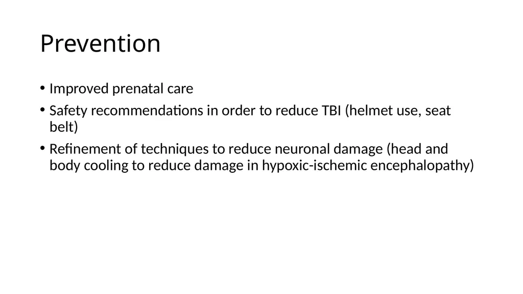
slide22. Prevention Improved prenatal care
Safety recommendations in order to reduce TBI (helmet use, seat belt)
Refinement of techniques to reduce neuronal damage (head and body cooling to reduce damage in hypoxic-ischemic encephalopathy)<br>
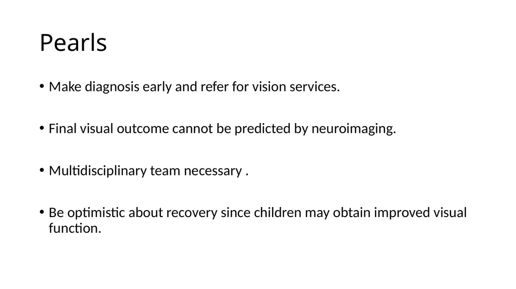
slide23. Pearls Make diagnosis early and refer for vision services.
Final visual outcome cannot be predicted by neuroimaging.
Multidisciplinary team necessary .
Be optimistic about recovery since children may obtain improved visual function.<br>

slide2. Case History Six month old ex 24-week premature infant has been recently discharged from the NICU with tracheostomy, feeding tube S/P NEC and has increased tone and back arching.
Mother of infant is concerned that child looks past her, stares at lights and ceiling fans and looks away when reaching for an object.<br>
slide3. Definition Bilateral visual impairment due to retrogeniculate brain damage
Eye exam typically normal
Displays atypical behaviors
May see optic atrophy and nystagmus<br>
slide4. Causes Structural: brain malformations, tumors
Vascular: hypoxic/ischemic event, perinatal stroke
Infectious: meningitis, encephalitis
Inflammatory: vasculitis
Trauma: TBI, shaken baby
Metabolic: neonatal hypoglycemia, mitochondrial disease, lysosomal disorders
Neurologic disease: seizure, hydrocephalus<br>
slide5. Pathophysiology in full term infants (more superficial damage) Hypoxia
↓
Hypercarbia
↓
Abnormal cerebral blood flow in watershed areas of cortex and subcortex
↓
Infarctions
↓
Cortical thinning with loss of gray matter<br>
slide6. Pathophysiology in preterm infants (deeper damage) Hypoxia/ischemia
↓
Hemorrhage
↓
Subcortex damage in periventricular deep white matter
↓
Periventricular leukomalacia (PVL)
↓
Optic radiation and corticospinal tract damage<br>
slide7. Dorsal stream function – getting there Connect occipital area to parietal area
Responsible for:
Finding objects in space
Figure/background
Extremity movement
Examples of deficits:
Difficulty with steps or changes in surfaces
Inaccurate reach
Difficulty with complexity<br>
slide8. Ventral stream function – who or what is there Connect occipital area to temporal lobe
Responsible for:
Form recognition
Visual memory
Examples of deficits:
Forget location of objects
Difficulty with recognition of faces, shapes, objects<br>
slide9. Visual acuity Variable
Acuity may be normal
Deficit may be higher level
Processing or interpretation of visual information
May be called cognitive visual dysfunction<br>
slide10. Visual field Various types may be seen:
Swiss cheese or islands
Hemianopsia
Inferior field loss<br>
slide11. Characteristics in infants/delayed children Variable, poor or atypical response
Eccentric viewing
Latency
Preference for familiarity
Light gazing
Color preference
Difficulty with complexity
Preference for certain visual fields
Better visual performance with movement<br>
slide12. Characteristics in older children with higher function Slow and inefficient visual performance
Deficiency of visual perception and integration
Contrast sensitivity impairment
Difficulty with complexity
Short visual attention
Poor visual memory
Difficulty with object recognition/face recognition
Expressive delay<br>
slide13. CVI Treatment Systemic therapy
disease process may cause or prevent improvement
treat underlying medical conditions (eg: seizures, hydrocephalus)<br>
slide14. CVI Treatment Correct refractive error
Treat accommodative insufficiency
Treat amblyopia
Refer for vision services<br>
slide15. CVI Treatment Strabismus
alignment may be variable and improve
alignment may improve as acuity improves
observation for improvement of visual function and stability of alignment is suggested prior to surgical intervention<br>
slide16. General principles for intervention Must take into account other developmental issues
Recommendations need to be specific and aimed at child's requirements
Need to do timely reassessments and alter plan/interventions since improvement in CVI occurs<br>
slide17. Multidisciplinary team approach parents/family
primary care physician
pediatric ophthalmologist
pediatric neurologist
educational specialist
teacher of visually impaired
occupational therapist
physical therapist
speech therapist
teacher of the hearing impaired<br>
slide18. Patient and family centered care listen to and validate parent's concerns
ask about parental expectations
provide education
encourage parents to communicate observations of progress or problems<br>
slide19. Recovery of vision in CVI Most children have some degree of improvement
Recovery can occur over months to years
Degree of recovery cannot be predicted from imaging studies<br>
slide20. Basic research Some studies suggest more mature visual systems may retain a degree of plasticity
Functional MRI may be used to differentiate specific areas of function<br>
slide21. Education Medical professionals
importance of early diagnosis
final acuity cannot be predicted by neuroimaging
importance of early referral for vision services
need for evaluations based on function and impact on ADL with appropriate interventions<br>
slide22. Prevention Improved prenatal care
Safety recommendations in order to reduce TBI (helmet use, seat belt)
Refinement of techniques to reduce neuronal damage (head and body cooling to reduce damage in hypoxic-ischemic encephalopathy)<br>
slide23. Pearls Make diagnosis early and refer for vision services.
Final visual outcome cannot be predicted by neuroimaging.
Multidisciplinary team necessary .
Be optimistic about recovery since children may obtain improved visual function.<br>