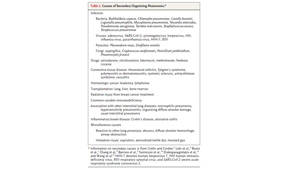
Cryptogenic Organizing Pneumonia Talmadge E. king and joyce S. Lee NEJM Fazil k JR2 Organizing Pneumonia (COP) Organizing pneumonia is a pattern of lung-tissue repair after injury. Formerly called BOOP Cryptogenic Secondary
"Cryptogenic Organizing Pneumonia Talmadge E. king" is the property of its rightful owner. Permission is granted to
download and print the materials on this website for personal, non-commercial use only, and to display it
on your personal computer provided you do not modify the materials and that you retain all copyright
notices contained in the materials. By downloading content from our website, you accept the terms of this
agreement.
Presentation Transcript
01
Cryptogenic Organizing Pneumonia Talmadge E. king and joyce S. Lee
NEJM
Fazil k
JR2<br>
02
Organizing Pneumonia (COP) Organizing pneumonia is a pattern of lung-tissue repair after injury.
Formerly called BOOP
Cryptogenic & Secondary<br>
03
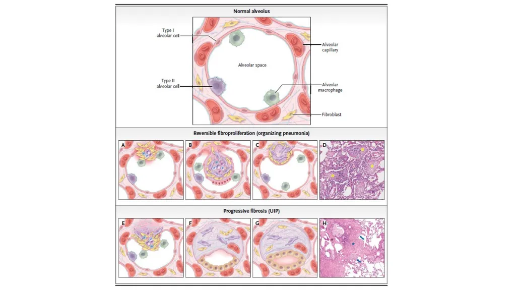
Pathophysiology Inflammatory and fibroproliferative process characterized
by intraalveolar fibroproliferation that is reversible<br>
04
Alveolar Injury
The alveolar epithelial injury is followed by leakage of
plasma proteins, fibrin formation, and migration of
inflammatory cells
Alveolar Organization
Fibroblasts are recruited to the alveolar lumen ->
myofibroblast-> fibroinflammatory buds (Masson’s
bodies )<br>
05
Remodeling
The inflammatory cells and fibrin deposits begin to disappear from the alveolar buds and are replaced by myofibroblasts organized in concentric rings alternating with layers of collagen bundles
The intraalveolar buds are remodeled into the interstitium, and collagen globules are formed
Alveolar epithelial cells proliferate and restore the continuity of the alveolar capillary membrane<br>
06
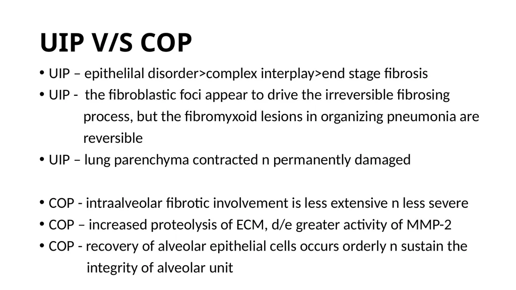
UIP V/S COP UIP – epithelilal disorder>complex interplay>end stage fibrosis
UIP - the fibroblastic foci appear to drive the irreversible fibrosing
process, but the fibromyxoid lesions in organizing pneumonia are
reversible
UIP – lung parenchyma contracted n permanently damaged
COP - intraalveolar fibrotic involvement is less extensive n less severe
COP – increased proteolysis of ECM, d/e greater activity of MMP-2
COP - recovery of alveolar epithelial cells occurs orderly n sustain the
integrity of alveolar unit<br>
07
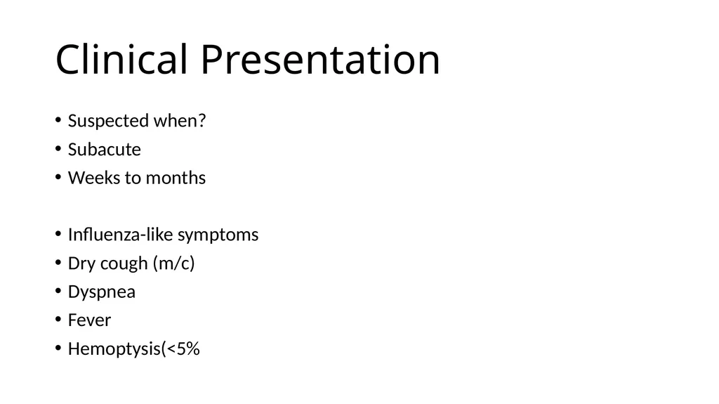
Clinical Presentation Suspected when?
Subacute
Weeks to months
Pulmonary-Function Testing Restrictive ventilatory defect and a reduced diffusing capacity for carbon monoxide.<br>
11
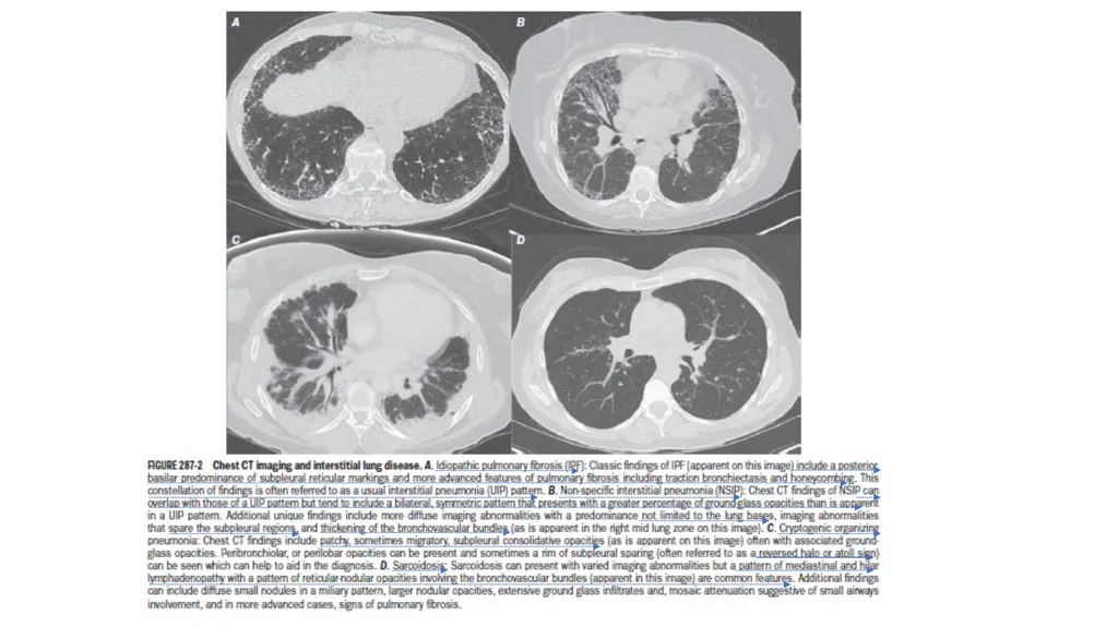
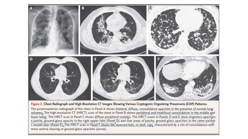
Radiologic Studies Radiogaph – Distinctive, b/l opacities (patchy or diffuse)
with normal lung volume
HRCT - peripheral and multifocal consolidation (+/- air bronchograms)
- unilateral or bilateral
- slightly predominant subpleural and lower-lung zone
distribution
- ground-glass opacities, nodules
- peribronchovascular pattern, with areas of consolidation
surrounding the bronchovascular bundles<br>
12
Migratory opacities
The reversed halo (or atoll) sign - a rim of consolidation with more central clearing or ground-glass opacities specific
Presence of bilateral alveolar opacities in a relevant clinical context can support a diagnosis of organizing pneumonia<br>
13
Bronchoalveolar Lavage Analysis
shows a lymphocytic alveolitis associated with an increase in neutrophil and eosinophil counts
rule out infection and other disorders, such as eosinophilic pneumonia and alveolar hemorrhage<br>
14
Histopathological Assessment
Often be managed without histopathological confirmation
Confirmation purposes
In cases managed without a biopsy, if the clinical follow-up is not consistent with the diagnosis of COP, the diagnosis should be reevaluated, and a biopsy may be necessary<br>
15
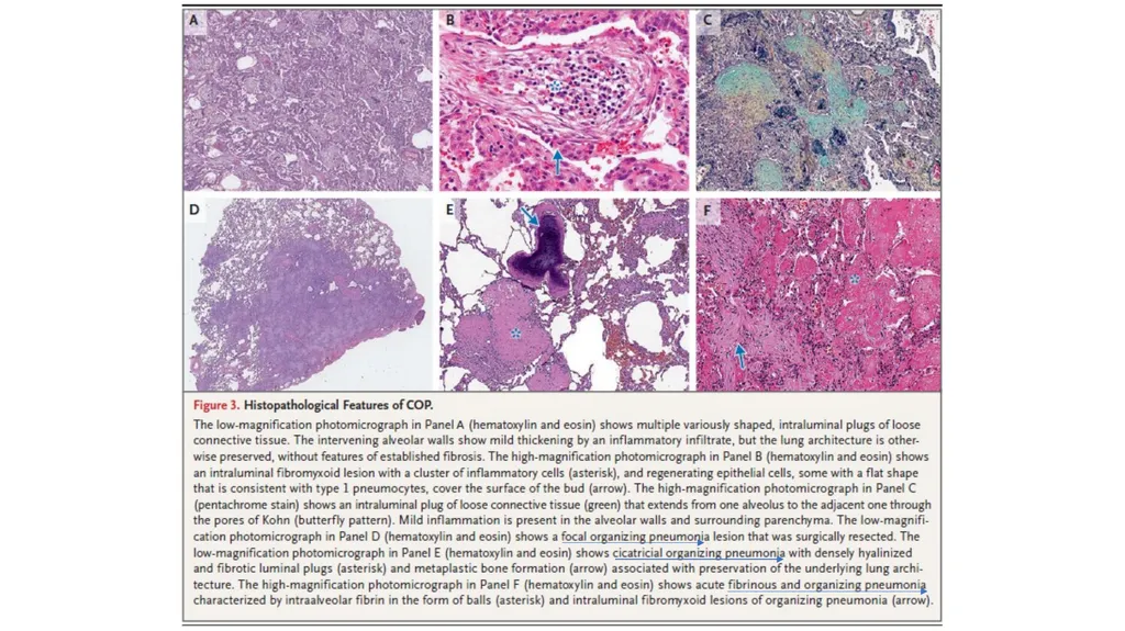
Intraluminal plugs of loose connective tissue(MASON BODIES) that involve alveolar spaces and alveolar ducts and may also involve bronchioles
The underlying architecture of the lung is preserved<br>
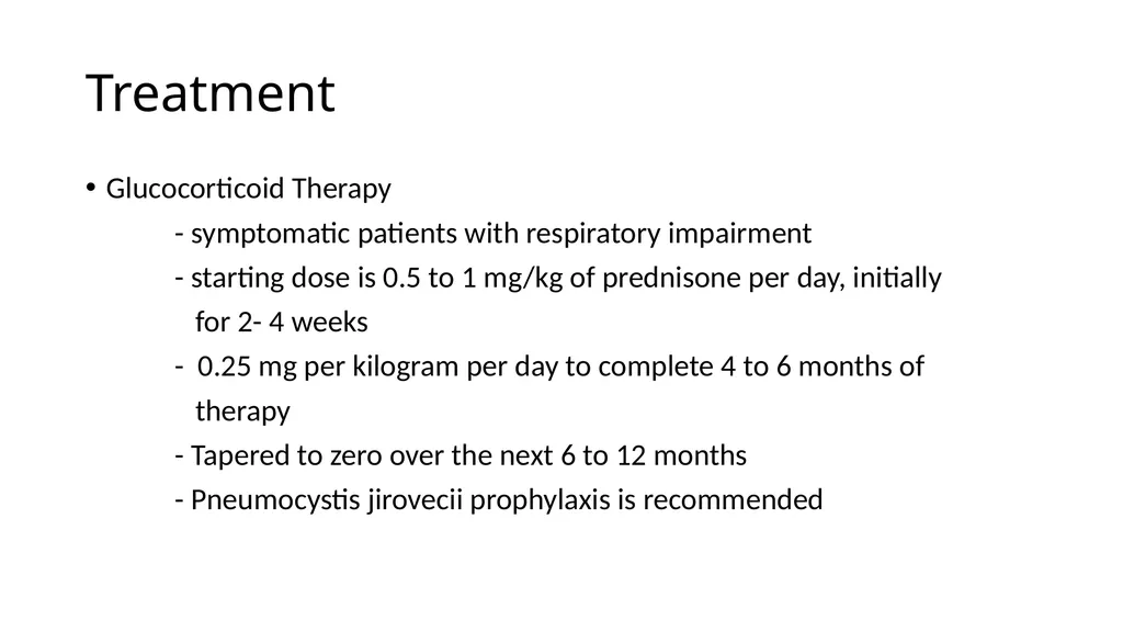
Treatment Glucocorticoid Therapy
- symptomatic patients with respiratory impairment
- starting dose is 0.5 to 1 mg/kg of prednisone per day, initially
for 2- 4 weeks
- 0.25 mg per kilogram per day to complete 4 to 6 months of
therapy
- Tapered to zero over the next 6 to 12 months
- Pneumocystis jirovecii prophylaxis is recommended<br>
18
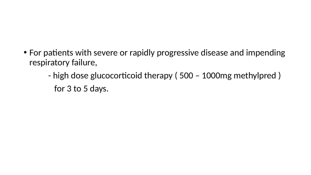
For patients with severe or rapidly progressive disease and impending respiratory failure,
- high dose glucocorticoid therapy ( 500 – 1000mg methylpred )
for 3 to 5 days.<br>
19
Other therapies Macrolide Antibiotics
- 3 to 6 months
- less effective than glucocorticoids
Cytotoxic Therapy
- azathioprine / cyclophosphamide
- Mycophenolate Mofetil<br>
20
Prognosis Improvement is generally excellent
Patients with radiologic airspace opacities have much better outcomes than those with reticular opacities
Progressive respiratory failure requiring mechanical ventilation and resulting in death is rare
Death more in secondary organizing pneumonia , d/e underlying illnes<br>
21
Future directions The incidence and prevalence of COP remain poorly defined
More studies are needed to distinguish primary COP from secondary organizing pneumonia
The usefulness of immunosuppressive therapy as primary or secondary treatment is unknown<br>