04
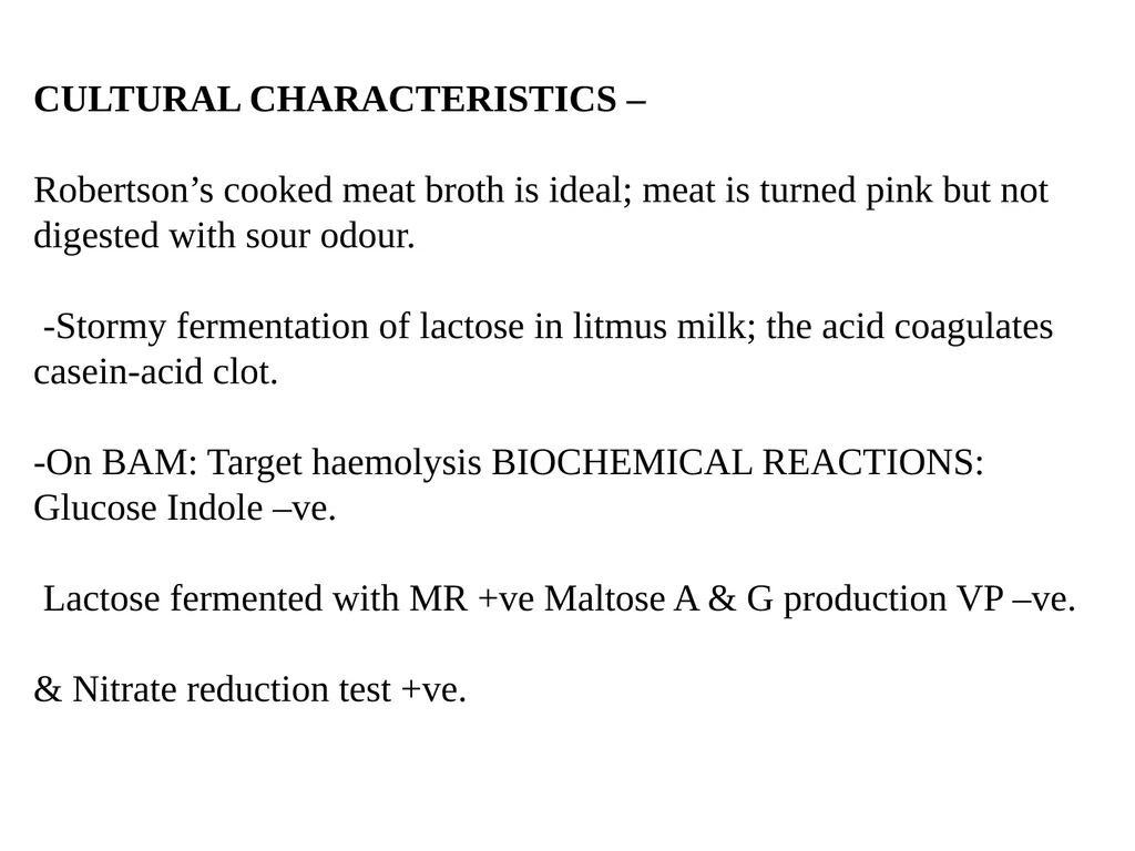
CULTURAL CHARACTERISTICS –
Robertson’s cooked meat broth is ideal; meat is turned pink but not digested with sour odour.
-Stormy fermentation of lactose in litmus milk; the acid coagulates casein-acid clot.
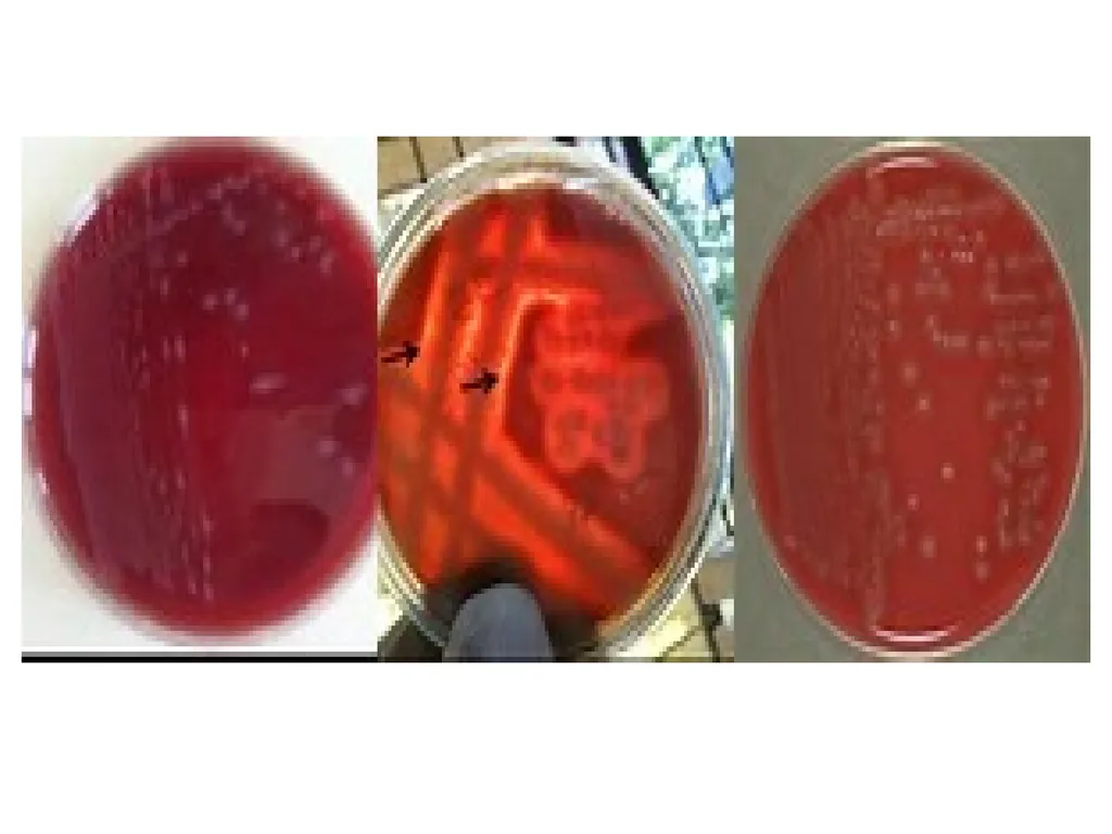
-On BAM: Target haemolysis BIOCHEMICAL REACTIONS: Glucose Indole –ve.
Lactose fermented with MR +ve Maltose A & G production VP –ve.
& Nitrate reduction test +ve.<br>
05
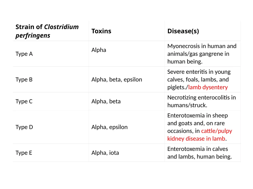
Toxins
Clostridium perfringens Type A
Clostridium perfringens Type B
Clostridium perfringens Type C
Clostridium perfringens Type D
Clostridium perfringens Type E<br>
06
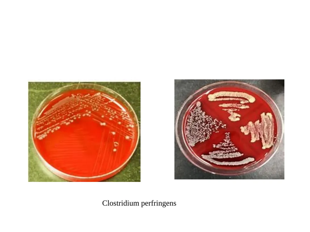
Clostridium perfringens<br>
07
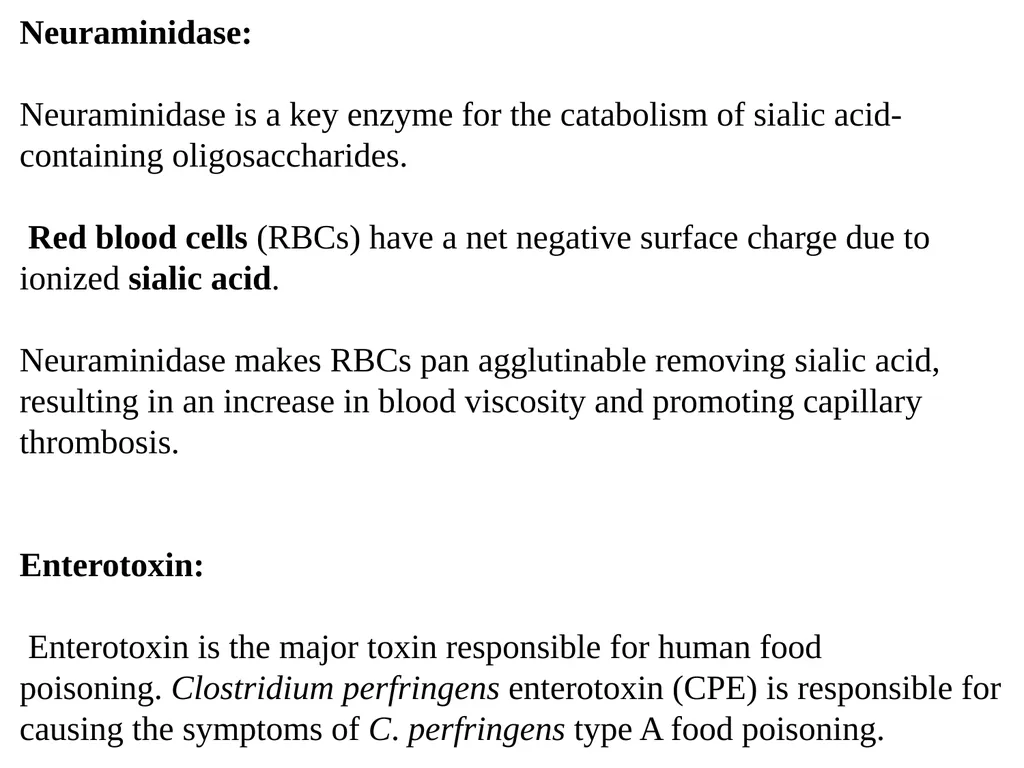
Neuraminidase:
Neuraminidase is a key enzyme for the catabolism of sialic acid-containing oligosaccharides.
Red blood cells (RBCs) have a net negative surface charge due to ionized sialic acid.
Neuraminidase makes RBCs pan agglutinable removing sialic acid, resulting in an increase in blood viscosity and promoting capillary thrombosis.
Enterotoxin:
Enterotoxin is the major toxin responsible for human food poisoning. Clostridium perfringens enterotoxin (CPE) is responsible for causing the symptoms of C. perfringens type A food poisoning.<br>
08
Sources of infection can be either:
Exogenous: injury, contamination of wound by animal faeces.
Endogenous: from own GIT after disruption of mucous membrane, often due to a mixed infection which can enhances an anaerobic environment, thereby encouraging the organism to grow.
Infections caused by C. perfringens can lead to:
Skin/Wound infections: superficial skin infection, anaerobic cellulitis, myonecrosis or gas gangrene and puerperal infections. A pus swab is required to confirm the cause of infection.<br>
09
Septicemia (can lead to Disseminated Intravascular Coagulation).
A Blood culture specimen will be required to confirm this.
Self-limiting food poisoning (watery diarrhea) where enterotoxin was produced in the meat.
Incubation period is 6 to 24 hours after ingestion.
If large dose is ingested it can lead to Necrotizing Enteritis.
A Stool specimen is required to confirm the diagnosis.<br>
10
Pathogenesis of Clostridium perfringens
Invasive infection and Gas gangrenes
In invasive clostridial infections, spores reach tissue either by contamination of traumatized areas (soil, feces) or from the intestinal tract.
The spores germinate at low oxidation-reduction potential; vegetative cells multiply, ferment carbohydrates present in the tissue, and produce gas.
The distention of tissue and interference with blood supply, together with the secretion of necrotizing toxin and hyaluronidase, favor the spread of infection.<br>
11
Tissue necrosis extends, providing an opportunity for increased bacterial growth; hemolytic anemia; and, ultimately, severe toxemia and death.
The toxin involved in gas gangrene is known as α-toxin, which inserts into the plasma membrane of cells, producing gaps in the membrane that disrupt normal cellular function. Clinical Features of Clostridium perfringens
Gas gangrene
Clostridium perfringens is the most common strain of clostridia
associated with trauma-induced gas gangrene in humans and is also
a major cause of spontaneous (nontraumatic) gas gangrene.
The classic features of gas gangrene are extensive local tissue destruction progressing to profound shock and death.<br>
12
From a contaminated wound (eg, a compound fracture, postpartum uterus), the infection spreads in 1–3 days to produce crepitation in the subcutaneous tissue and muscle, foul-smelling discharge, rapidly progressing necrosis, fever, hemolysis, toxemia, shock, and death.
At times, the infection results only in anaerobic fasciitis or cellulitis.<br>
13
Food Poisoning
C.perfringens food poisoning usually follows the ingestion of large numbers of clostridia that have grown in warmed meat dishes.
The toxin forms when the organisms sporulate in the gut, with the onset of diarrhea and abdominal cramps—usually without vomiting or fever—in 7–30 hours.
The illness suddenly begins and lasts only 1–2 days.<br>
14
Nagler’s Test
C. perfringens can be diagnosed by Nagler’s reaction, where the suspect organism is cultured on an egg yolk media plate.
One side of the plate contains anti-alpha-toxin, while the other side does not.
A streak of a suspect organism is placed through both sides.
An area of turbidity will form around the side that does not have the anti-alpha-toxin, indicating uninhibited lecithinase activity.<br>
15
Prevention of Clostridium perfringens Infection
The growth of C. perfringens spores can be prevented by most importantly cooking food, especially beef, and poultry, thoroughly, to the recommended temperatures.
Leftover food should be refrigerated to a temperature below 4 °C within two hours of preparation.
Large pots of food such as soup or stew with meats should be divided into small quantities and covered for refrigeration.<br>
16
Leftovers should be reheated to at least 74 °C before serving.
A rule of thumb is that if the food tastes, smells, or looks different from what it is supposed to, then the food should be avoided.
Even if it looks safe, food that has been out for a long time can also be dangerous to eat.
In case of tissue infections, early and adequate cleansing of contaminated wounds and surgical debridement, together with the administration of antimicrobial drugs directed against clostridia (eg, penicillin), are the best available preventive measures.<br>
17
Clostridium chauvoei
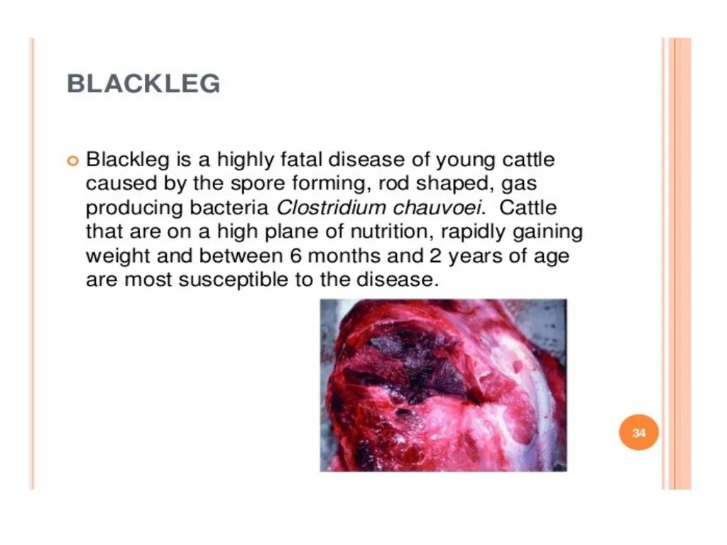
Clostridium chauvoei is an anaerobic, motile, Gram-positive bacterium.
It is a soil-borne pathogen that can cause blackleg in cattle and sheep.
In cattle affects young stage (6 months to 2years ) during rainy season.
Affect specially in hind and fore quarters of animals.
In sheep due to wound, lambing docking, shearing and castration.<br>
18
Disease –Blackleg/blackleg baillus /Black quarter (B.Q.) in cattle
and sheep.
Symptoms of Blackleg in cattle
Depression, anorexia, rumen stasis, high fever (41-42 degrees), and tachycardia.
These signs are usually followed by marked lameness with noticeable muscle swelling of the affected leg’s upper part.
Edematous and crepitate swelling develops in the hip, shoulder, chest, back, neck, or elsewhere.
The swelling is small, hot, and painful, and then swelling enlarges, and there is crepitation on palpation. The skin is often discolored in affected areas, and on the cut section, a serosanguineous rancid fluid appears and gets escapes.<br>
19
How to Diagnosis the Black Quarter in Cattle? A rapidly fatal, febrile disease in well-nourished
young cattle with the massive muscles’ crepitant swellings suggests Blackleg.
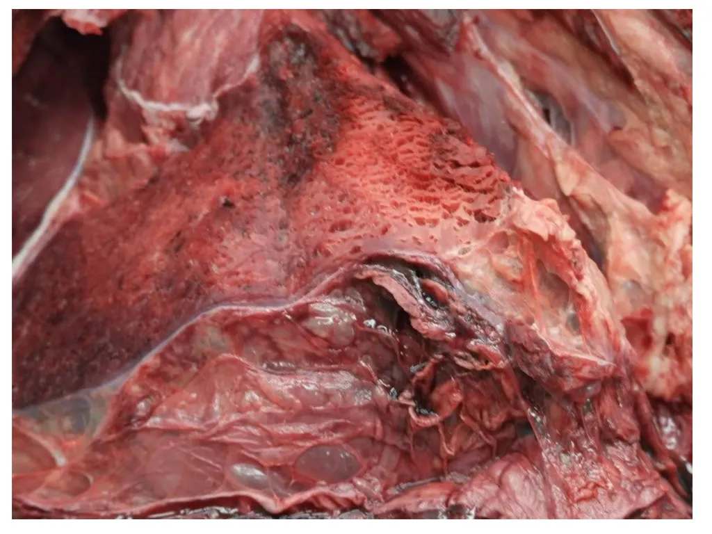
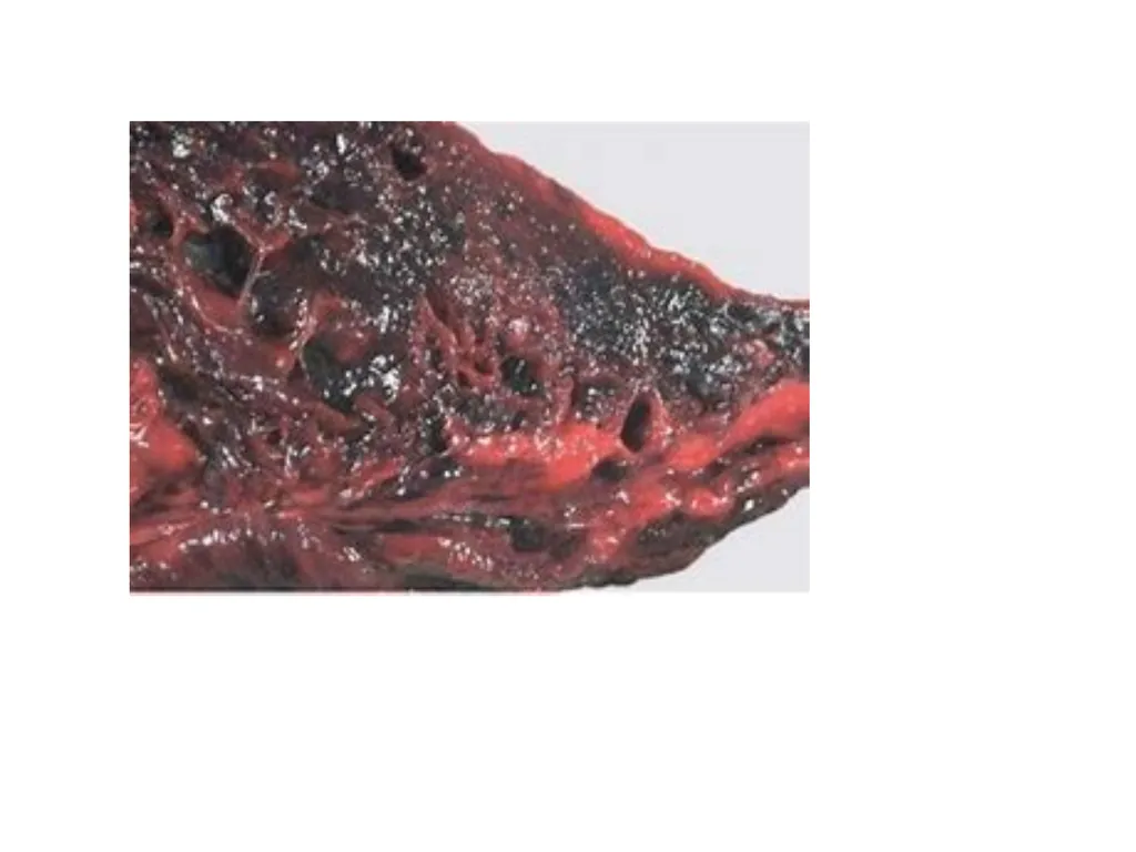
The affected muscle is a dark red to black, dry and spongy, it has a sweetish odor and is filtered with small bubbles,
but with edema, suggests Blackleg.
Field diagnosis can be made on history, clinical findings and necropsy examination, history of vaccination,
history of recent transferring of the vicinity soil inside and outside the barn, course of the disease is
per acute and acute, and outbreak six months to 2 years of age in cattle.<br>