
Diabetic Foot Ulcers and When to Revascularize
TA
Published · 23 slides · 0 views
1 / 1





Description
Diabetic Foot Ulcers and When to Revascularize Wednesday, August 14, 2024 Agenda What is a diabetic foot ulcer (DFU)? What do diabetic foot ulcers look like? What causes a diabetic foot ulcer? What is the Ankle Brachial Index (ABI)? When is
Related Topics
Share
Embed code
Download this presentation From Below
"Diabetic Foot Ulcers and When to Revascularize" is the property of its rightful owner. Permission is granted to download and print the materials on this website for personal, non-commercial use only, and to display it on your personal computer provided you do not modify the materials and that you retain all copyright notices contained in the materials. By downloading content from our website, you accept the terms of this agreement.
Presentation Transcript
01
Diabetic Foot Ulcers and When to Revascularize
Wednesday, August 14, 2024<br>
02
Agenda What is a diabetic foot ulcer (DFU)?
What do diabetic foot ulcers look like?
What causes a diabetic foot ulcer?
What is the Ankle Brachial Index (ABI)?
When is the ABI Measured?
How is the ABI measured?
What are the ABI guidelines?
What is revascularization?
When should a diabetic foot ulcer be revascularized? What do the studies suggest?
What is the algorithm for patient presenting with DFU?<br>
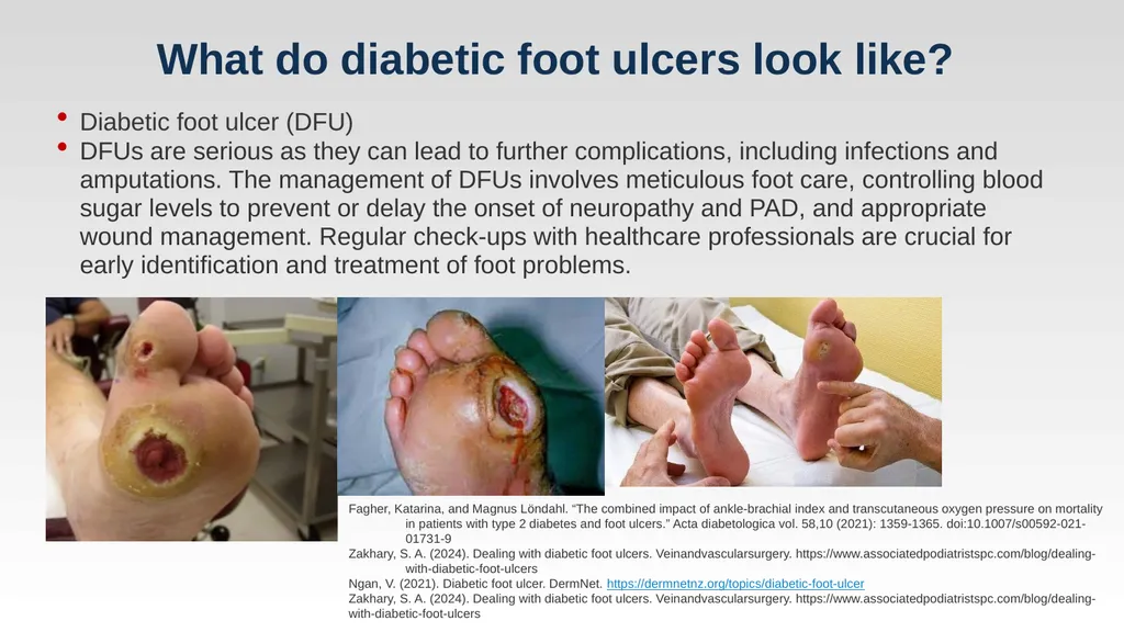
What do diabetic foot ulcers look like?
What causes a diabetic foot ulcer?
What is the Ankle Brachial Index (ABI)?
When is the ABI Measured?
How is the ABI measured?
What are the ABI guidelines?
What is revascularization?
When should a diabetic foot ulcer be revascularized? What do the studies suggest?
What is the algorithm for patient presenting with DFU?<br>
03
What is a diabetic foot ulcer? Gallagher KA, Mills JL, Armstrong DG, Conte MS, Kirsner RS, Minc SD, Plutzky J, Southerland KW, Tomic-Canic M; on behalf of the American Heart Association Council on Peripheral Vascular Disease, Council on Cardiovascular and Stroke Nursing, Council on Clinical Cardiology, and Council on Lifestyle and Cardiometabolic Health. Current status and principles for the treatment and prevention of diabetic foot ulcers in the cardiovascular patient population: a scientific statement from the American Heart Association. Circulation. 2024;149:e232–e253. doi: 10.1161/CIR.0000000000001192 A diabetic foot ulcer (DFU) is a prevalent complication of diabetes, marked by an open sore or wound that typically occurs on the bottom of the foot in patients with diabetes. The pathophysiology of DFUs involves a combination of neuropathy, peripheral artery disease (PAD), and immune system inefficiencies, which are common in individuals with diabetes.<br>
04
What is a diabetic foot ulcer? Oliver TI, Mutluoglu M. Diabetic Foot Ulcer (archived) [Updated 2023 Aug 8]. In: StatPearls [Internet]. Treasure Island (FL): StatPearls Publishing; 2024 Jan-. Available from: https://www.ncbi.nlm.nih.gov/books/NBK537328/ About 60% of diabetic patients will develop neuropathy, eventually leading to a foot ulcer. The risk of a foot ulcer is increased in individuals with a flat foot as they have disproportionate stress across the foot, leading to tissue inflammation in high risk areas of the foot.
Diabetic foot ulcers are responsible for more admissions than any other diabetic complication. Today, diabetes is the leading cause of non-traumatic amputations in the US. Overall, about 5% of patients with diabetes mellitus develop foot ulcers and 1% end up with an amputation.<br>
Diabetic foot ulcers are responsible for more admissions than any other diabetic complication. Today, diabetes is the leading cause of non-traumatic amputations in the US. Overall, about 5% of patients with diabetes mellitus develop foot ulcers and 1% end up with an amputation.<br>
05
What do diabetic foot ulcers look like? Fagher, Katarina, and Magnus Löndahl. “The combined impact of ankle-brachial index and transcutaneous oxygen pressure on mortality in patients with type 2 diabetes and foot ulcers.” Acta diabetologica vol. 58,10 (2021): 1359-1365. doi:10.1007/s00592-021-01731-9
Zakhary, S. A. (2024). Dealing with diabetic foot ulcers. Veinandvascularsurgery. https://www.associatedpodiatristspc.com/blog/dealing-with-diabetic-foot-ulcers
Ngan, V. (2021). Diabetic foot ulcer. DermNet. https://dermnetnz.org/topics/diabetic-foot-ulcer
Zakhary, S. A. (2024). Dealing with diabetic foot ulcers. Veinandvascularsurgery. https://www.associatedpodiatristspc.com/blog/dealing-with-diabetic-foot-ulcers Diabetic foot ulcer (DFU)
DFUs are serious as they can lead to further complications, including infections and amputations. The management of DFUs involves meticulous foot care, controlling blood sugar levels to prevent or delay the onset of neuropathy and PAD, and appropriate wound management. Regular check-ups with healthcare professionals are crucial for early identification and treatment of foot problems.<br>
Zakhary, S. A. (2024). Dealing with diabetic foot ulcers. Veinandvascularsurgery. https://www.associatedpodiatristspc.com/blog/dealing-with-diabetic-foot-ulcers
Ngan, V. (2021). Diabetic foot ulcer. DermNet. https://dermnetnz.org/topics/diabetic-foot-ulcer
Zakhary, S. A. (2024). Dealing with diabetic foot ulcers. Veinandvascularsurgery. https://www.associatedpodiatristspc.com/blog/dealing-with-diabetic-foot-ulcers Diabetic foot ulcer (DFU)
DFUs are serious as they can lead to further complications, including infections and amputations. The management of DFUs involves meticulous foot care, controlling blood sugar levels to prevent or delay the onset of neuropathy and PAD, and appropriate wound management. Regular check-ups with healthcare professionals are crucial for early identification and treatment of foot problems.<br>
06
What causes a diabetic foot ulcers? Diabetic foot ulcers (DFUs) are a significant and common complication in individuals with diabetes, typically arising from a combination of factors including neuropathy, peripheral artery disease (PAD), and immunological weaknesses, often exacerbated by poor glycemic control and mechanical stress on the feet.
Neuropathy plays a critical role by impairing sensation in the feet, which can prevent early detection of cuts, blisters, and other foot injuries. Without the ability to feel pain, minor injuries may become severely infected or develop into ulcers without the patient's awareness Viadé, J., Nicolás, M., Bundó, M. et al. A novel assessment, diagnostic and treatment system for diabetic foot. J Foot Ankle Res 16, 84 (2023). https://doi.org/10.1186/s13047-023-00687-z<br>
Neuropathy plays a critical role by impairing sensation in the feet, which can prevent early detection of cuts, blisters, and other foot injuries. Without the ability to feel pain, minor injuries may become severely infected or develop into ulcers without the patient's awareness Viadé, J., Nicolás, M., Bundó, M. et al. A novel assessment, diagnostic and treatment system for diabetic foot. J Foot Ankle Res 16, 84 (2023). https://doi.org/10.1186/s13047-023-00687-z<br>
07
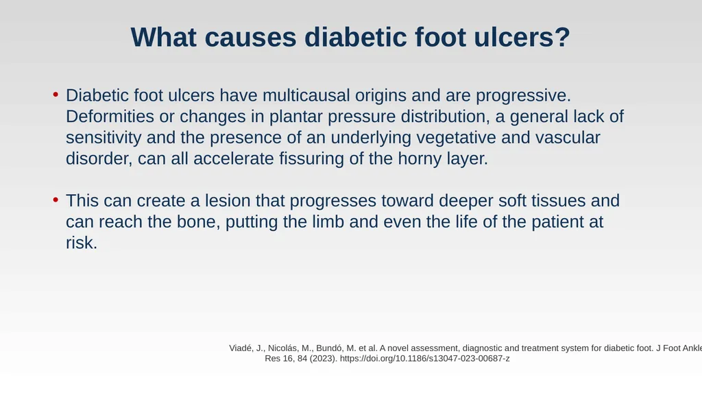
What causes diabetic foot ulcers? Viadé, J., Nicolás, M., Bundó, M. et al. A novel assessment, diagnostic and treatment system for diabetic foot. J Foot Ankle Res 16, 84 (2023). https://doi.org/10.1186/s13047-023-00687-z Diabetic foot ulcers have multicausal origins and are progressive. Deformities or changes in plantar pressure distribution, a general lack of sensitivity and the presence of an underlying vegetative and vascular disorder, can all accelerate fissuring of the horny layer.
This can create a lesion that progresses toward deeper soft tissues and can reach the bone, putting the limb and even the life of the patient at risk.<br>
This can create a lesion that progresses toward deeper soft tissues and can reach the bone, putting the limb and even the life of the patient at risk.<br>
08
What causes diabetic foot ulcers? Mahmoudvand, G., Karimi Rouzbahani, A., Razavi, Z. S., Mahjoor, M., & Afkhami, H. (2023). Mesenchymal stem cell therapy for non-healing diabetic foot ulcer infection: New insight. Frontiers in Bioengineering and Biotechnology, 11. https://doi.org/10.3389/fbioe.2023.1158484
Pomatto, Margherita et al. “Differential Therapeutic Effect of Extracellular Vesicles Derived by Bone Marrow and Adipose Mesenchymal Stem Cells on Wound Healing of Diabetic Ulcers and Correlation to Their Cargoes.” International journal of molecular sciences vol. 22,8 3851. 8 Apr. 2021, doi:10.3390/ijms22083851 Constant inflammatory factor release from neutrophil infiltration, macrophage imbalance, mast cell degranulation, and dendritic cell and T cell dysregulation in a diabetic ulcer causes inflammatory cascades, vascular maturation disorders, and decreased collagen deposition, all of which contribute to a diabetic ulcer’s protracted or non-healing course.<br>
Pomatto, Margherita et al. “Differential Therapeutic Effect of Extracellular Vesicles Derived by Bone Marrow and Adipose Mesenchymal Stem Cells on Wound Healing of Diabetic Ulcers and Correlation to Their Cargoes.” International journal of molecular sciences vol. 22,8 3851. 8 Apr. 2021, doi:10.3390/ijms22083851 Constant inflammatory factor release from neutrophil infiltration, macrophage imbalance, mast cell degranulation, and dendritic cell and T cell dysregulation in a diabetic ulcer causes inflammatory cascades, vascular maturation disorders, and decreased collagen deposition, all of which contribute to a diabetic ulcer’s protracted or non-healing course.<br>
09
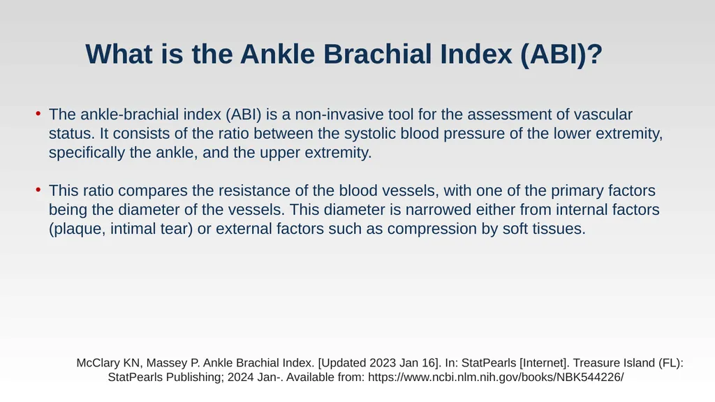
What is the Ankle Brachial Index (ABI)? McClary KN, Massey P. Ankle Brachial Index. [Updated 2023 Jan 16]. In: StatPearls [Internet]. Treasure Island (FL): StatPearls Publishing; 2024 Jan-. Available from: https://www.ncbi.nlm.nih.gov/books/NBK544226/ The ankle-brachial index (ABI) is a non-invasive tool for the assessment of vascular status. It consists of the ratio between the systolic blood pressure of the lower extremity, specifically the ankle, and the upper extremity.
This ratio compares the resistance of the blood vessels, with one of the primary factors being the diameter of the vessels. This diameter is narrowed either from internal factors (plaque, intimal tear) or external factors such as compression by soft tissues.<br>
This ratio compares the resistance of the blood vessels, with one of the primary factors being the diameter of the vessels. This diameter is narrowed either from internal factors (plaque, intimal tear) or external factors such as compression by soft tissues.<br>
10
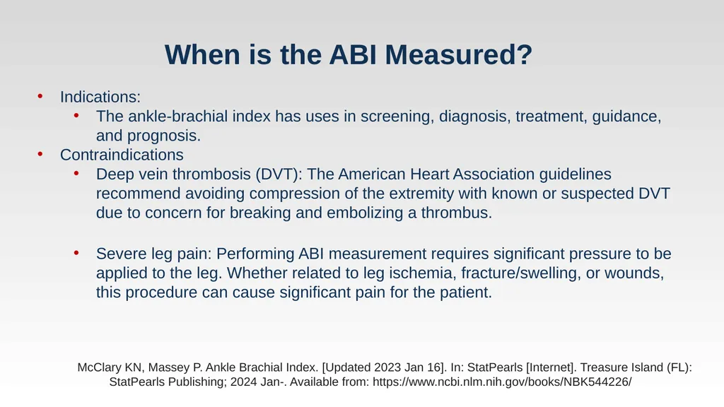
When is the ABI Measured? McClary KN, Massey P. Ankle Brachial Index. [Updated 2023 Jan 16]. In: StatPearls [Internet]. Treasure Island (FL): StatPearls Publishing; 2024 Jan-. Available from: https://www.ncbi.nlm.nih.gov/books/NBK544226/ Indications:
The ankle-brachial index has uses in screening, diagnosis, treatment, guidance, and prognosis.
Contraindications
Deep vein thrombosis (DVT): The American Heart Association guidelines recommend avoiding compression of the extremity with known or suspected DVT due to concern for breaking and embolizing a thrombus.
Severe leg pain: Performing ABI measurement requires significant pressure to be applied to the leg. Whether related to leg ischemia, fracture/swelling, or wounds, this procedure can cause significant pain for the patient.<br>
The ankle-brachial index has uses in screening, diagnosis, treatment, guidance, and prognosis.
Contraindications
Deep vein thrombosis (DVT): The American Heart Association guidelines recommend avoiding compression of the extremity with known or suspected DVT due to concern for breaking and embolizing a thrombus.
Severe leg pain: Performing ABI measurement requires significant pressure to be applied to the leg. Whether related to leg ischemia, fracture/swelling, or wounds, this procedure can cause significant pain for the patient.<br>
11
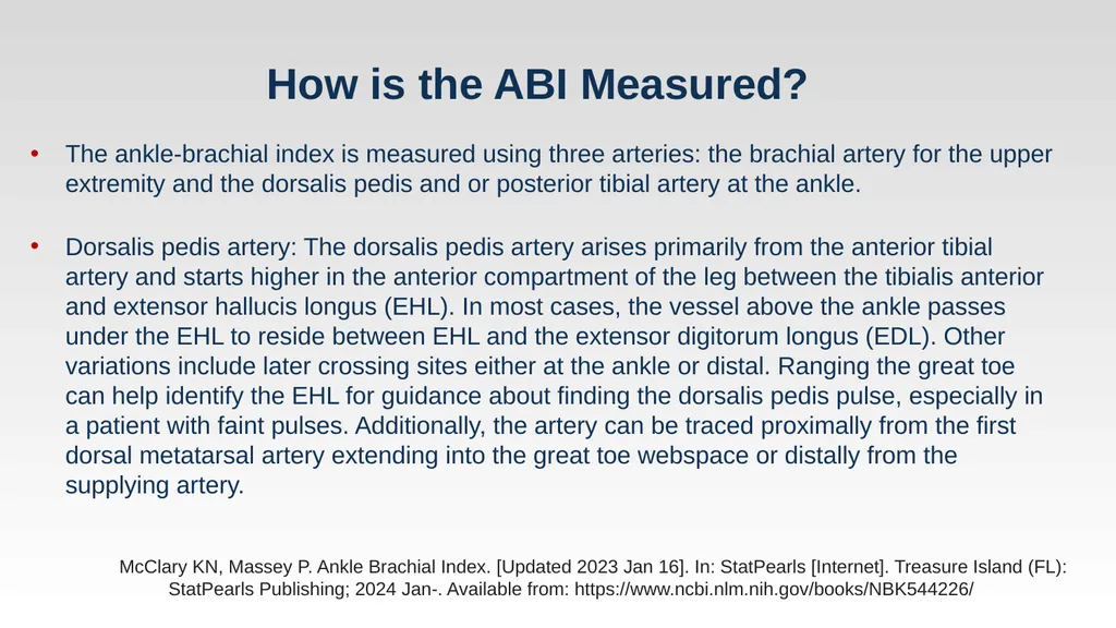
How is the ABI Measured? McClary KN, Massey P. Ankle Brachial Index. [Updated 2023 Jan 16]. In: StatPearls [Internet]. Treasure Island (FL): StatPearls Publishing; 2024 Jan-. Available from: https://www.ncbi.nlm.nih.gov/books/NBK544226/ The ankle-brachial index is measured using three arteries: the brachial artery for the upper extremity and the dorsalis pedis and or posterior tibial artery at the ankle.
Dorsalis pedis artery: The dorsalis pedis artery arises primarily from the anterior tibial artery and starts higher in the anterior compartment of the leg between the tibialis anterior and extensor hallucis longus (EHL). In most cases, the vessel above the ankle passes under the EHL to reside between EHL and the extensor digitorum longus (EDL). Other variations include later crossing sites either at the ankle or distal. Ranging the great toe can help identify the EHL for guidance about finding the dorsalis pedis pulse, especially in a patient with faint pulses. Additionally, the artery can be traced proximally from the first dorsal metatarsal artery extending into the great toe webspace or distally from the supplying artery.<br>
Dorsalis pedis artery: The dorsalis pedis artery arises primarily from the anterior tibial artery and starts higher in the anterior compartment of the leg between the tibialis anterior and extensor hallucis longus (EHL). In most cases, the vessel above the ankle passes under the EHL to reside between EHL and the extensor digitorum longus (EDL). Other variations include later crossing sites either at the ankle or distal. Ranging the great toe can help identify the EHL for guidance about finding the dorsalis pedis pulse, especially in a patient with faint pulses. Additionally, the artery can be traced proximally from the first dorsal metatarsal artery extending into the great toe webspace or distally from the supplying artery.<br>
12
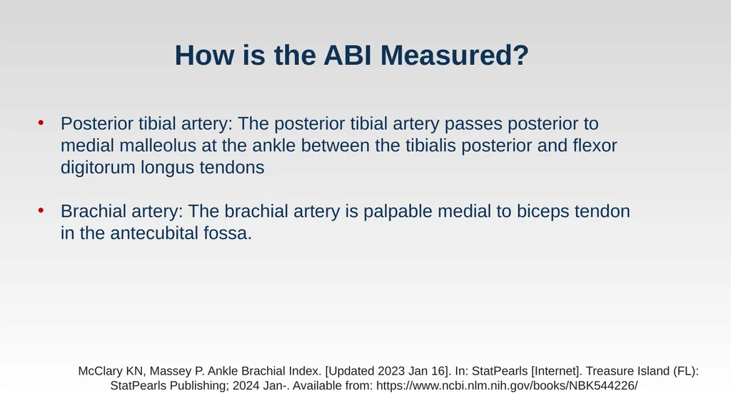
How is the ABI Measured? McClary KN, Massey P. Ankle Brachial Index. [Updated 2023 Jan 16]. In: StatPearls [Internet]. Treasure Island (FL): StatPearls Publishing; 2024 Jan-. Available from: https://www.ncbi.nlm.nih.gov/books/NBK544226/ Posterior tibial artery: The posterior tibial artery passes posterior to medial malleolus at the ankle between the tibialis posterior and flexor digitorum longus tendons
Brachial artery: The brachial artery is palpable medial to biceps tendon in the antecubital fossa.<br>
Brachial artery: The brachial artery is palpable medial to biceps tendon in the antecubital fossa.<br>
13
What does the ABI indicate? Akkus, Gamze, and Murat Sert. “Diabetic foot ulcers: A devastating complication of diabetes mellitus continues non-stop in spite of new medical treatment modalities.” World journal of diabetes vol. 13,12 (2022): 1106-1121. doi:10.4239/wjd.v13.i12.1106 Ankle-brachial index (ABI) has emerged as a relatively non-invasive tool for the diagnosis of peripheral arterial disease. The measurement of ABI (ratio of systolic blood pressure on the ankle to the systolic blood pressure in the upper arm) is normal in the 1.0-1.4 range, obviously pathologic under 0.9. An ABI over than 1.4 is also considered as abnormal, reflecting calcified and stiffed arteries.<br>
14
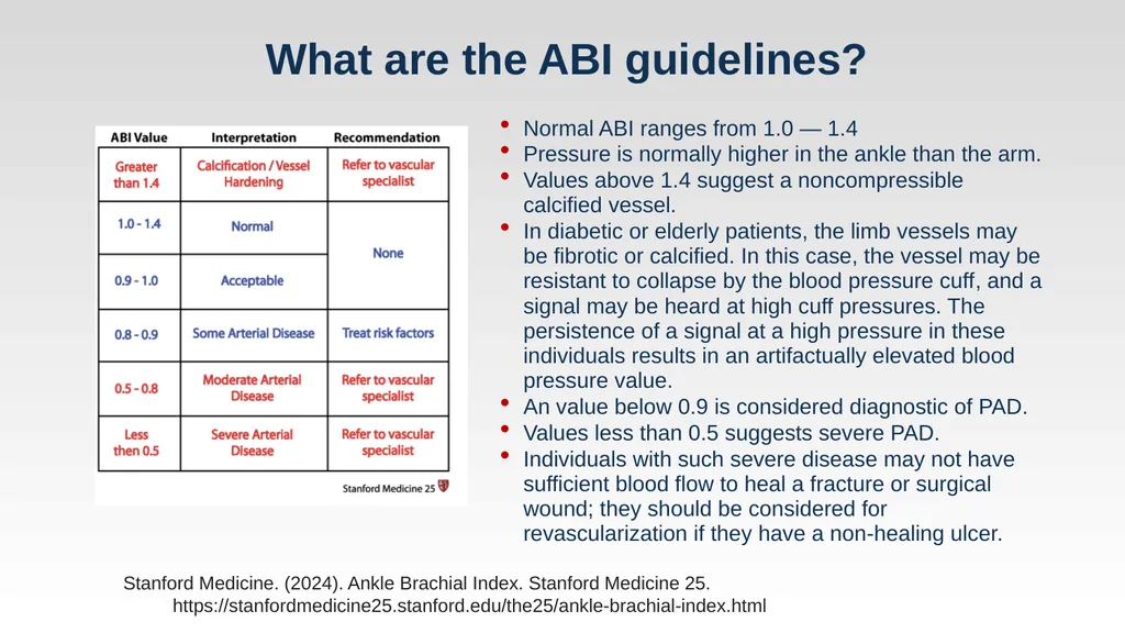
What are the ABI guidelines? Stanford Medicine. (2024). Ankle Brachial Index. Stanford Medicine 25. https://stanfordmedicine25.stanford.edu/the25/ankle-brachial-index.html Normal ABI ranges from 1.0 — 1.4
Pressure is normally higher in the ankle than the arm.
Values above 1.4 suggest a noncompressible calcified vessel.
In diabetic or elderly patients, the limb vessels may be fibrotic or calcified. In this case, the vessel may be resistant to collapse by the blood pressure cuff, and a signal may be heard at high cuff pressures. The persistence of a signal at a high pressure in these individuals results in an artifactually elevated blood pressure value.
An value below 0.9 is considered diagnostic of PAD.
Values less than 0.5 suggests severe PAD.
Individuals with such severe disease may not have sufficient blood flow to heal a fracture or surgical wound; they should be considered for revascularization if they have a non-healing ulcer.<br>
Pressure is normally higher in the ankle than the arm.
Values above 1.4 suggest a noncompressible calcified vessel.
In diabetic or elderly patients, the limb vessels may be fibrotic or calcified. In this case, the vessel may be resistant to collapse by the blood pressure cuff, and a signal may be heard at high cuff pressures. The persistence of a signal at a high pressure in these individuals results in an artifactually elevated blood pressure value.
An value below 0.9 is considered diagnostic of PAD.
Values less than 0.5 suggests severe PAD.
Individuals with such severe disease may not have sufficient blood flow to heal a fracture or surgical wound; they should be considered for revascularization if they have a non-healing ulcer.<br>
15
What is revascularization? Perez-Favila, A.; Martinez-Fierro, M.L.; Rodriguez-Lazalde, J.G.; Cid-Baez, M.A.; Zamudio-Osuna, M.d.J.; Martinez-Blanco, M.d.R.; Mollinedo-Montaño, F.E.; Rodriguez-Sanchez, I.P.; Castañeda-Miranda, R.; Garza-Veloz, I. Current Therapeutic Strategies in Diabetic Foot Ulcers. Medicina 2019, 55, 714. https://doi.org/10.3390/medicina55110714 Revascularization of diabetic foot ulcers (DFUs) is a critical procedure aimed at restoring adequate blood flow to the foot and ankle in patients with severe peripheral arterial disease (PAD), especially when non-healing ulcers are present.
This procedure is essential because it significantly enhances the chances of ulcer healing and helps prevent major amputations.<br>
This procedure is essential because it significantly enhances the chances of ulcer healing and helps prevent major amputations.<br>
16
What is revascularization? Perez-Favila, A.; Martinez-Fierro, M.L.; Rodriguez-Lazalde, J.G.; Cid-Baez, M.A.; Zamudio-Osuna, M.d.J.; Martinez-Blanco, M.d.R.; Mollinedo-Montaño, F.E.; Rodriguez-Sanchez, I.P.; Castañeda-Miranda, R.; Garza-Veloz, I. Current Therapeutic Strategies in Diabetic Foot Ulcers. Medicina 2019, 55, 714. https://doi.org/10.3390/medicina55110714 In the context of diabetic foot ulcers, revascularization often involves procedures like angioplasty or bypass surgery targeting the lower extremity arteries that are obstructed. The choice of procedure largely depends on the location and extent of arterial blockages.
For DFUs associated with ischemic conditions, where there is a significant reduction in blood flow due to PAD, revascularization becomes particularly crucial.
It is indicated when there are signs of critical limb ischemia, which include rest pain, gangrene, or non-healing ulcers, typically when conservative treatments have failed to improve the condition<br>
For DFUs associated with ischemic conditions, where there is a significant reduction in blood flow due to PAD, revascularization becomes particularly crucial.
It is indicated when there are signs of critical limb ischemia, which include rest pain, gangrene, or non-healing ulcers, typically when conservative treatments have failed to improve the condition<br>
17
What is revascularization? Perez-Favila, A.; Martinez-Fierro, M.L.; Rodriguez-Lazalde, J.G.; Cid-Baez, M.A.; Zamudio-Osuna, M.d.J.; Martinez-Blanco, M.d.R.; Mollinedo-Montaño, F.E.; Rodriguez-Sanchez, I.P.; Castañeda-Miranda, R.; Garza-Veloz, I. Current Therapeutic Strategies in Diabetic Foot Ulcers. Medicina 2019, 55, 714. https://doi.org/10.3390/medicina55110714 The revascularization techniques used as a first-line strategy are open bypass or endovascular techniques. Bypass is usually more effective and ensures long patency in obstruction of the common femoral artery and its bifurcation or in the case of long occlusion of femoral–popliteal and infrapopliteal vessels; however, PAD may be treated by the endovascular approach in centers with a long experience of angioplasty.
Alternative techniques in the treatment of PAD are angioplasty in the lower extremities, in which a small balloon is passed into a narrow section of an artery, and inflated to open up the artery in order to improve blood flow. To be effective, this procedure must be performed on a permeable distal vessel.<br>
Alternative techniques in the treatment of PAD are angioplasty in the lower extremities, in which a small balloon is passed into a narrow section of an artery, and inflated to open up the artery in order to improve blood flow. To be effective, this procedure must be performed on a permeable distal vessel.<br>
18
What is revascularization? Perez-Favila, A.; Martinez-Fierro, M.L.; Rodriguez-Lazalde, J.G.; Cid-Baez, M.A.; Zamudio-Osuna, M.d.J.; Martinez-Blanco, M.d.R.; Mollinedo-Montaño, F.E.; Rodriguez-Sanchez, I.P.; Castañeda-Miranda, R.; Garza-Veloz, I. Current Therapeutic Strategies in Diabetic Foot Ulcers. Medicina 2019, 55, 714. https://doi.org/10.3390/medicina55110714 Atherectomy is another technique, in which the atheroma is excised by a rotating cutting blade. However, there is no evidence for superiority of atherectomy over angioplasty on any outcome.
Once any of the aforementioned procedures is performed, the patient must receive multidisciplinary care to make the treatment effective. This includes pharmacological treatment for hypertension, hypercholesterolemia, and bleeding as a complication<br>
Once any of the aforementioned procedures is performed, the patient must receive multidisciplinary care to make the treatment effective. This includes pharmacological treatment for hypertension, hypercholesterolemia, and bleeding as a complication<br>
19
When should a diabetic foot ulcer be revascularized? Andrew J.M. Boulton, David G. Armstrong, Magnus Löndahl, Robert G. Frykberg, Frances L. Game, Michael E. Edmonds, Dennis P. Orgill, Kimberly Kramer, Geoffrey C. Gurtner, Michael Januszyk, Loretta Vileikyte; New Evidence-Based Therapies for Complex Diabetic Foot Wounds. ADA Clinical Compendia 1 February 2022; 2022 (2): 1–23. https://doi.org/10.2337/db2022-02 The decision to revascularize a diabetic foot ulcer (DFU) should be guided by several clinical indicators and diagnostic assessments to ascertain the presence and severity of peripheral artery disease (PAD), which significantly influences ulcer healing.<br>
20
What do the studies say? Recommendations from The International Working Group on the Diabetic Foot (2020)
The decision to revascularize a diabetic foot ulcer (DFU) should be informed by a comprehensive assessment of the patient's vascular status. Revascularization is generally recommended when there is significant peripheral artery disease (PAD) impacting blood flow to the lower extremities, which is critical for healing DFUs. The Ankle-Brachial Index (ABI) is a diagnostic tool used to measure this. A normal ABI ranges from 1.0 to 1.4.
Values below 0.9 usually indicate PAD, and in the context of DFUs, suggest that revascularization might be necessary to improve blood flow and facilitate healing. Hinchliffe, Robert J et al. “Guidelines on diagnosis, prognosis, and management of peripheral artery disease in patients with foot ulcers and diabetes (IWGDF 2019 update).” Diabetes/metabolism research and reviews vol. 36 Suppl 1 (2020): e3276. doi:10.1002/dmrr.3276<br>
The decision to revascularize a diabetic foot ulcer (DFU) should be informed by a comprehensive assessment of the patient's vascular status. Revascularization is generally recommended when there is significant peripheral artery disease (PAD) impacting blood flow to the lower extremities, which is critical for healing DFUs. The Ankle-Brachial Index (ABI) is a diagnostic tool used to measure this. A normal ABI ranges from 1.0 to 1.4.
Values below 0.9 usually indicate PAD, and in the context of DFUs, suggest that revascularization might be necessary to improve blood flow and facilitate healing. Hinchliffe, Robert J et al. “Guidelines on diagnosis, prognosis, and management of peripheral artery disease in patients with foot ulcers and diabetes (IWGDF 2019 update).” Diabetes/metabolism research and reviews vol. 36 Suppl 1 (2020): e3276. doi:10.1002/dmrr.3276<br>
21
What do the studies say? Recommendations from The International Working Group on the Diabetic Foot (2020)
Always consider revascularization in a patient with a diabetic foot ulcer and PAD, irrespective of the results of bedside tests, when the ulcer is not healing within 4 to 6 weeks despite optimal management
Always consider urgent vascular imaging, and revascularization, in a patient with a diabetic foot ulcer and an ankle pressure of <50 mmHg, ABI of <0.5, a toe pressure of <30 mmHg, or a TcPO2 of <25 mmHg
The ABI has very little value in predicting ulcer healing, but an ABI (<0.5) and/or an ankle pressure (<50 mmHg) does confer a higher risk of amputation. Hinchliffe, Robert J et al. “Guidelines on diagnosis, prognosis, and management of peripheral artery disease in patients with foot ulcers and diabetes (IWGDF 2019 update).” Diabetes/metabolism research and reviews vol. 36 Suppl 1 (2020): e3276. doi:10.1002/dmrr.3276<br>
Always consider revascularization in a patient with a diabetic foot ulcer and PAD, irrespective of the results of bedside tests, when the ulcer is not healing within 4 to 6 weeks despite optimal management
Always consider urgent vascular imaging, and revascularization, in a patient with a diabetic foot ulcer and an ankle pressure of <50 mmHg, ABI of <0.5, a toe pressure of <30 mmHg, or a TcPO2 of <25 mmHg
The ABI has very little value in predicting ulcer healing, but an ABI (<0.5) and/or an ankle pressure (<50 mmHg) does confer a higher risk of amputation. Hinchliffe, Robert J et al. “Guidelines on diagnosis, prognosis, and management of peripheral artery disease in patients with foot ulcers and diabetes (IWGDF 2019 update).” Diabetes/metabolism research and reviews vol. 36 Suppl 1 (2020): e3276. doi:10.1002/dmrr.3276<br>
22
What do the studies say? American Heart Association (2024)
In general, addressing infection should take precedence over any attempts at revascularization. However, close communication with vascular specialists as part of a multidisciplinary limb preservation team is critical for avoiding any potential delays in care once source control of the infection has been obtained.
Diabetes is associated with an increase in both the incidence and severity of PAD, as well as differences in anatomic disease distribution compared with patients who have PAD without diabetes. Concomitant diabetes and PAD pose a grave threat to life and limb, where patients with both disease processes have a risk of limb loss that is 4 times the national average. Hence, evidence-based, effective revascularization represents a critical component of limb salvage in patients with DFUs.
In everyday practice, revascularization decisions in diabetic patients with Chronic limb-threatening ischemia (CLTI) are heavily influenced by the fragility of the patient, the concomitant degree of tissue loss and infection (limb severity), and the anatomic complexity of disease encountered. These decisions are complex and often require shared decision-making between patient and physician. Gallagher, Katherine A et al. “Current Status and Principles for the Treatment and Prevention of Diabetic Foot Ulcers in the Cardiovascular Patient Population: A Scientific Statement From the American Heart Association.” Circulation vol. 149,4 (2024): e232-e253. doi:10.1161/CIR.0000000000001192<br>
In general, addressing infection should take precedence over any attempts at revascularization. However, close communication with vascular specialists as part of a multidisciplinary limb preservation team is critical for avoiding any potential delays in care once source control of the infection has been obtained.
Diabetes is associated with an increase in both the incidence and severity of PAD, as well as differences in anatomic disease distribution compared with patients who have PAD without diabetes. Concomitant diabetes and PAD pose a grave threat to life and limb, where patients with both disease processes have a risk of limb loss that is 4 times the national average. Hence, evidence-based, effective revascularization represents a critical component of limb salvage in patients with DFUs.
In everyday practice, revascularization decisions in diabetic patients with Chronic limb-threatening ischemia (CLTI) are heavily influenced by the fragility of the patient, the concomitant degree of tissue loss and infection (limb severity), and the anatomic complexity of disease encountered. These decisions are complex and often require shared decision-making between patient and physician. Gallagher, Katherine A et al. “Current Status and Principles for the Treatment and Prevention of Diabetic Foot Ulcers in the Cardiovascular Patient Population: A Scientific Statement From the American Heart Association.” Circulation vol. 149,4 (2024): e232-e253. doi:10.1161/CIR.0000000000001192<br>
23
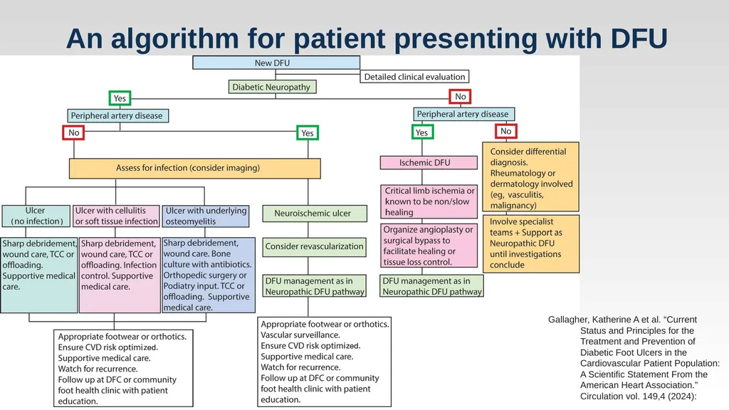
An algorithm for patient presenting with DFU Gallagher, Katherine A et al. “Current Status and Principles for the Treatment and Prevention of Diabetic Foot Ulcers in the Cardiovascular Patient Population: A Scientific Statement From the American Heart Association.” Circulation vol. 149,4 (2024):<br>