
ECLIPSE: A Large-scale, Randomized Trial of
DJ
Published · 26 slides · 0 views
1 / 1
Description
ECLIPSE: A Large-scale, Randomized Trial of Orbital Atherectomy vs. Conventional Balloon Angioplasty in Severely Calcified Coronary Arteries Prior to DES Implantation For the ECLIPSE Investigators Ajay J. Kirtane, MD, SM Financial Conflict
Related Topics
Share
Embed code
Download this presentation From Below
"ECLIPSE: A Large-scale, Randomized Trial of" is the property of its rightful owner. Permission is granted to download and print the materials on this website for personal, non-commercial use only, and to display it on your personal computer provided you do not modify the materials and that you retain all copyright notices contained in the materials. By downloading content from our website, you accept the terms of this agreement.
Presentation Transcript
01
ECLIPSE: A Large-scale, Randomized Trial of Orbital Atherectomy vs. Conventional Balloon Angioplasty in Severely Calcified Coronary Arteries Prior to DES Implantation For the ECLIPSE Investigators Ajay J. Kirtane, MD, SM<br>
02
Financial Conflict of Interest Disclosure Dr. Kirtane reports Institutional funding to Columbia University and/or Cardiovascular Research Foundation from Medtronic, Boston Scientific, Abbott Vascular, Amgen, CathWorks, Concept Medical, Philips, ReCor Medical, Neurotronic, Biotronik, Chiesi, Bolt Medical, Magenta Medical, SoniVie, and Shockwave Medical. In addition to research grants, institutional funding includes fees paid to Columbia University and/or Cardiovascular Research Foundation for consulting and/or speaking engagements in which Dr. Kirtane controlled the content. Personal: Equity options in Bolt Medical, Airiver; Travel Expenses/Meals from Amgen, Medtronic, Biotronik, Boston Scientific, Abbott Vascular, CathWorks, Concept Medical, Novartis, Philips, Abiomed, ReCor Medical, Chiesi, Zoll, Shockwave, and Regeneron<br>
03
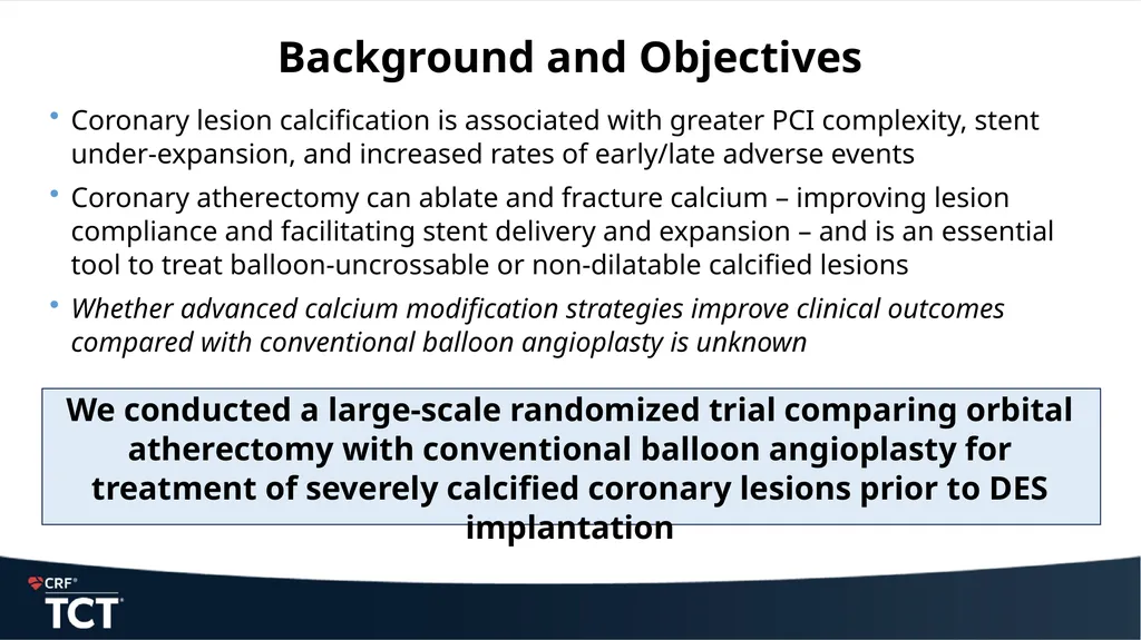
Background and Objectives Coronary lesion calcification is associated with greater PCI complexity, stent under-expansion, and increased rates of early/late adverse events
Coronary atherectomy can ablate and fracture calcium – improving lesion compliance and facilitating stent delivery and expansion – and is an essential tool to treat balloon-uncrossable or non-dilatable calcified lesions
Whether advanced calcium modification strategies improve clinical outcomes compared with conventional balloon angioplasty is unknown
We conducted a large-scale randomized trial comparing orbital atherectomy with conventional balloon angioplasty for treatment of severely calcified coronary lesions prior to DES implantation<br>
Coronary atherectomy can ablate and fracture calcium – improving lesion compliance and facilitating stent delivery and expansion – and is an essential tool to treat balloon-uncrossable or non-dilatable calcified lesions
Whether advanced calcium modification strategies improve clinical outcomes compared with conventional balloon angioplasty is unknown
We conducted a large-scale randomized trial comparing orbital atherectomy with conventional balloon angioplasty for treatment of severely calcified coronary lesions prior to DES implantation<br>
04
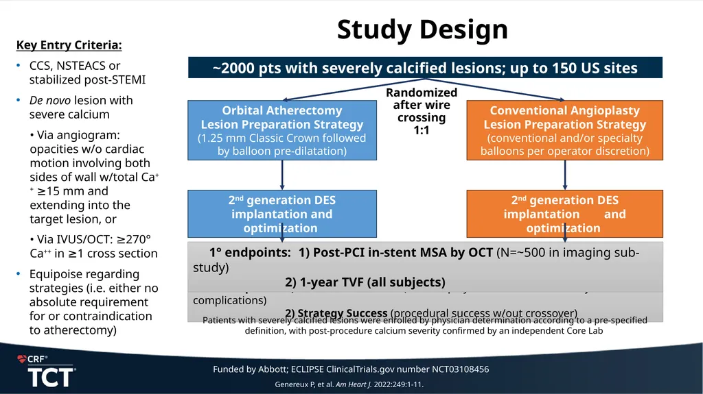
Study Design ~2000 pts with severely calcified lesions; up to 150 US sites Orbital Atherectomy
Lesion Preparation Strategy
(1.25 mm Classic Crown followed by balloon pre-dilatation) Conventional Angioplasty
Lesion Preparation Strategy
(conventional and/or specialty balloons per operator discretion) 2° endpoints: 1) Procedural Success (stent deployed w/RS≤20% & no major complications)
2) Strategy Success (procedural success w/out crossover) Key Entry Criteria:
CCS, NSTEACS or stabilized post-STEMI
De novo lesion with severe calcium
• Via angiogram: opacities w/o cardiac motion involving both sides of wall w/total Ca++ ≥15 mm and extending into the target lesion, or
• Via IVUS/OCT: ≥270° Ca++ in ≥1 cross section
Equipoise regarding strategies (i.e. either no absolute requirement for or contraindication to atherectomy) Randomized
after wire crossing
1:1 Patients with severely calcified lesions were enrolled by physician determination according to a pre-specified definition, with post-procedure calcium severity confirmed by an independent Core Lab Genereux P, et al. Am Heart J. 2022:249:1-11. Funded by Abbott; ECLIPSE ClinicalTrials.gov number NCT03108456<br>
Lesion Preparation Strategy
(1.25 mm Classic Crown followed by balloon pre-dilatation) Conventional Angioplasty
Lesion Preparation Strategy
(conventional and/or specialty balloons per operator discretion) 2° endpoints: 1) Procedural Success (stent deployed w/RS≤20% & no major complications)
2) Strategy Success (procedural success w/out crossover) Key Entry Criteria:
CCS, NSTEACS or stabilized post-STEMI
De novo lesion with severe calcium
• Via angiogram: opacities w/o cardiac motion involving both sides of wall w/total Ca++ ≥15 mm and extending into the target lesion, or
• Via IVUS/OCT: ≥270° Ca++ in ≥1 cross section
Equipoise regarding strategies (i.e. either no absolute requirement for or contraindication to atherectomy) Randomized
after wire crossing
1:1 Patients with severely calcified lesions were enrolled by physician determination according to a pre-specified definition, with post-procedure calcium severity confirmed by an independent Core Lab Genereux P, et al. Am Heart J. 2022:249:1-11. Funded by Abbott; ECLIPSE ClinicalTrials.gov number NCT03108456<br>
05
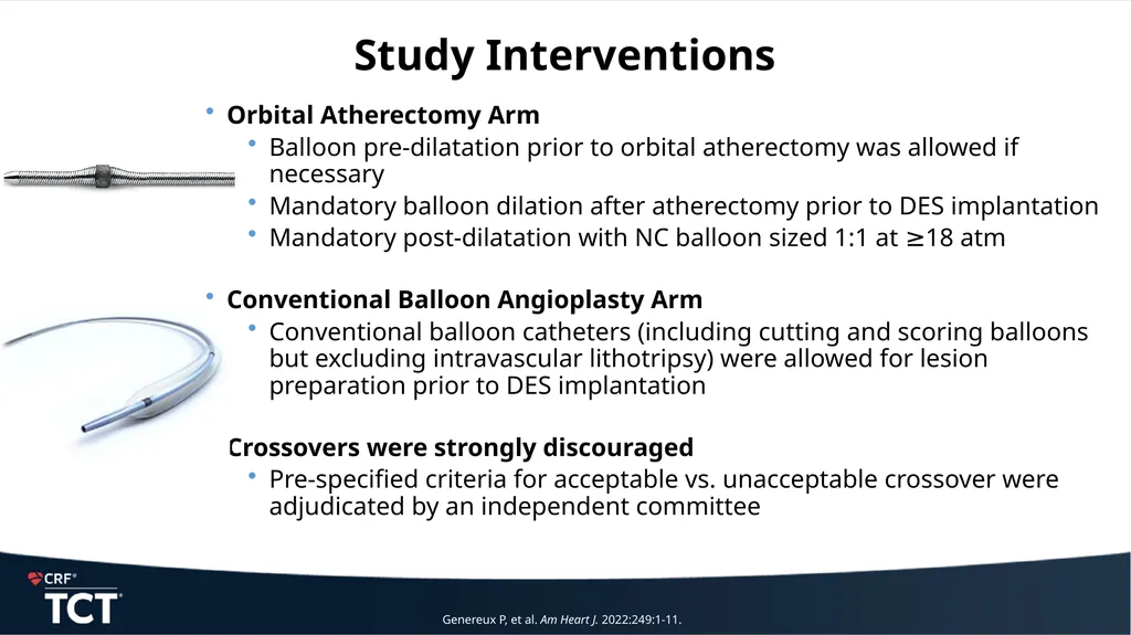
Orbital Atherectomy Arm
Balloon pre-dilatation prior to orbital atherectomy was allowed if necessary
Mandatory balloon dilation after atherectomy prior to DES implantation
Mandatory post-dilatation with NC balloon sized 1:1 at ≥18 atm
Conventional Balloon Angioplasty Arm
Conventional balloon catheters (including cutting and scoring balloons but excluding intravascular lithotripsy) were allowed for lesion preparation prior to DES implantation
Crossovers were strongly discouraged
Pre-specified criteria for acceptable vs. unacceptable crossover were adjudicated by an independent committee Study Interventions Genereux P, et al. Am Heart J. 2022:249:1-11.<br>
Balloon pre-dilatation prior to orbital atherectomy was allowed if necessary
Mandatory balloon dilation after atherectomy prior to DES implantation
Mandatory post-dilatation with NC balloon sized 1:1 at ≥18 atm
Conventional Balloon Angioplasty Arm
Conventional balloon catheters (including cutting and scoring balloons but excluding intravascular lithotripsy) were allowed for lesion preparation prior to DES implantation
Crossovers were strongly discouraged
Pre-specified criteria for acceptable vs. unacceptable crossover were adjudicated by an independent committee Study Interventions Genereux P, et al. Am Heart J. 2022:249:1-11.<br>
06
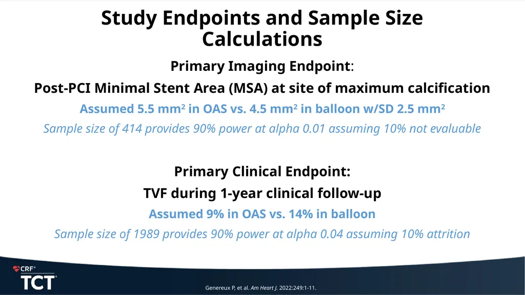
Study Endpoints and Sample Size Calculations Primary Imaging Endpoint:
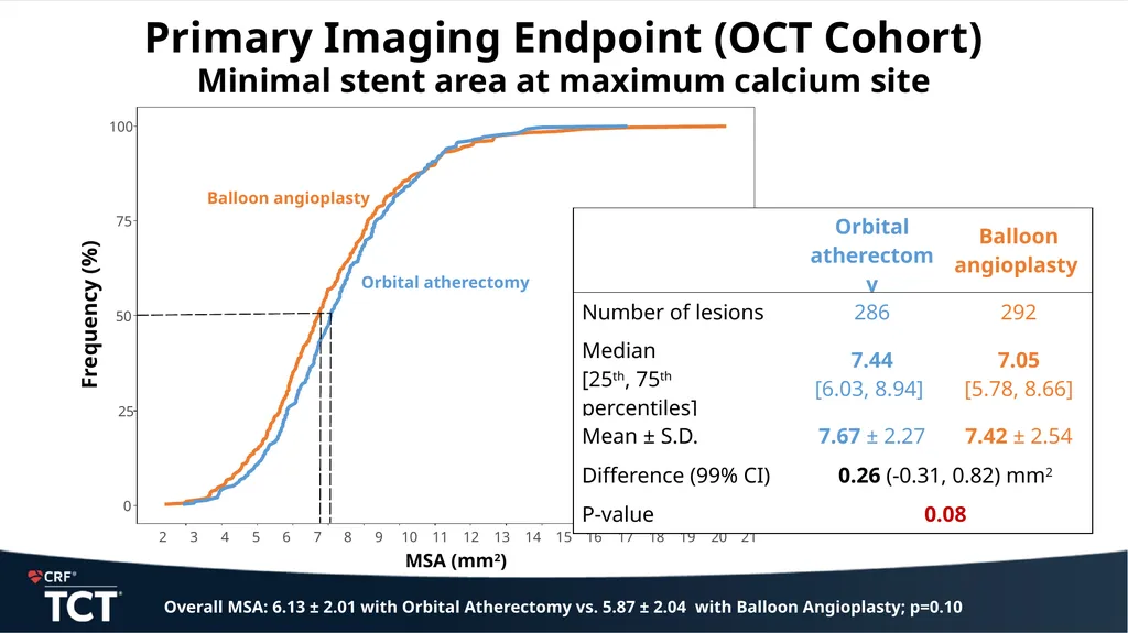
Post-PCI Minimal Stent Area (MSA) at site of maximum calcification
Assumed 5.5 mm2 in OAS vs. 4.5 mm2 in balloon w/SD 2.5 mm2
Sample size of 414 provides 90% power at alpha 0.01 assuming 10% not evaluable
Primary Clinical Endpoint:
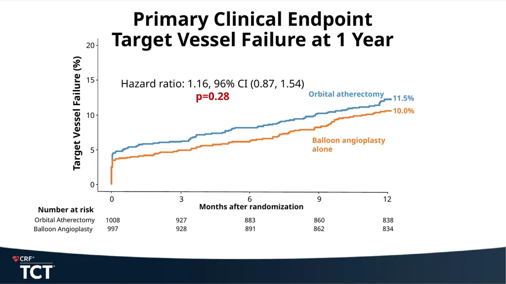
TVF during 1-year clinical follow-up
Assumed 9% in OAS vs. 14% in balloon
Sample size of 1989 provides 90% power at alpha 0.04 assuming 10% attrition Genereux P, et al. Am Heart J. 2022:249:1-11.<br>
Post-PCI Minimal Stent Area (MSA) at site of maximum calcification
Assumed 5.5 mm2 in OAS vs. 4.5 mm2 in balloon w/SD 2.5 mm2
Sample size of 414 provides 90% power at alpha 0.01 assuming 10% not evaluable
Primary Clinical Endpoint:
TVF during 1-year clinical follow-up
Assumed 9% in OAS vs. 14% in balloon
Sample size of 1989 provides 90% power at alpha 0.04 assuming 10% attrition Genereux P, et al. Am Heart J. 2022:249:1-11.<br>
07
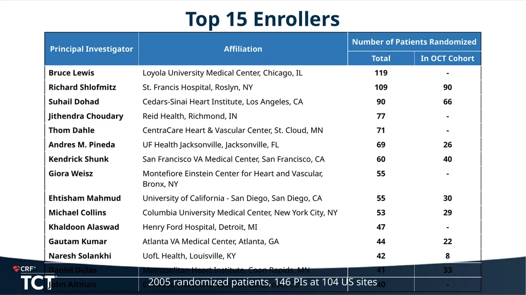
Top 15 Enrollers 2005 randomized patients, 146 PIs at 104 US sites<br>
08
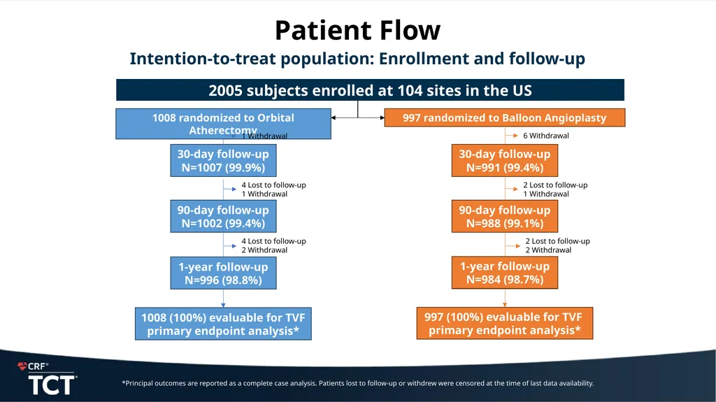
Patient Flow Intention-to-treat population: Enrollment and follow-up 2005 subjects enrolled at 104 sites in the US 1008 randomized to Orbital Atherectomy 30-day follow-up
N=1007 (99.9%) 997 randomized to Balloon Angioplasty 30-day follow-up
N=991 (99.4%) 90-day follow-up
N=1002 (99.4%) 1-year follow-up
N=996 (98.8%) 1008 (100%) evaluable for TVF
primary endpoint analysis* *Principal outcomes are reported as a complete case analysis. Patients lost to follow-up or withdrew were censored at the time of last data availability. 1 Withdrawal 90-day follow-up
N=988 (99.1%) 1-year follow-up
N=984 (98.7%) 997 (100%) evaluable for TVF
primary endpoint analysis* 4 Lost to follow-up
1 Withdrawal 4 Lost to follow-up
2 Withdrawal 6 Withdrawal 2 Lost to follow-up
1 Withdrawal 2 Lost to follow-up
2 Withdrawal<br>
N=1007 (99.9%) 997 randomized to Balloon Angioplasty 30-day follow-up
N=991 (99.4%) 90-day follow-up
N=1002 (99.4%) 1-year follow-up
N=996 (98.8%) 1008 (100%) evaluable for TVF
primary endpoint analysis* *Principal outcomes are reported as a complete case analysis. Patients lost to follow-up or withdrew were censored at the time of last data availability. 1 Withdrawal 90-day follow-up
N=988 (99.1%) 1-year follow-up
N=984 (98.7%) 997 (100%) evaluable for TVF
primary endpoint analysis* 4 Lost to follow-up
1 Withdrawal 4 Lost to follow-up
2 Withdrawal 6 Withdrawal 2 Lost to follow-up
1 Withdrawal 2 Lost to follow-up
2 Withdrawal<br>
09
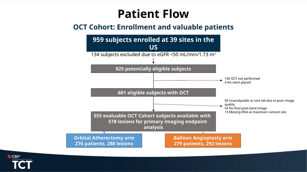
Patient Flow OCT Cohort: Enrollment and valuable patients 959 subjects enrolled at 39 sites in the US 825 potentially eligible subjects 681 eligible subjects with OCT 555 evaluable OCT Cohort subjects available with 578 lesions for primary imaging endpoint analysis Orbital Atherectomy arm
276 patients, 286 lesions Balloon Angioplasty arm
279 patients, 292 lesions 140 OCT not performed
4 No stent placed 59 Unanalyzable at core lab due to poor image quality
54 No final post-stent image
13 Missing MSA at maximum calcium site 134 subjects excluded due to eGFR <50 mL/min/1.73 m2<br>
276 patients, 286 lesions Balloon Angioplasty arm
279 patients, 292 lesions 140 OCT not performed
4 No stent placed 59 Unanalyzable at core lab due to poor image quality
54 No final post-stent image
13 Missing MSA at maximum calcium site 134 subjects excluded due to eGFR <50 mL/min/1.73 m2<br>
10
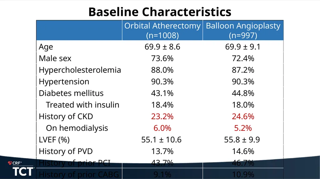
Baseline Characteristics<br>
11
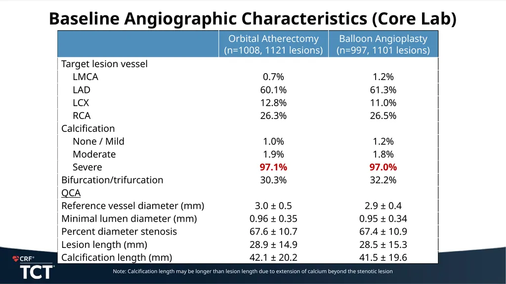
Baseline Angiographic Characteristics (Core Lab) Note: Calcification length may be longer than lesion length due to extension of calcium beyond the stenotic lesion<br>
12
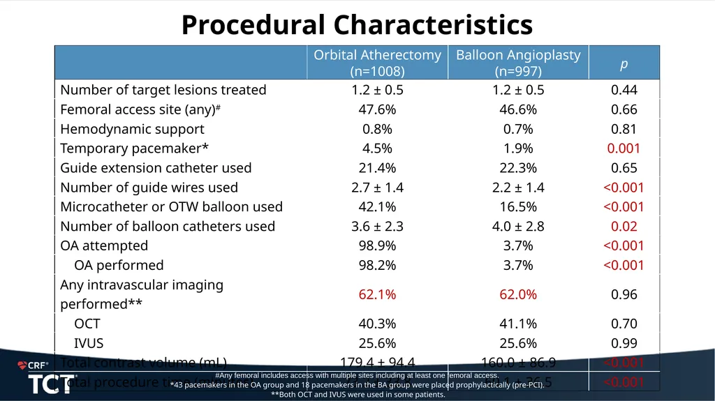
Procedural Characteristics #Any femoral includes access with multiple sites including at least one femoral access.
*43 pacemakers in the OA group and 18 pacemakers in the BA group were placed prophylactically (pre-PCI).
**Both OCT and IVUS were used in some patients.<br>
*43 pacemakers in the OA group and 18 pacemakers in the BA group were placed prophylactically (pre-PCI).
**Both OCT and IVUS were used in some patients.<br>
13
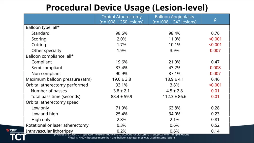
Procedural Device Usage (Lesion-level) p-values are based on repeated measures modeling to account for clustering in subjects with multiple lesions
*Total is >100% because more than one balloon catheter type was used in some lesions<br>
*Total is >100% because more than one balloon catheter type was used in some lesions<br>
14
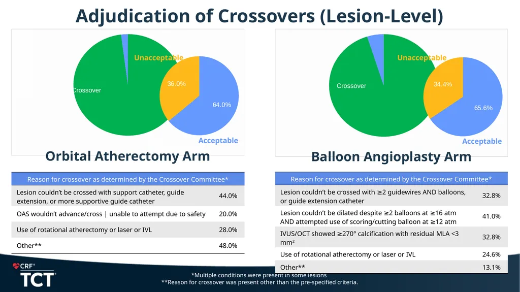
Adjudication of Crossovers (Lesion-Level) *Multiple conditions were present in some lesions
**Reason for crossover was present other than the pre-specified criteria. Crossover Crossover<br>
**Reason for crossover was present other than the pre-specified criteria. Crossover Crossover<br>
15
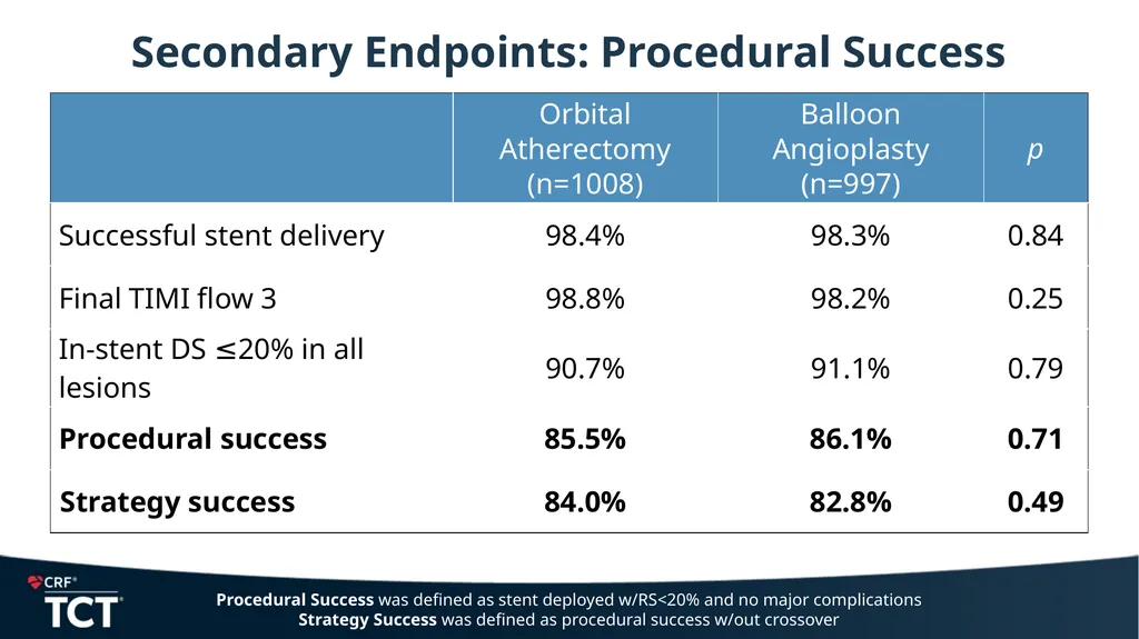
Secondary Endpoints: Procedural Success Procedural Success was defined as stent deployed w/RS<20% and no major complications
Strategy Success was defined as procedural success w/out crossover<br>
Strategy Success was defined as procedural success w/out crossover<br>
16
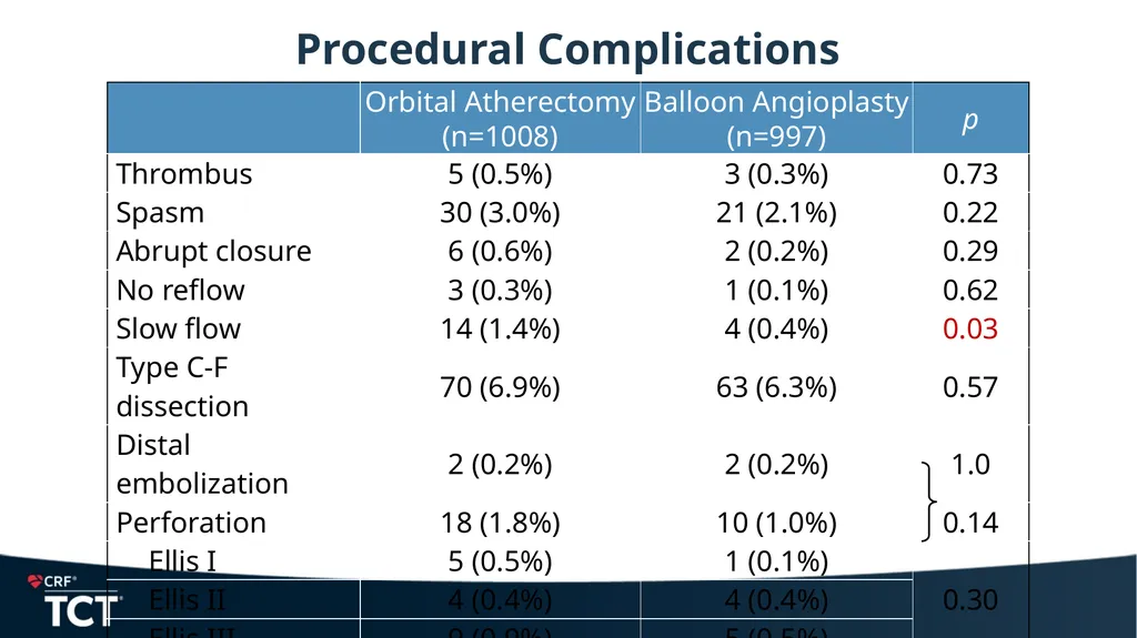
Procedural Complications<br>
17
Primary Imaging Endpoint (OCT Cohort) 0 25 50 75 100 2 3 4 5 6 7 8 9 10 11 12 13 14 15 16 17 18 19 20 21 MSA (mm2) Frequency (%) Balloon angioplasty Orbital atherectomy Minimal stent area at maximum calcium site Overall MSA: 6.13 ± 2.01 with Orbital Atherectomy vs. 5.87 ± 2.04 with Balloon Angioplasty; p=0.10<br>
18
Primary Clinical Endpoint
Target Vessel Failure at 1 Year 0 3 6 9 12 0 5 10 15 20 Target Vessel Failure (%) Hazard ratio: 1.16, 96% CI (0.87, 1.54)
p=0.28<br>
p=0.28<br>
19
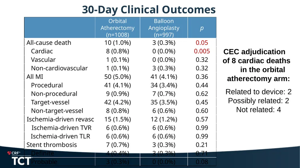
30-Day Clinical Outcomes CEC adjudication of 8 cardiac deaths in the orbital atherectomy arm:
Related to device: 2
Possibly related: 2
Not related: 4<br>
Related to device: 2
Possibly related: 2
Not related: 4<br>
20
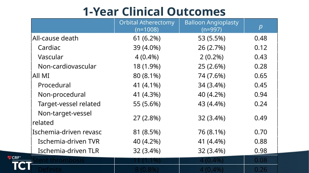
1-Year Clinical Outcomes<br>
21
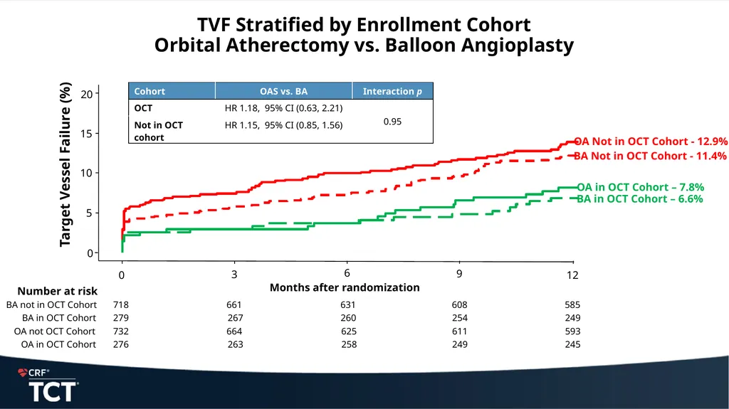
718 661 631 608 585 BA not in OCT Cohort 279 267 260 254 249 BA in OCT Cohort Number at risk Months after randomization 732 664 625 611 593 OA not OCT Cohort 276 263 258 249 245 OA in OCT Cohort TVF Stratified by Enrollment Cohort
Orbital Atherectomy vs. Balloon Angioplasty Target Vessel Failure (%) 20 15 10 5 12 9 6 3 0 0<br>
22
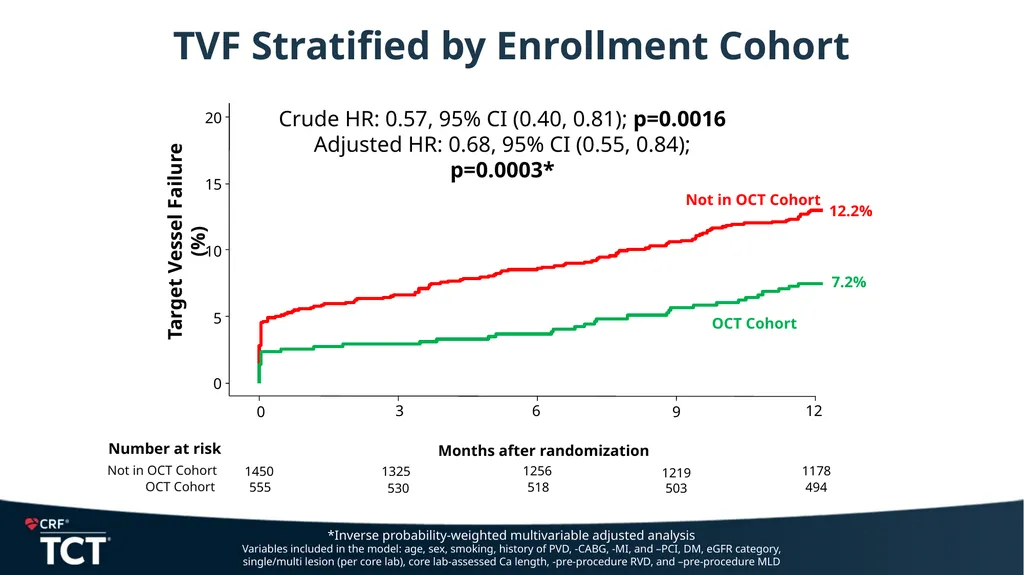
1450 1325 1256 1219 1178 555 530 518 503 494 OCT Cohort Not OCT Cohort Number at risk Months after randomization Target Vessel Failure (%) 0 5 10 15 20 0 3 6 9 12 OCT Cohort Not OCT Cohort OCT Cohort Not in OCT Cohort Crude HR: 0.57, 95% CI (0.40, 0.81); p=0.0016
Adjusted HR: 0.68, 95% CI (0.55, 0.84); p=0.0003* *Inverse probability-weighted multivariable adjusted analysis
Variables included in the model: age, sex, smoking, history of PVD, -CABG, -MI, and –PCI, DM, eGFR category, single/multi lesion (per core lab), core lab-assessed Ca length, -pre-procedure RVD, and –pre-procedure MLD TVF Stratified by Enrollment Cohort<br>
Adjusted HR: 0.68, 95% CI (0.55, 0.84); p=0.0003* *Inverse probability-weighted multivariable adjusted analysis
Variables included in the model: age, sex, smoking, history of PVD, -CABG, -MI, and –PCI, DM, eGFR category, single/multi lesion (per core lab), core lab-assessed Ca length, -pre-procedure RVD, and –pre-procedure MLD TVF Stratified by Enrollment Cohort<br>
23
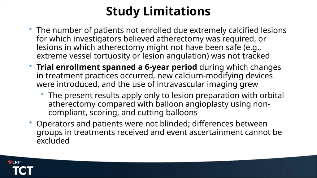
Study Limitations The number of patients not enrolled due extremely calcified lesions for which investigators believed atherectomy was required, or lesions in which atherectomy might not have been safe (e.g., extreme vessel tortuosity or lesion angulation) was not tracked
Trial enrollment spanned a 6-year period during which changes in treatment practices occurred, new calcium-modifying devices were introduced, and the use of intravascular imaging grew
The present results apply only to lesion preparation with orbital atherectomy compared with balloon angioplasty using non-compliant, scoring, and cutting balloons
Operators and patients were not blinded; differences between groups in treatments received and event ascertainment cannot be excluded<br>
Trial enrollment spanned a 6-year period during which changes in treatment practices occurred, new calcium-modifying devices were introduced, and the use of intravascular imaging grew
The present results apply only to lesion preparation with orbital atherectomy compared with balloon angioplasty using non-compliant, scoring, and cutting balloons
Operators and patients were not blinded; differences between groups in treatments received and event ascertainment cannot be excluded<br>
24
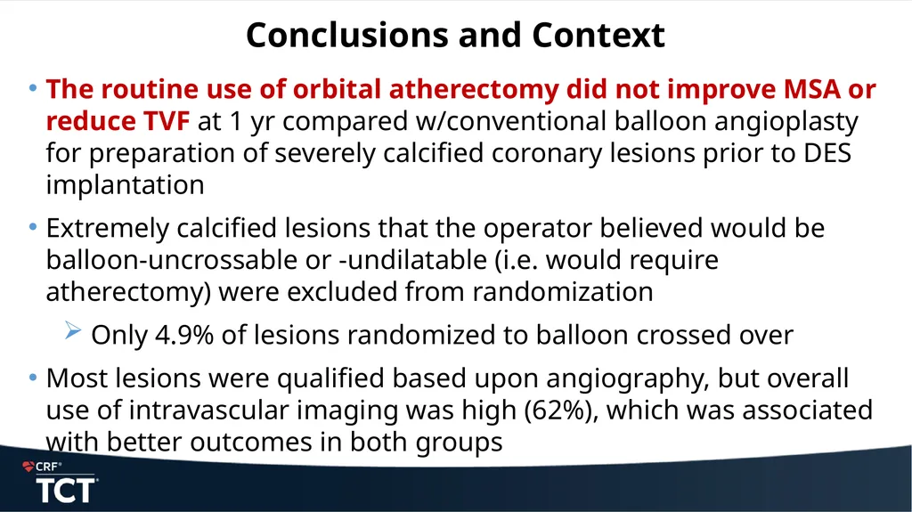
Conclusions and Context The routine use of orbital atherectomy did not improve MSA or reduce TVF at 1 yr compared w/conventional balloon angioplasty for preparation of severely calcified coronary lesions prior to DES implantation
Extremely calcified lesions that the operator believed would be balloon-uncrossable or -undilatable (i.e. would require atherectomy) were excluded from randomization
Only 4.9% of lesions randomized to balloon crossed over
Most lesions were qualified based upon angiography, but overall use of intravascular imaging was high (62%), which was associated with better outcomes in both groups<br>
Extremely calcified lesions that the operator believed would be balloon-uncrossable or -undilatable (i.e. would require atherectomy) were excluded from randomization
Only 4.9% of lesions randomized to balloon crossed over
Most lesions were qualified based upon angiography, but overall use of intravascular imaging was high (62%), which was associated with better outcomes in both groups<br>
25
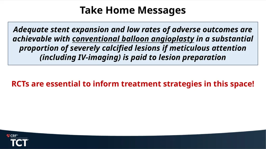
Take Home Messages Adequate stent expansion and low rates of adverse outcomes are achievable with conventional balloon angioplasty in a substantial proportion of severely calcified lesions if meticulous attention (including IV-imaging) is paid to lesion preparation
RCTs are essential to inform treatment strategies in this space!<br>
RCTs are essential to inform treatment strategies in this space!<br>
26
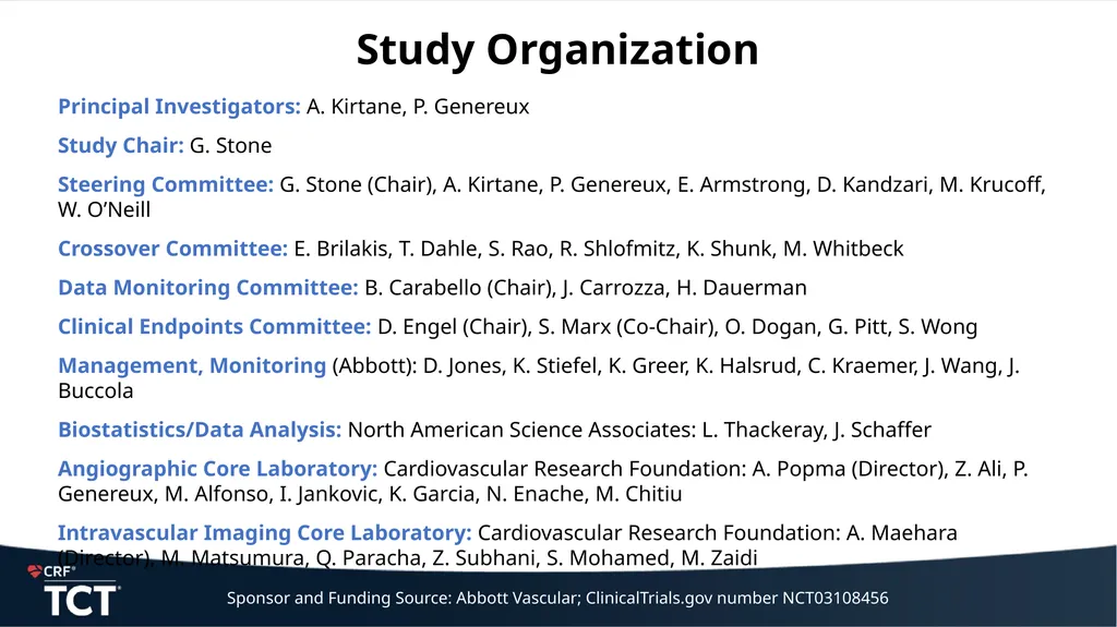
Study Organization Principal Investigators: A. Kirtane, P. Genereux
Study Chair: G. Stone
Steering Committee: G. Stone (Chair), A. Kirtane, P. Genereux, E. Armstrong, D. Kandzari, M. Krucoff, W. O’Neill
Crossover Committee: E. Brilakis, T. Dahle, S. Rao, R. Shlofmitz, K. Shunk, M. Whitbeck
Data Monitoring Committee: B. Carabello (Chair), J. Carrozza, H. Dauerman
Clinical Endpoints Committee: D. Engel (Chair), S. Marx (Co-Chair), O. Dogan, G. Pitt, S. Wong
Management, Monitoring (Abbott): D. Jones, K. Stiefel, K. Greer, K. Halsrud, C. Kraemer, J. Wang, J. Buccola
Biostatistics/Data Analysis: North American Science Associates: L. Thackeray, J. Schaffer
Angiographic Core Laboratory: Cardiovascular Research Foundation: A. Popma (Director), Z. Ali, P. Genereux, M. Alfonso, I. Jankovic, K. Garcia, N. Enache, M. Chitiu
Intravascular Imaging Core Laboratory: Cardiovascular Research Foundation: A. Maehara (Director), M. Matsumura, Q. Paracha, Z. Subhani, S. Mohamed, M. Zaidi Sponsor and Funding Source: Abbott Vascular; ClinicalTrials.gov number NCT03108456<br>
Study Chair: G. Stone
Steering Committee: G. Stone (Chair), A. Kirtane, P. Genereux, E. Armstrong, D. Kandzari, M. Krucoff, W. O’Neill
Crossover Committee: E. Brilakis, T. Dahle, S. Rao, R. Shlofmitz, K. Shunk, M. Whitbeck
Data Monitoring Committee: B. Carabello (Chair), J. Carrozza, H. Dauerman
Clinical Endpoints Committee: D. Engel (Chair), S. Marx (Co-Chair), O. Dogan, G. Pitt, S. Wong
Management, Monitoring (Abbott): D. Jones, K. Stiefel, K. Greer, K. Halsrud, C. Kraemer, J. Wang, J. Buccola
Biostatistics/Data Analysis: North American Science Associates: L. Thackeray, J. Schaffer
Angiographic Core Laboratory: Cardiovascular Research Foundation: A. Popma (Director), Z. Ali, P. Genereux, M. Alfonso, I. Jankovic, K. Garcia, N. Enache, M. Chitiu
Intravascular Imaging Core Laboratory: Cardiovascular Research Foundation: A. Maehara (Director), M. Matsumura, Q. Paracha, Z. Subhani, S. Mohamed, M. Zaidi Sponsor and Funding Source: Abbott Vascular; ClinicalTrials.gov number NCT03108456<br>