
Electron Microscopy What is Electron Microscopy?

Description: Electron Microscopy What is Electron Microscopy? Electron microscopy (EM) is a technique for obtaining high resolution images of biological and non-biological specimens. It is used in biomedical research to investigate the detailed
Related Topics
Download Presentation
"Electron Microscopy What is Electron Microscopy?" is the property of its rightful owner. Permission is granted to download and print the materials on this website for personal, non-commercial use only, and to display it on your personal computer provided you do not modify the materials and that you retain all copyright notices contained in the materials. By downloading content from our website, you accept the terms of this agreement.
Presentation Transcript
slide1. Electron Microscopy<br>
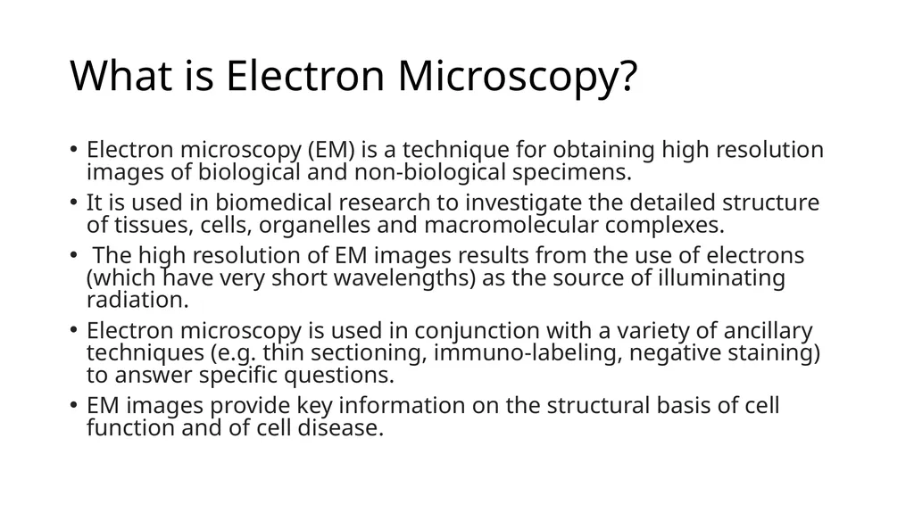
slide2. What is Electron Microscopy? Electron microscopy (EM) is a technique for obtaining high resolution images of biological and non-biological specimens.
It is used in biomedical research to investigate the detailed structure of tissues, cells, organelles and macromolecular complexes.
The high resolution of EM images results from the use of electrons (which have very short wavelengths) as the source of illuminating radiation.
Electron microscopy is used in conjunction with a variety of ancillary techniques (e.g. thin sectioning, immuno-labeling, negative staining) to answer specific questions.
EM images provide key information on the structural basis of cell function and of cell disease.<br>
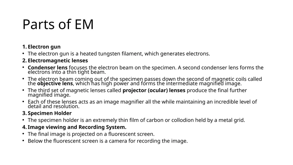
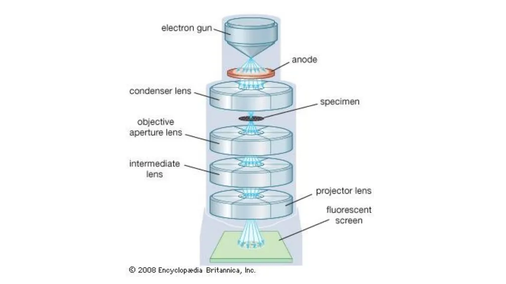
slide3. Parts of EM Electron gun
The electron gun is a heated tungsten filament, which generates electrons.
Electromagnetic lenses
Condenser lens focuses the electron beam on the specimen. A second condenser lens forms the electrons into a thin tight beam.
The electron beam coming out of the specimen passes down the second of magnetic coils called the objective lens, which has high power and forms the intermediate magnified image.
The third set of magnetic lenses called projector (ocular) lenses produce the final further magnified image.
Each of these lenses acts as an image magnifier all the while maintaining an incredible level of detail and resolution.
Specimen Holder
The specimen holder is an extremely thin film of carbon or collodion held by a metal grid.
Image viewing and Recording System.
The final image is projected on a fluorescent screen.
Below the fluorescent screen is a camera for recording the image.<br>
slide4. Types of Electron Microscope There are two main types of electron microscope – the transmission EM (TEM) and the scanning EM (SEM).
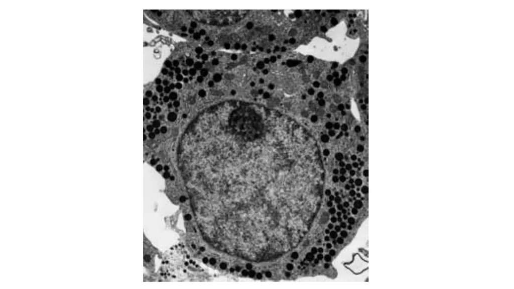
TEM: The transmission electron microscope is used to view thin specimens (tissue sections, molecules, etc) through which electrons can pass generating a projection image.
The TEM is analogous in many ways to the conventional (compound) light microscope.
TEM is used, among other things, to image the interior of cells (in thin sections), the structure of protein molecules (contrasted by metal shadowing), the organization of molecules in viruses and cytoskeletal filaments (prepared by the negative staining technique), and the arrangement of protein molecules in cell membranes (by freeze-fracture).<br>
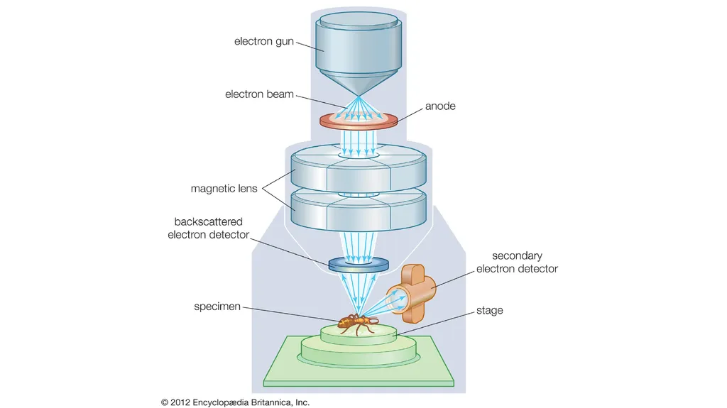
slide7. SEM: Scanning electron microscopy depends on the emission of secondary electrons from the surface of a specimen.
The image by SEM is formed by scanning a focused electron beam onto the surface of the specimen in a raster pattern. The interaction of the primary electron beam with the atoms near the surface causes the emission of particles at each point in the raster (e.g., low energy secondary electrons, high energy back scatter electrons, X-rays and even photons). These can be collected with a variety of detectors, and their relative number translated to on a cathode ray tube that gives a magnified the final picture of the specimen.
Because of its great depth of focus, a scanning electron microscope is the EM analog of a stereo light microscope.
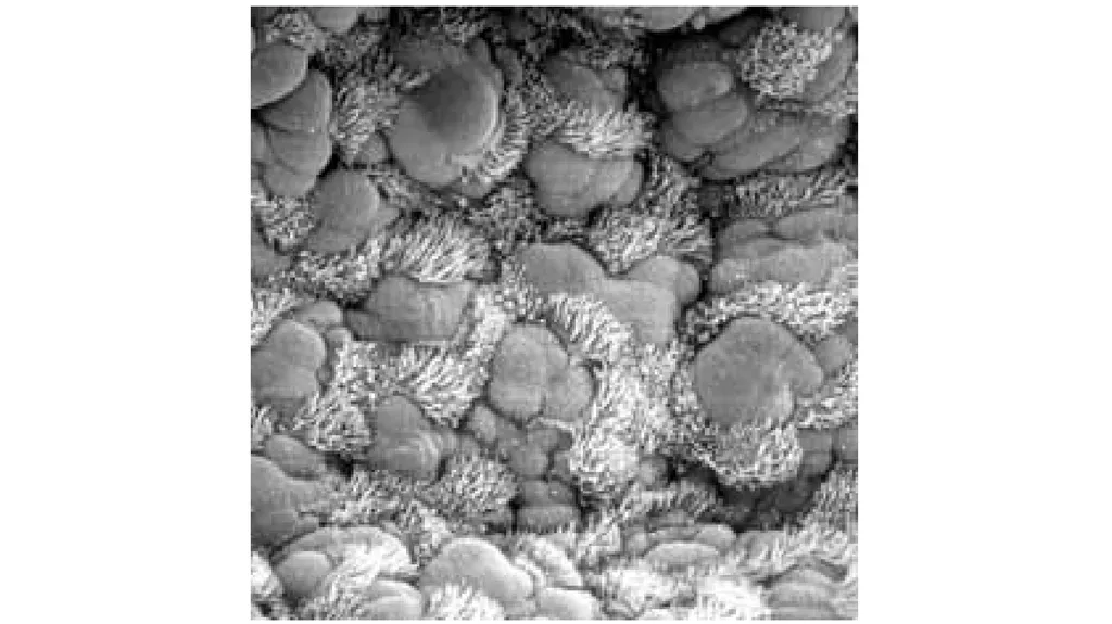
It provides detailed images of the surfaces of cells and whole organisms that are not possible by TEM.
It can also be used for particle counting and size determination, and for process control. Appropriately equipped SEMs (with secondary, backscatter and X-ray detectors) can be used to study the topography and atomic composition of specimens, and also, for example, the surface distribution of immuno-labels.<br>
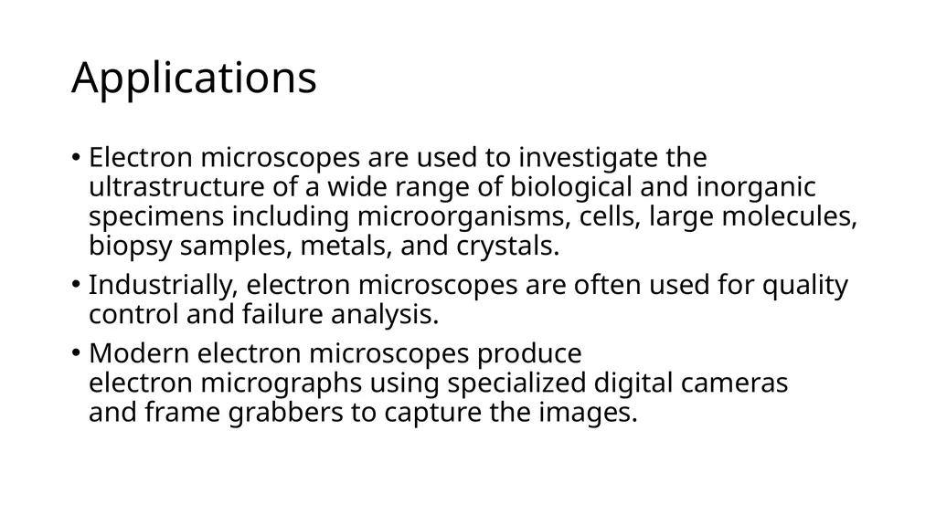
slide10. Applications Electron microscopes are used to investigate the ultrastructure of a wide range of biological and inorganic specimens including microorganisms, cells, large molecules, biopsy samples, metals, and crystals.
Industrially, electron microscopes are often used for quality control and failure analysis.
Modern electron microscopes produce electron micrographs using specialized digital cameras and frame grabbers to capture the images.<br>
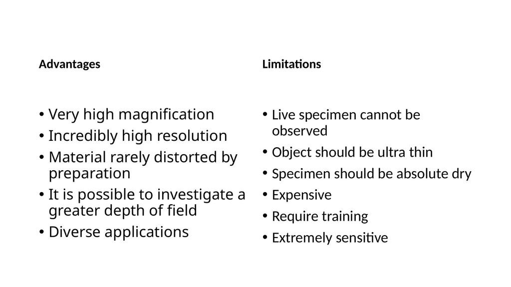
slide11. Advantages Very high magnification
Incredibly high resolution
Material rarely distorted by preparation
It is possible to investigate a greater depth of field
Diverse applications Limitations Live specimen cannot be observed
Object should be ultra thin
Specimen should be absolute dry
Expensive
Require training
Extremely sensitive<br>
slide12. https://www.youtube.com/watch?v=t4hhdgJADE8
https://www.youtube.com/watch?v=ljTEG-B-kGc<br>
slide2. What is Electron Microscopy? Electron microscopy (EM) is a technique for obtaining high resolution images of biological and non-biological specimens.
It is used in biomedical research to investigate the detailed structure of tissues, cells, organelles and macromolecular complexes.
The high resolution of EM images results from the use of electrons (which have very short wavelengths) as the source of illuminating radiation.
Electron microscopy is used in conjunction with a variety of ancillary techniques (e.g. thin sectioning, immuno-labeling, negative staining) to answer specific questions.
EM images provide key information on the structural basis of cell function and of cell disease.<br>
slide3. Parts of EM Electron gun
The electron gun is a heated tungsten filament, which generates electrons.
Electromagnetic lenses
Condenser lens focuses the electron beam on the specimen. A second condenser lens forms the electrons into a thin tight beam.
The electron beam coming out of the specimen passes down the second of magnetic coils called the objective lens, which has high power and forms the intermediate magnified image.
The third set of magnetic lenses called projector (ocular) lenses produce the final further magnified image.
Each of these lenses acts as an image magnifier all the while maintaining an incredible level of detail and resolution.
Specimen Holder
The specimen holder is an extremely thin film of carbon or collodion held by a metal grid.
Image viewing and Recording System.
The final image is projected on a fluorescent screen.
Below the fluorescent screen is a camera for recording the image.<br>
slide4. Types of Electron Microscope There are two main types of electron microscope – the transmission EM (TEM) and the scanning EM (SEM).
TEM: The transmission electron microscope is used to view thin specimens (tissue sections, molecules, etc) through which electrons can pass generating a projection image.
The TEM is analogous in many ways to the conventional (compound) light microscope.
TEM is used, among other things, to image the interior of cells (in thin sections), the structure of protein molecules (contrasted by metal shadowing), the organization of molecules in viruses and cytoskeletal filaments (prepared by the negative staining technique), and the arrangement of protein molecules in cell membranes (by freeze-fracture).<br>
slide7. SEM: Scanning electron microscopy depends on the emission of secondary electrons from the surface of a specimen.
The image by SEM is formed by scanning a focused electron beam onto the surface of the specimen in a raster pattern. The interaction of the primary electron beam with the atoms near the surface causes the emission of particles at each point in the raster (e.g., low energy secondary electrons, high energy back scatter electrons, X-rays and even photons). These can be collected with a variety of detectors, and their relative number translated to on a cathode ray tube that gives a magnified the final picture of the specimen.
Because of its great depth of focus, a scanning electron microscope is the EM analog of a stereo light microscope.
It provides detailed images of the surfaces of cells and whole organisms that are not possible by TEM.
It can also be used for particle counting and size determination, and for process control. Appropriately equipped SEMs (with secondary, backscatter and X-ray detectors) can be used to study the topography and atomic composition of specimens, and also, for example, the surface distribution of immuno-labels.<br>
slide10. Applications Electron microscopes are used to investigate the ultrastructure of a wide range of biological and inorganic specimens including microorganisms, cells, large molecules, biopsy samples, metals, and crystals.
Industrially, electron microscopes are often used for quality control and failure analysis.
Modern electron microscopes produce electron micrographs using specialized digital cameras and frame grabbers to capture the images.<br>
slide11. Advantages Very high magnification
Incredibly high resolution
Material rarely distorted by preparation
It is possible to investigate a greater depth of field
Diverse applications Limitations Live specimen cannot be observed
Object should be ultra thin
Specimen should be absolute dry
Expensive
Require training
Extremely sensitive<br>
slide12. https://www.youtube.com/watch?v=t4hhdgJADE8
https://www.youtube.com/watch?v=ljTEG-B-kGc<br>