
EMBRYOLOGY-I ا.م.د.ورقاء محمود علي INTRODUCTION:
A
Published · 25 slides · 0 views
1 / 1

Description
EMBRYOLOGY-I ا.م.د.ورقاء محمود علي INTRODUCTION: The human embryo is developed from fertilization of the ovum with sperm which has 23 chromosomes for each After fertilization of the ovum(ZYGOTE); there will be restore to the normal
Related Topics
Share
Embed code
Download this presentation From Below
"EMBRYOLOGY-I ا.م.د.ورقاء محمود علي INTRODUCTION:" is the property of its rightful owner. Permission is granted to download and print the materials on this website for personal, non-commercial use only, and to display it on your personal computer provided you do not modify the materials and that you retain all copyright notices contained in the materials. By downloading content from our website, you accept the terms of this agreement.
Presentation Transcript
01
EMBRYOLOGY-I ا.م.د.ورقاء محمود علي<br>
02
INTRODUCTION: The human embryo is developed from fertilization of the ovum with sperm which has 23 chromosomes for each
After fertilization of the ovum(ZYGOTE); there will be restore to the normal chromosome number of somatic cells which is 46 , a series of cell divisions gives rise to an egg cell mass known as the morula in mammals<br>
After fertilization of the ovum(ZYGOTE); there will be restore to the normal chromosome number of somatic cells which is 46 , a series of cell divisions gives rise to an egg cell mass known as the morula in mammals<br>
03
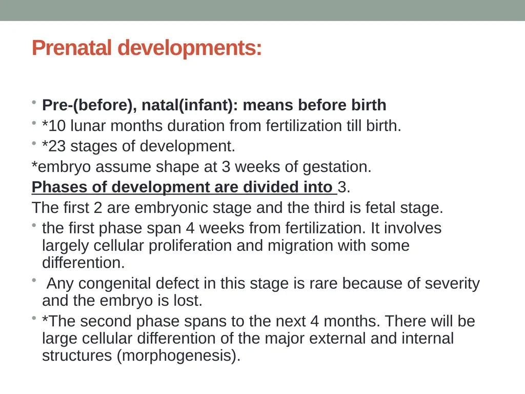
Prenatal developments: Pre-(before), natal(infant): means before birth
*10 lunar months duration from fertilization till birth.
*23 stages of development.
*embryo assume shape at 3 weeks of gestation.
Phases of development are divided into 3.
The first 2 are embryonic stage and the third is fetal stage.
the first phase span 4 weeks from fertilization. It involves largely cellular proliferation and migration with some differention.
Any congenital defect in this stage is rare because of severity and the embryo is lost.
*The second phase spans to the next 4 months. There will be large cellular differention of the major external and internal structures (morphogenesis).<br>
*10 lunar months duration from fertilization till birth.
*23 stages of development.
*embryo assume shape at 3 weeks of gestation.
Phases of development are divided into 3.
The first 2 are embryonic stage and the third is fetal stage.
the first phase span 4 weeks from fertilization. It involves largely cellular proliferation and migration with some differention.
Any congenital defect in this stage is rare because of severity and the embryo is lost.
*The second phase spans to the next 4 months. There will be large cellular differention of the major external and internal structures (morphogenesis).<br>
04
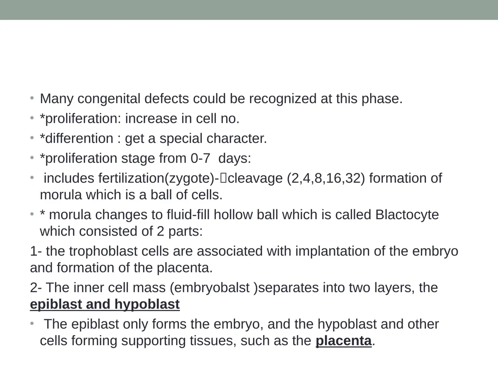
Many congenital defects could be recognized at this phase.
*proliferation: increase in cell no.
*differention : get a special character.
*proliferation stage from 0-7 days:
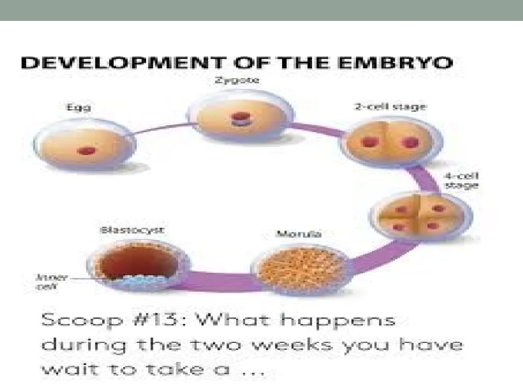
includes fertilization(zygote)-cleavage (2,4,8,16,32) formation of morula which is a ball of cells.
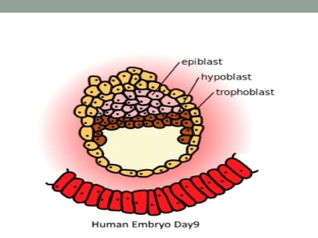
* morula changes to fluid-fill hollow ball which is called Blactocyte which consisted of 2 parts:
1- the trophoblast cells are associated with implantation of the embryo and formation of the placenta.
2- The inner cell mass (embryobalst )separates into two layers, the epiblast and hypoblast
The epiblast only forms the embryo, and the hypoblast and other cells forming supporting tissues, such as the placenta.<br>
*proliferation: increase in cell no.
*differention : get a special character.
*proliferation stage from 0-7 days:
includes fertilization(zygote)-cleavage (2,4,8,16,32) formation of morula which is a ball of cells.
* morula changes to fluid-fill hollow ball which is called Blactocyte which consisted of 2 parts:
1- the trophoblast cells are associated with implantation of the embryo and formation of the placenta.
2- The inner cell mass (embryobalst )separates into two layers, the epiblast and hypoblast
The epiblast only forms the embryo, and the hypoblast and other cells forming supporting tissues, such as the placenta.<br>
05
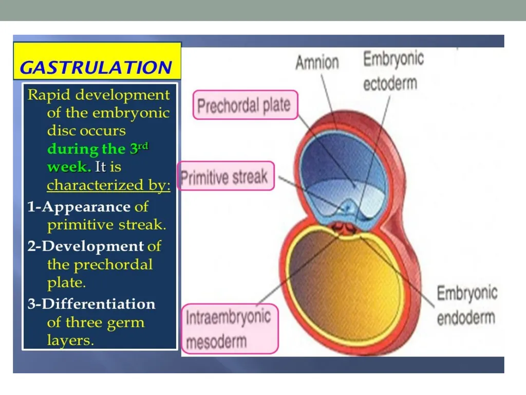
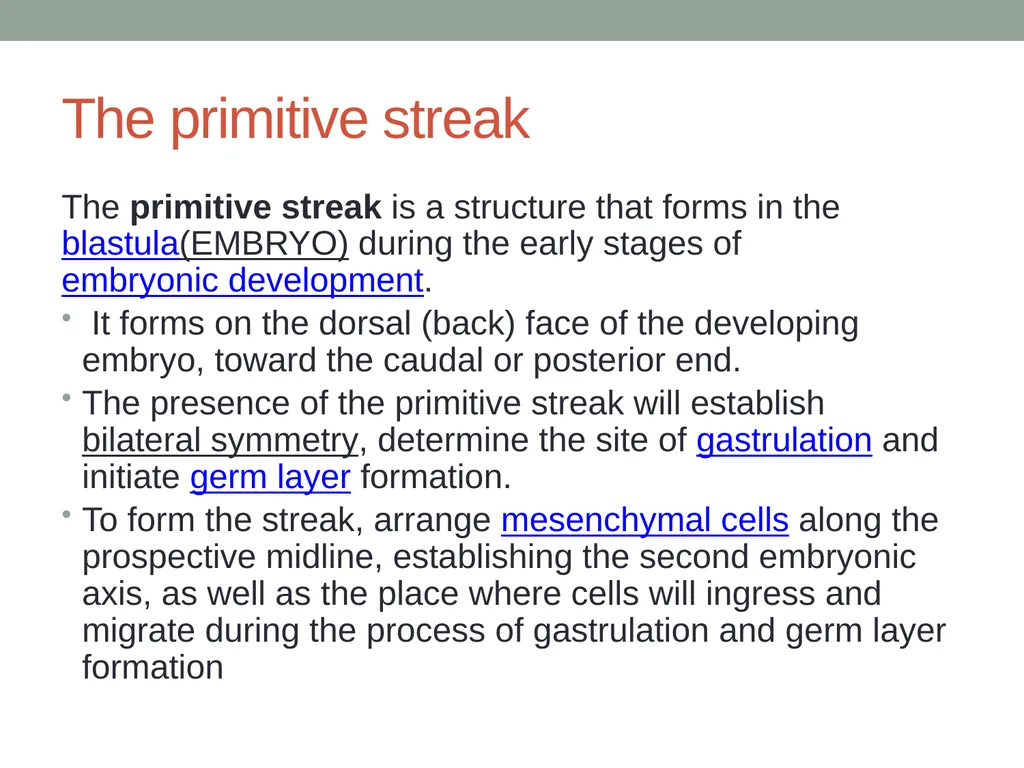
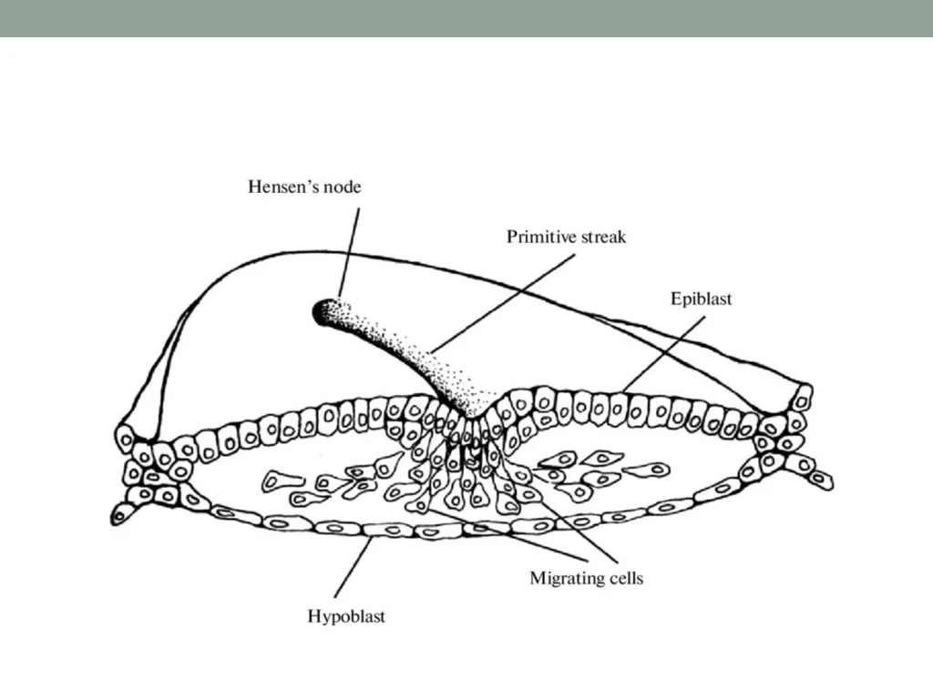
The primitive streak The primitive streak is a structure that forms in the blastula(EMBRYO) during the early stages of embryonic development.
It forms on the dorsal (back) face of the developing embryo, toward the caudal or posterior end.
The presence of the primitive streak will establish bilateral symmetry, determine the site of gastrulation and initiate germ layer formation.
To form the streak, arrange mesenchymal cells along the prospective midline, establishing the second embryonic axis, as well as the place where cells will ingress and migrate during the process of gastrulation and germ layer formation<br>
It forms on the dorsal (back) face of the developing embryo, toward the caudal or posterior end.
The presence of the primitive streak will establish bilateral symmetry, determine the site of gastrulation and initiate germ layer formation.
To form the streak, arrange mesenchymal cells along the prospective midline, establishing the second embryonic axis, as well as the place where cells will ingress and migrate during the process of gastrulation and germ layer formation<br>
06
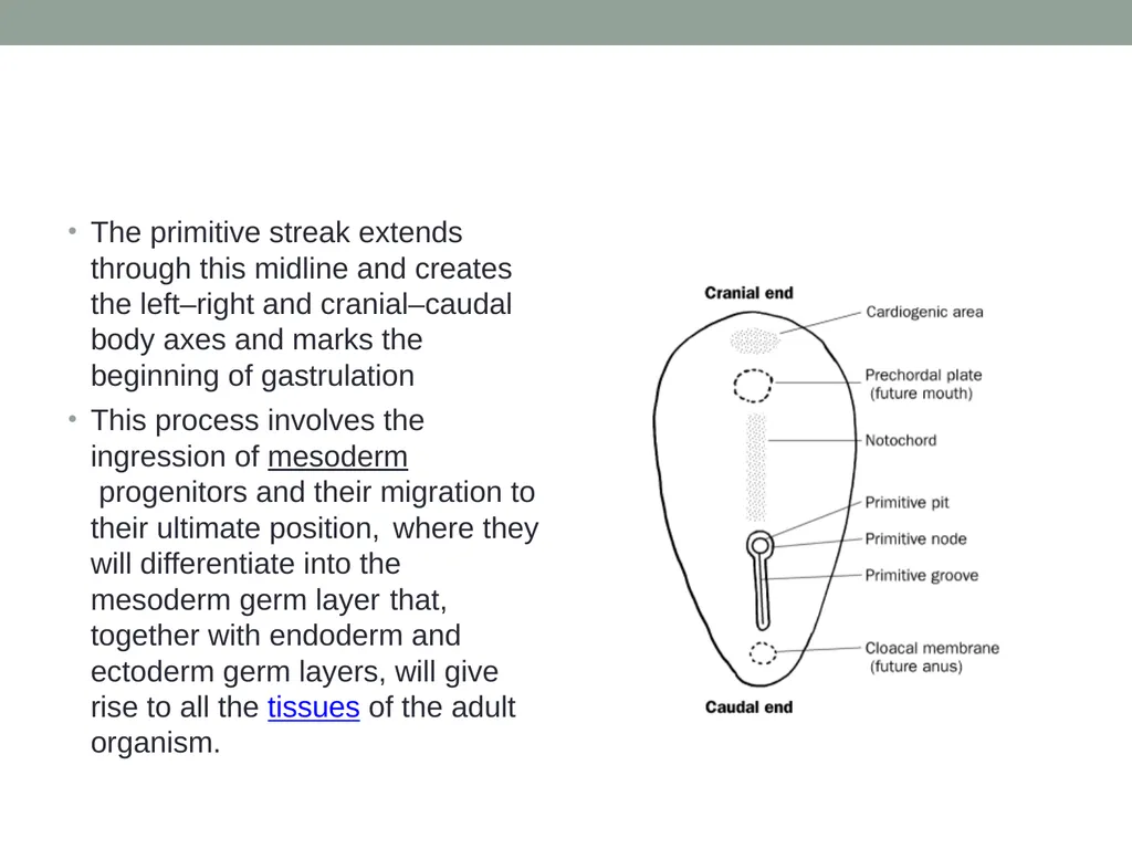
The primitive streak extends through this midline and creates the left–right and cranial–caudal body axes and marks the beginning of gastrulation
This process involves the ingression of mesoderm progenitors and their migration to their ultimate position, where they will differentiate into the mesoderm germ layer that, together with endoderm and ectoderm germ layers, will give rise to all the tissues of the adult organism.<br>
This process involves the ingression of mesoderm progenitors and their migration to their ultimate position, where they will differentiate into the mesoderm germ layer that, together with endoderm and ectoderm germ layers, will give rise to all the tissues of the adult organism.<br>
07
The anterior (rostral) end of the primitive streak forms the lower germ layer, the endoderm, in which are embedded the midline notochordal and prechordal plates.
primitive node: is a small depression locates in the head end of primitive streak from which a population of ectodermal cells will migrate toward the endoderm.
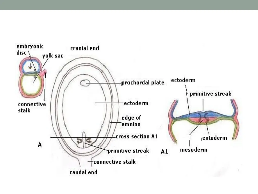
Prospective mesodermal cells migrate from the epiblast through the primitive streak to form the middle germ layer, the mesoderm.
Cells remaining in the epiblast form the ectoderm, completing formation of the three germ layers. Thus, at this stage, three distinct populations of embryonic cells have arisen largely through division and migration.
They follow distinctly separate courses during later development.
Migrations, create new associations between cells, which, in turn, allow unique possibilities for subsequent development through interactions between the cell<br>
primitive node: is a small depression locates in the head end of primitive streak from which a population of ectodermal cells will migrate toward the endoderm.
Prospective mesodermal cells migrate from the epiblast through the primitive streak to form the middle germ layer, the mesoderm.
Cells remaining in the epiblast form the ectoderm, completing formation of the three germ layers. Thus, at this stage, three distinct populations of embryonic cells have arisen largely through division and migration.
They follow distinctly separate courses during later development.
Migrations, create new associations between cells, which, in turn, allow unique possibilities for subsequent development through interactions between the cell<br>
08
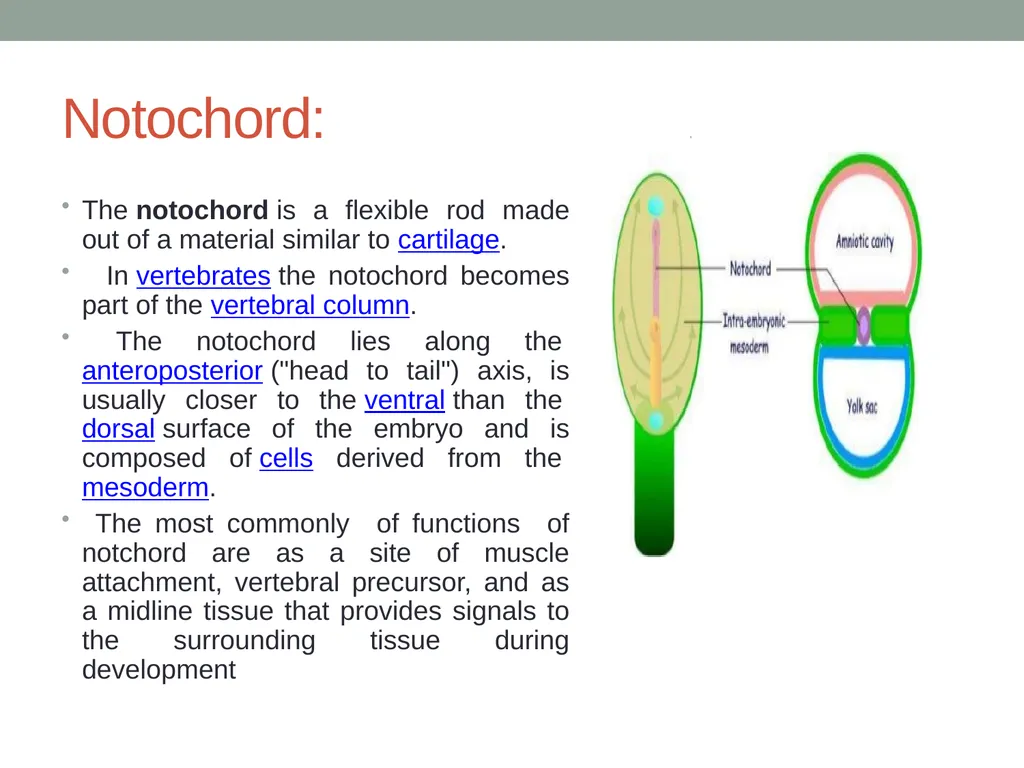
Notochord: The notochord is a flexible rod made out of a material similar to cartilage.
In vertebrates the notochord becomes part of the vertebral column.
The notochord lies along the anteroposterior ("head to tail") axis, is usually closer to the ventral than the dorsal surface of the embryo and is composed of cells derived from the mesoderm.
The most commonly of functions of notchord are as a site of muscle attachment, vertebral precursor, and as a midline tissue that provides signals to the surrounding tissue during development<br>
In vertebrates the notochord becomes part of the vertebral column.
The notochord lies along the anteroposterior ("head to tail") axis, is usually closer to the ventral than the dorsal surface of the embryo and is composed of cells derived from the mesoderm.
The most commonly of functions of notchord are as a site of muscle attachment, vertebral precursor, and as a midline tissue that provides signals to the surrounding tissue during development<br>
09
prechordal plate The prechordal plate is a "uniquely thickened portion" of the endoderm that is in contact with ectoderm immediately rostral to the cephalic tip of the notochord
A median strip of mesoderm cells (the chordamesoderm) extending throughout the length of the embryo induces neural plate formation within the overlying ectoderm.
The prechordal plate is thought to have a similar role in the anterior neural plate region. It is the most likely origin of the rostral cranial mesoderm.
The nature of such inductive stimuli is presently unknown.
Sometimes cell-to-cell contact appears to be necessary, whereas in other cases (as in neural plate induction) the inductive influences appear to be able to act between cells separated by considerable distances and to consist of diffusible substances.<br>
A median strip of mesoderm cells (the chordamesoderm) extending throughout the length of the embryo induces neural plate formation within the overlying ectoderm.
The prechordal plate is thought to have a similar role in the anterior neural plate region. It is the most likely origin of the rostral cranial mesoderm.
The nature of such inductive stimuli is presently unknown.
Sometimes cell-to-cell contact appears to be necessary, whereas in other cases (as in neural plate induction) the inductive influences appear to be able to act between cells separated by considerable distances and to consist of diffusible substances.<br>
10
It is known that inductive influences need only be present for a short time, after which the responding tissue is capable of independent development.
For example, an induced neural plate isolated will roll up into a tube, which then differentiates into the
brain, spinal cord, and other structures.
In addition to inducing neural plate formation, the chordamesoderm appears to be responsible for developing the organizational plan of the head.<br>
For example, an induced neural plate isolated will roll up into a tube, which then differentiates into the
brain, spinal cord, and other structures.
In addition to inducing neural plate formation, the chordamesoderm appears to be responsible for developing the organizational plan of the head.<br>
11
Nervous system development: It starts as:
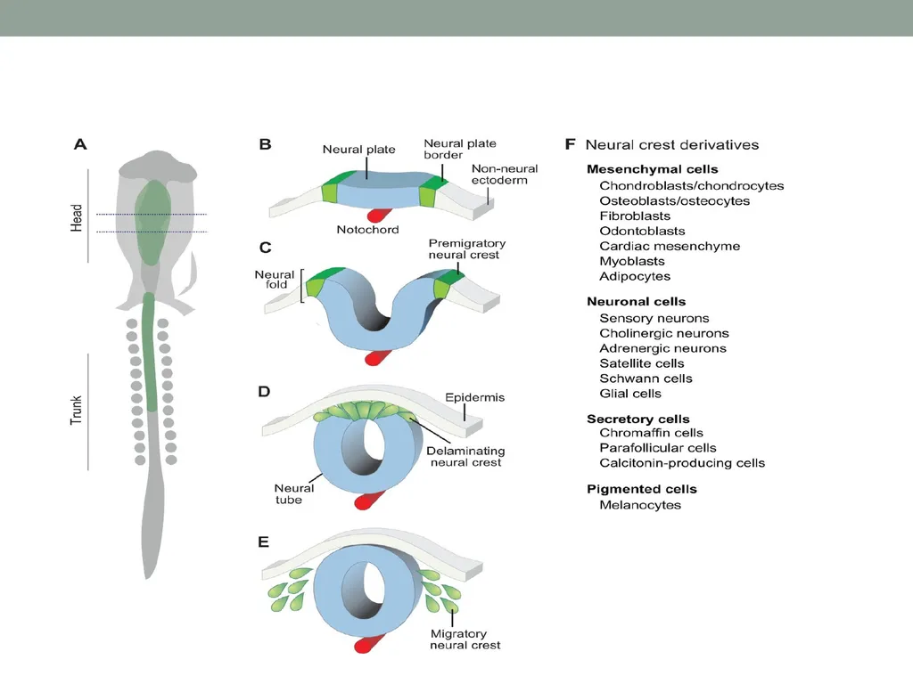
1-Formation of neural plate which is thickening in the ectoderm at cephalic end of the embryo.
2-Neural folds formation which are raising margins from neural plate.
3-Neural groove formation from the approximation of the folds toward the midline and then complete fusion to form the neural tube.
4-The neural tube gives rise brain and spinal cord.
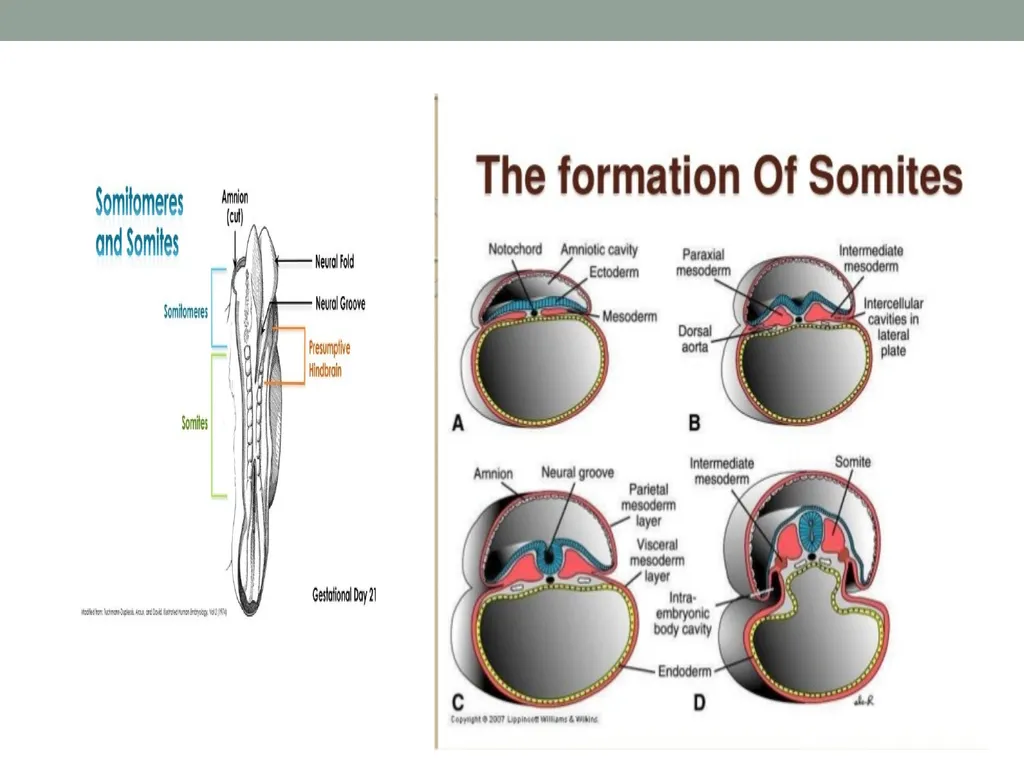
3-Somites are condensed masses of cells derived from mesoderm located adjacent to the neural tube
Neural crest cells. a unique population of cells develops from the ectoderm along the lateral margins of the neural plate They undergo extensive migrations, usually beginning at about the time of tube closure , and give rise to a variety of different cells that form components of many tissues.<br>
1-Formation of neural plate which is thickening in the ectoderm at cephalic end of the embryo.
2-Neural folds formation which are raising margins from neural plate.
3-Neural groove formation from the approximation of the folds toward the midline and then complete fusion to form the neural tube.
4-The neural tube gives rise brain and spinal cord.
3-Somites are condensed masses of cells derived from mesoderm located adjacent to the neural tube
Neural crest cells. a unique population of cells develops from the ectoderm along the lateral margins of the neural plate They undergo extensive migrations, usually beginning at about the time of tube closure , and give rise to a variety of different cells that form components of many tissues.<br>
12
DIRIVITIVE OF N.C.C.: 1- The crest cells that migrate in the trunk region form mostly neural, endocrine, and pigment cells, whereas those that migrate in the head and neck also contribute extensively to skeletal and connective tissues (i.e., cartilage, bone, dentin, dermis, etc.).
The supporting connective tissue found in facial muscles is derived from neural crest cells. The connective tissue components in these structures (e.g., fibroblasts, odontoblasts, and the cells of tooth-supporting tissues) are derived from neural crest cells.<br>
The supporting connective tissue found in facial muscles is derived from neural crest cells. The connective tissue components in these structures (e.g., fibroblasts, odontoblasts, and the cells of tooth-supporting tissues) are derived from neural crest cells.<br>
13
2- The supporting connective tissue found in facial muscles is derived from neural crest cells.
the connective tissue components in these structures (e.g., fibroblasts, odontoblasts, and the cells of tooth-supporting tissues) are derived from neural crest cells.
The enamel-forming cells are derived from ectoderm lining the oral cavity.
The migration routes that cephalic (head) neural crest cells move around the sides of the head beneath the surface ectoderm, as a sheet of cells.
They form all the mesenchyme in the upper facial region, whereas in the lower facial region they surround mesodermal cores already present in the visceral arches.<br>
the connective tissue components in these structures (e.g., fibroblasts, odontoblasts, and the cells of tooth-supporting tissues) are derived from neural crest cells.
The enamel-forming cells are derived from ectoderm lining the oral cavity.
The migration routes that cephalic (head) neural crest cells move around the sides of the head beneath the surface ectoderm, as a sheet of cells.
They form all the mesenchyme in the upper facial region, whereas in the lower facial region they surround mesodermal cores already present in the visceral arches.<br>
14
3- Toward the completion of migration, the irregular edge of the crest cell mass appears to attach itself to the neural tube at locations where sensory ganglia of the fifth, seventh, ninth, and tenth cranial nerves will form.
4-In the trunk sensory ganglia, supporting (e.g., Schwann) cells and all neurons are derived from neural crest cells. On the other hand, many of the sensory neurons of the cranial sensory ganglia originate from placodes in the surface ectoderm.<br>
4-In the trunk sensory ganglia, supporting (e.g., Schwann) cells and all neurons are derived from neural crest cells. On the other hand, many of the sensory neurons of the cranial sensory ganglia originate from placodes in the surface ectoderm.<br>
15
5-Eventually, capillary endothelial cells derived from mesoderm cells invade the crest cell mesenchyme, and it is from this mesenchyme that the supporting cells of the developing blood vessels are derived.
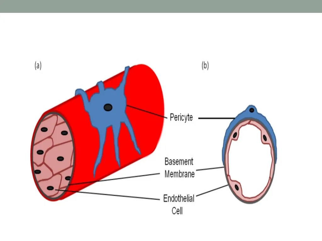
Initially, these supporting cells include only pericytes, which are closely opposed to the outer surfaces of endothelial cells.
Later, additional crest cells differentiate into the fibroblasts and smooth muscle cells that will form the vessel wall.
The developing blood vessels become interconnected to form vascular networks. These networks undergo a series of modifications, before they eventually form the mature vascular system. Besides inducing the neural plate from overlying ectoderm, the chordamesoderm organizes the positional relationships of various neural plate components, such as the initial primordium of the eye.<br>
Initially, these supporting cells include only pericytes, which are closely opposed to the outer surfaces of endothelial cells.
Later, additional crest cells differentiate into the fibroblasts and smooth muscle cells that will form the vessel wall.
The developing blood vessels become interconnected to form vascular networks. These networks undergo a series of modifications, before they eventually form the mature vascular system. Besides inducing the neural plate from overlying ectoderm, the chordamesoderm organizes the positional relationships of various neural plate components, such as the initial primordium of the eye.<br>
16
Mesoderm of the head The mesodermal portion differentiates into well-organized blocks of cells, called somites, caudal to the developing ear and less-organized somitomeres rostral to the ear.
Later these structures form myoblasts and some of the skeletal and connective tissues of the head.
Almost all the myoblasts that subsequently fuse with each other to form the multinucleated striated muscle fibers are derived from mesoderm.
The myoblasts that form the hypoglossal (tongue) muscles are derived from somites located beside the developing hindbrain.
The myoblasts of the extrinsic occular muscles originate from the prechordal plate.<br>
Later these structures form myoblasts and some of the skeletal and connective tissues of the head.
Almost all the myoblasts that subsequently fuse with each other to form the multinucleated striated muscle fibers are derived from mesoderm.
The myoblasts that form the hypoglossal (tongue) muscles are derived from somites located beside the developing hindbrain.
The myoblasts of the extrinsic occular muscles originate from the prechordal plate.<br>
17
A number of other structures in the facial region, such as the epithelial components or glands and the enamel organ of the tooth bud, are derived from epithelium that grows (invaginates) into underlying mesenchyme.
Again, the connective tissue components in these structures (e.g., fibroblasts, odontoblasts, and the cells of tooth-supporting tissues) are derived from neural crest cells.
In the trunk, all skeletal and connective tissues are formed by mesoderm<br>
Again, the connective tissue components in these structures (e.g., fibroblasts, odontoblasts, and the cells of tooth-supporting tissues) are derived from neural crest cells.
In the trunk, all skeletal and connective tissues are formed by mesoderm<br>