
Endorsed by the Society for Cardiovascular
YM
Published · 129 slides · 0 views

1 / 1








Description
Endorsed by the Society for Cardiovascular Angiography and Interventions 2021 ACCAHAAATSSTSSCAI Guideline for Coronary Artery Revascularization This slide set is adapted from the 2021 ACCAHAAATSSTSSCAI Guideline for Coronary Artery
Related Topics
Share
Embed code
Download this presentation From Below
"Endorsed by the Society for Cardiovascular" is the property of its rightful owner. Permission is granted to download and print the materials on this website for personal, non-commercial use only, and to display it on your personal computer provided you do not modify the materials and that you retain all copyright notices contained in the materials. By downloading content from our website, you accept the terms of this agreement.
Presentation Transcript
01
Endorsed by the Society for Cardiovascular Angiography and Interventions 2021 ACC/AHA/AATS/STS/SCAI Guideline for Coronary Artery Revascularization<br>
02
This slide set is adapted from the 2021 ACC/AHA/AATS/STS/SCAI Guideline for Coronary Artery Revascularization. Published online ahead of print December 3, 2021, available at: Circulation. https://www.ahajournals.org/doi/10.1161/CIR.0000000000001038 and Journal of the American College of Cardiology published online ahead of print December 3, 2021. J Am Coll Cardiol. https://doi.org/10.1016/j.jacc.2021.09.006 Citation<br>
03
3 2021 Writing Committee Members* Jennifer S. Lawton, MD, FAHA, Chair†
Jacqueline E. Tamis-Holland, MD, FAHA, FACC, FSCAI, Vice Chair‡ *Writing committee members are required to recuse themselves from voting on sections to which their specific relationships with industry may apply; see Appendix 1 for detailed information. †ACC/AHA Representative. ‡ACC/AHA Joint Committee on Clinical Practice Guidelines Liaison. § ACC/AHA Task Force on Data Standards Representative. ¶SCAI Representative. #AATS Representative. ||STS representative. **Former Joint Committee member; current member during the writing effort.<br>
Jacqueline E. Tamis-Holland, MD, FAHA, FACC, FSCAI, Vice Chair‡ *Writing committee members are required to recuse themselves from voting on sections to which their specific relationships with industry may apply; see Appendix 1 for detailed information. †ACC/AHA Representative. ‡ACC/AHA Joint Committee on Clinical Practice Guidelines Liaison. § ACC/AHA Task Force on Data Standards Representative. ¶SCAI Representative. #AATS Representative. ||STS representative. **Former Joint Committee member; current member during the writing effort.<br>
04
Top 10 Take-Home Messages 4 2021 Guideline for Coronary Artery Revascularization<br>
05
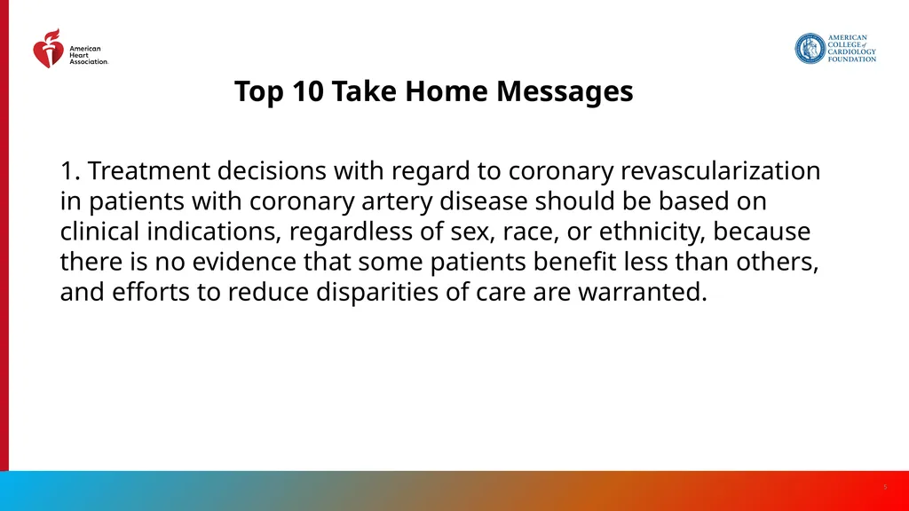
Top 10 Take Home Messages 5 1. Treatment decisions with regard to coronary revascularization in patients with coronary artery disease should be based on clinical indications, regardless of sex, race, or ethnicity, because there is no evidence that some patients benefit less than others, and efforts to reduce disparities of care are warranted.<br>
06
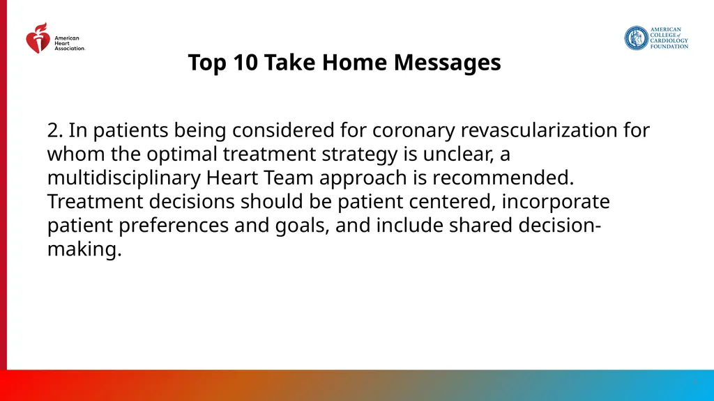
2. In patients being considered for coronary revascularization for whom the optimal treatment strategy is unclear, a multidisciplinary Heart Team approach is recommended. Treatment decisions should be patient centered, incorporate patient preferences and goals, and include shared decision-making. Top 10 Take Home Messages 6<br>
07
3. For patients with significant left main disease, surgical revascularization is indicated to improve survival relative to that likely to be achieved with medical therapy. Percutaneous revascularization is a reasonable option to improve survival, compared with medical therapy, in selected patients with low to medium anatomic complexity of coronary artery disease and left main disease that is equally suitable for surgical or percutaneous revascularization. Top 10 Take Home Messages 7<br>
08
4. Updated evidence from contemporary trials supplement older evidence with regard to mortality benefit of revascularization in patients with stable ischemic heart disease, normal left ventricular ejection fraction, and triple-vessel coronary artery disease. Surgical revascularization may be reasonable to improve survival. A survival benefit with percutaneous revascularization is uncertain. Revascularization decisions are based on consideration of disease complexity, technical feasibility of treatment, and a Heart Team discussion. Top 10 Take Home Messages 8<br>
09
5. The use of a radial artery as a surgical revascularization conduit is preferred to the use of a saphenous vein conduit to bypass the second most important target vessel with significant stenosis after the left anterior descending coronary artery. Benefits include superior patency, reduced adverse cardiac events, and improved survival. Top 10 Take Home Messages 9<br>
10
6. Radial artery access is recommended in patients undergoing percutaneous intervention who have acute coronary syndromes or stable ischemic heart disease, to reduce bleeding and vascular complications compared with a femoral approach. Patients with acute coronary syndromes also benefit from a reduction in mortality rate with this approach. Top 10 Take Home Messages 10<br>
11
7. A short duration of dual antiplatelet therapy after percutaneous revascularization in patients with stable ischemic heart disease is reasonable to reduce the risk of bleeding events. After consideration of recurrent ischemia and bleeding risks, select patients may safely transition to P2Y12 inhibitor monotherapy and stop aspirin after 1 to 3 months of dual antiplatelet therapy. Top 10 Take Home Messages 11<br>
12
8. Staged percutaneous intervention (while in hospital or after discharge) of a significantly stenosed non-culprit artery in patients presenting with an ST-segment–elevation myocardial infarction is recommended in select patients to improve outcomes. Percutaneous intervention of the non-culprit artery at the time of primary percutaneous coronary intervention is less clear and may be considered in stable patients with uncomplicated revascularization of the culprit artery, low-complexity non-culprit artery disease, and normal renal function. In contrast, percutaneous intervention of the non-culprit artery can be harmful in patients in cardiogenic shock. Top 10 Take Home Messages 12<br>
13
9. Revascularization decisions in patients with diabetes and multivessel coronary artery disease are optimized by the use of a Heart Team approach. Patients with diabetes who have triple-vessel disease should undergo surgical revascularization; percutaneous coronary intervention may be considered if they are poor candidates for surgery. Top 10 Take Home Messages 13<br>
14
10. Treatment decisions for patients undergoing surgical revascularization of coronary artery disease should include the calculation of a patient’s surgical risk with the Society of Thoracic Surgeons score. The usefulness of the SYNTAX score calculation in treatment decisions is less clear because of the interobserver variability in its calculation and its absence of clinical variables. Top 10 Take Home Messages 14<br>
15
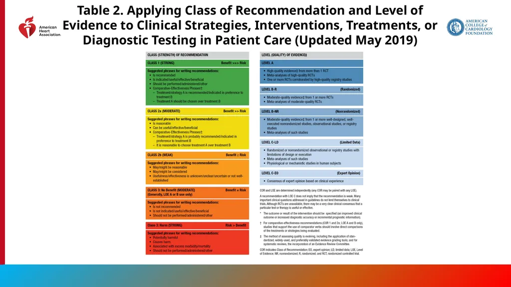
15 Table 2. Applying Class of Recommendation and Level of Evidence to Clinical Strategies, Interventions, Treatments, or Diagnostic Testing in Patient Care (Updated May 2019)<br>
16
16 Improving Equity of Care in Revascularization and Shared Decision-Making<br>
17
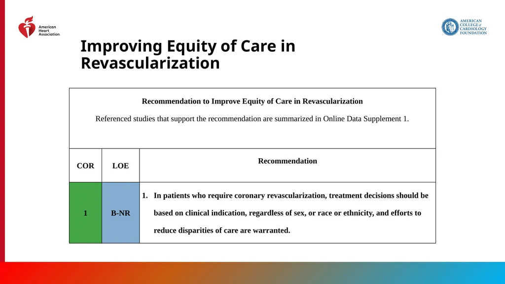
Improving Equity of Care in Revascularization 17<br>
18
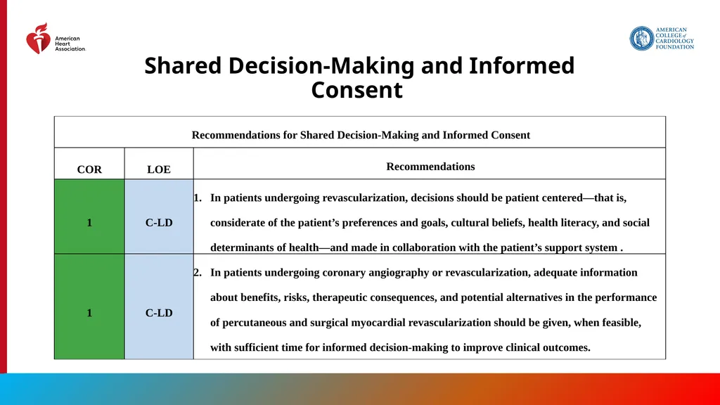
18 Shared Decision-Making and Informed Consent<br>
19
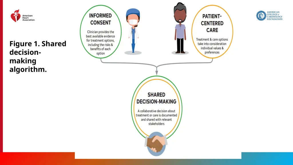
19 Figure 1. Shared decision-making algorithm.<br>
20
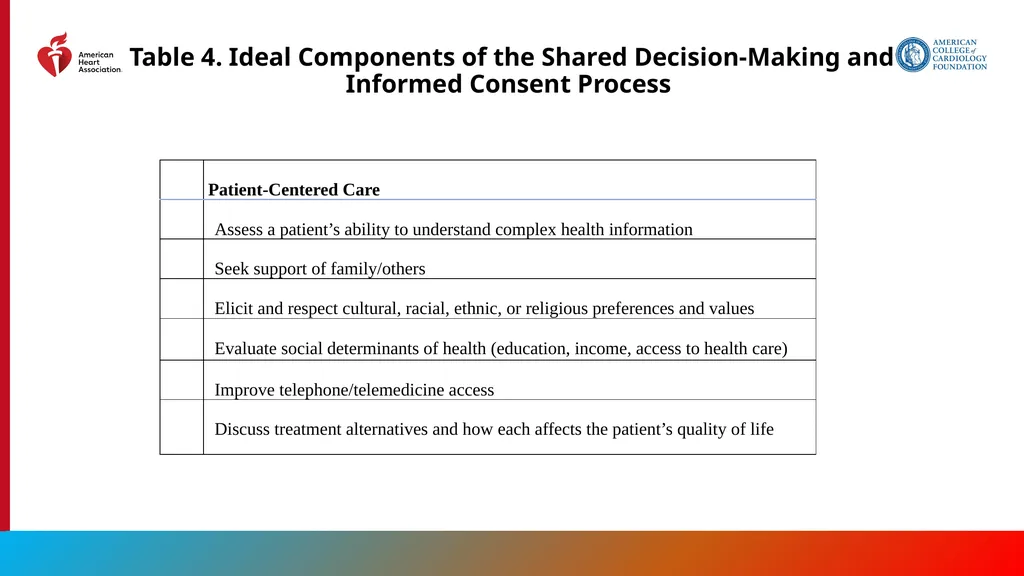
Table 4. Ideal Components of the Shared Decision-Making and Informed Consent Process 20<br>
21
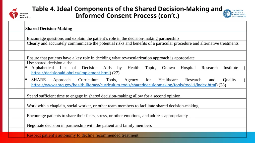
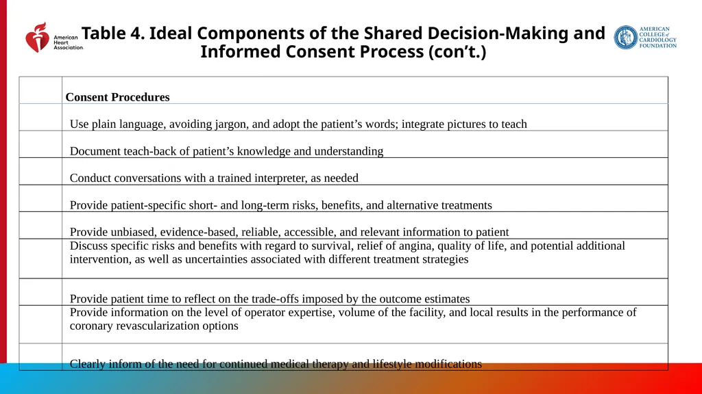
21 Table 4. Ideal Components of the Shared Decision-Making and Informed Consent Process (con’t.)<br>
22
22 Table 4. Ideal Components of the Shared Decision-Making and Informed Consent Process (con’t.)<br>
23
23 Preprocedural Assessment and the Heart Team<br>
24
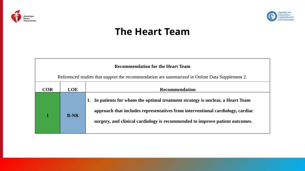
24 The Heart Team<br>
25
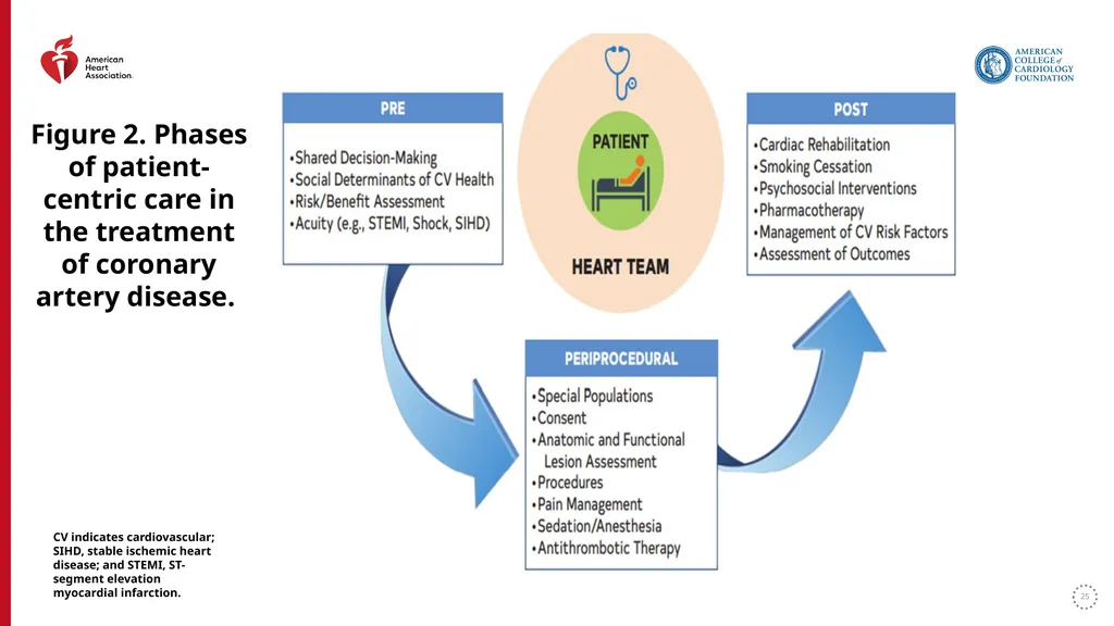
25 Figure 2. Phases of patient-centric care in the treatment of coronary artery disease. CV indicates cardiovascular; SIHD, stable ischemic heart disease; and STEMI, ST-segment elevation myocardial infarction.<br>
26
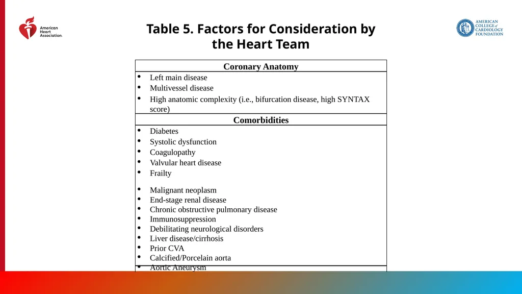
26 Table 5. Factors for Consideration by the Heart Team<br>
27
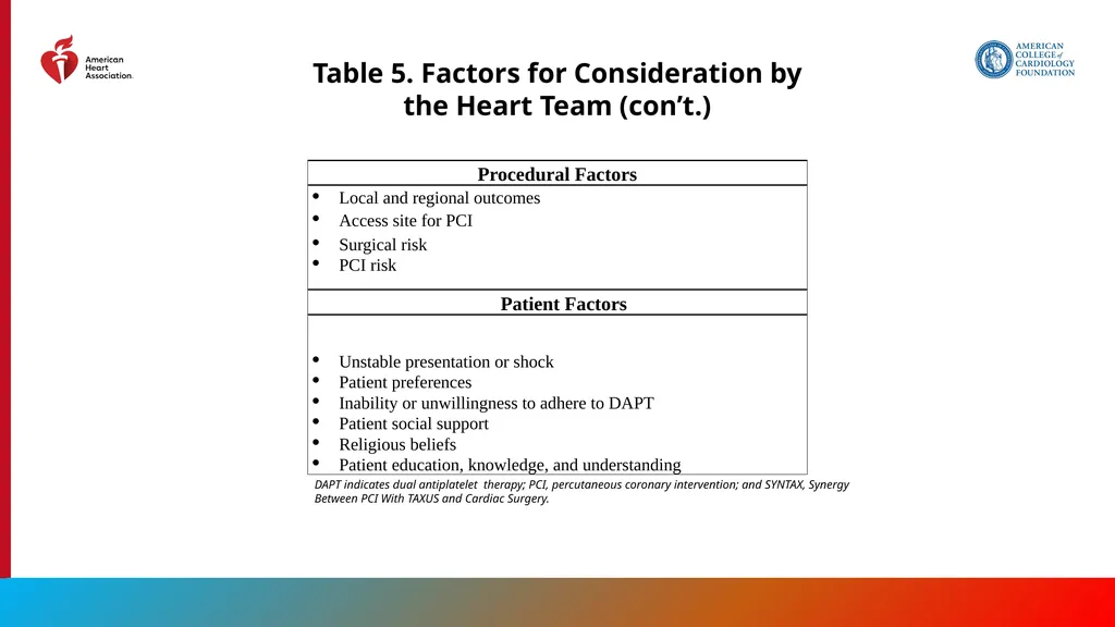
27 Table 5. Factors for Consideration by the Heart Team (con’t.) DAPT indicates dual antiplatelet therapy; PCI, percutaneous coronary intervention; and SYNTAX, Synergy Between PCI With TAXUS and Cardiac Surgery.<br>
28
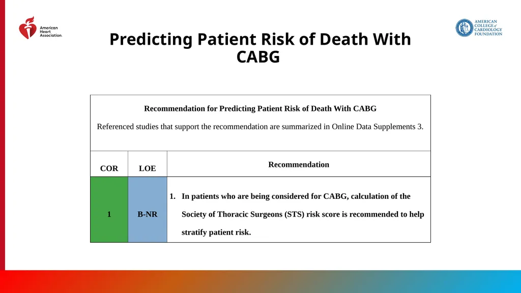
28 Predicting Patient Risk of Death With CABG<br>
29
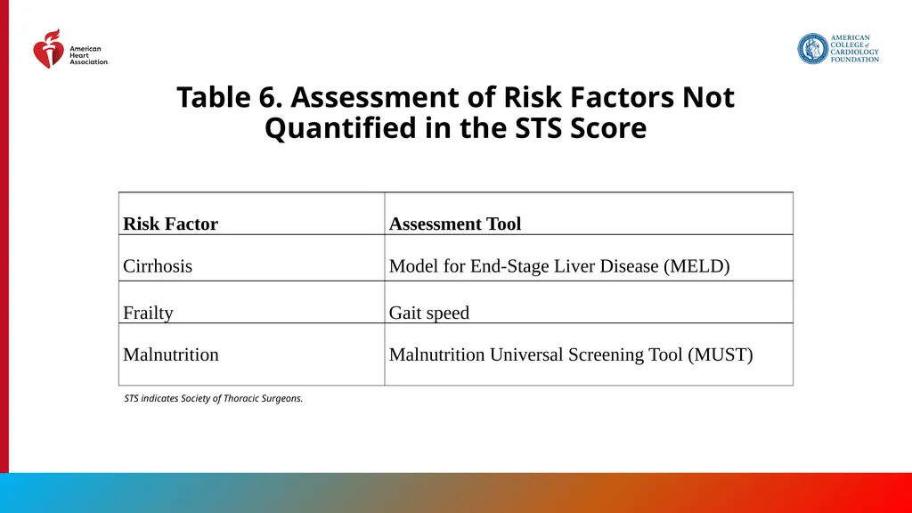
29 Table 6. Assessment of Risk Factors Not Quantified in the STS Score STS indicates Society of Thoracic Surgeons.<br>
30
30 Defining Lesion Severity<br>
31
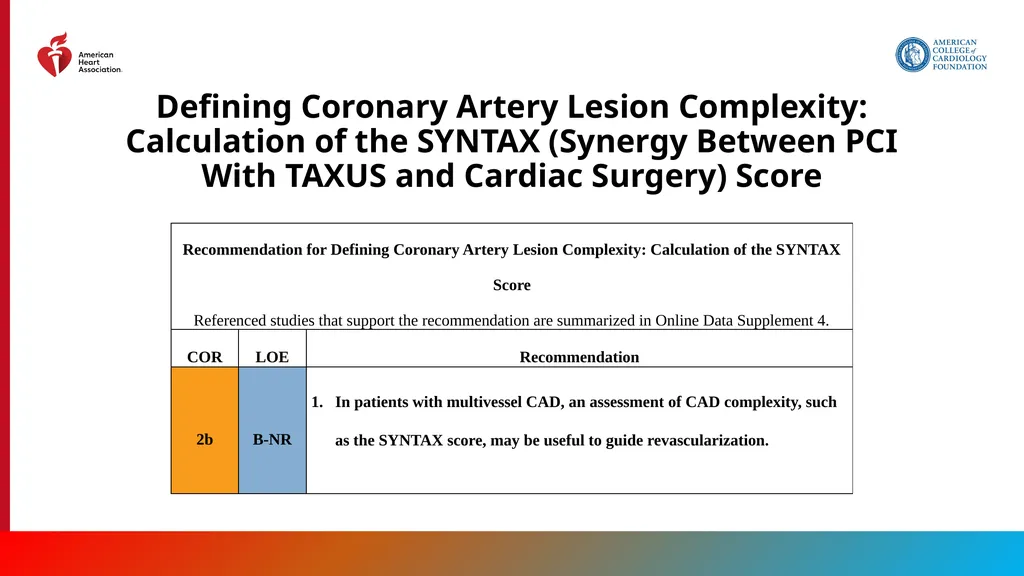
31 Defining Coronary Artery Lesion Complexity: Calculation of the SYNTAX (Synergy Between PCI With TAXUS and Cardiac Surgery) Score<br>
32
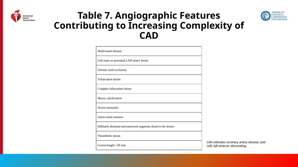
32 Table 7. Angiographic Features Contributing to Increasing Complexity of CAD CAD indicates coronary artery disease; and LAD, left anterior descending.<br>
33
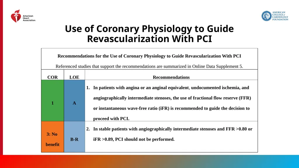
33 Use of Coronary Physiology to Guide Revascularization With PCI<br>
34
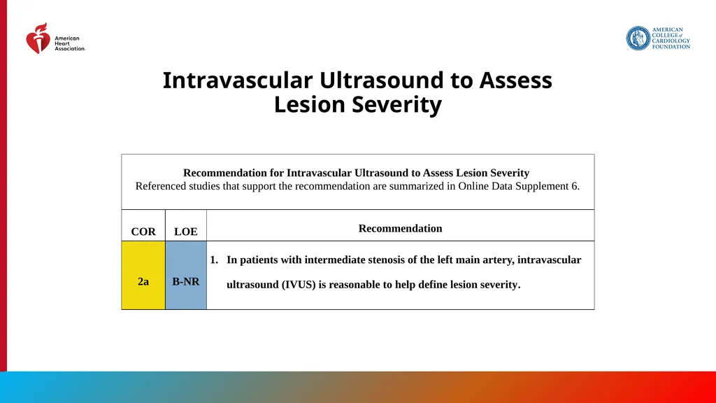
34 Intravascular Ultrasound to Assess Lesion Severity<br>
35
35 Revascularization in STEMI<br>
36
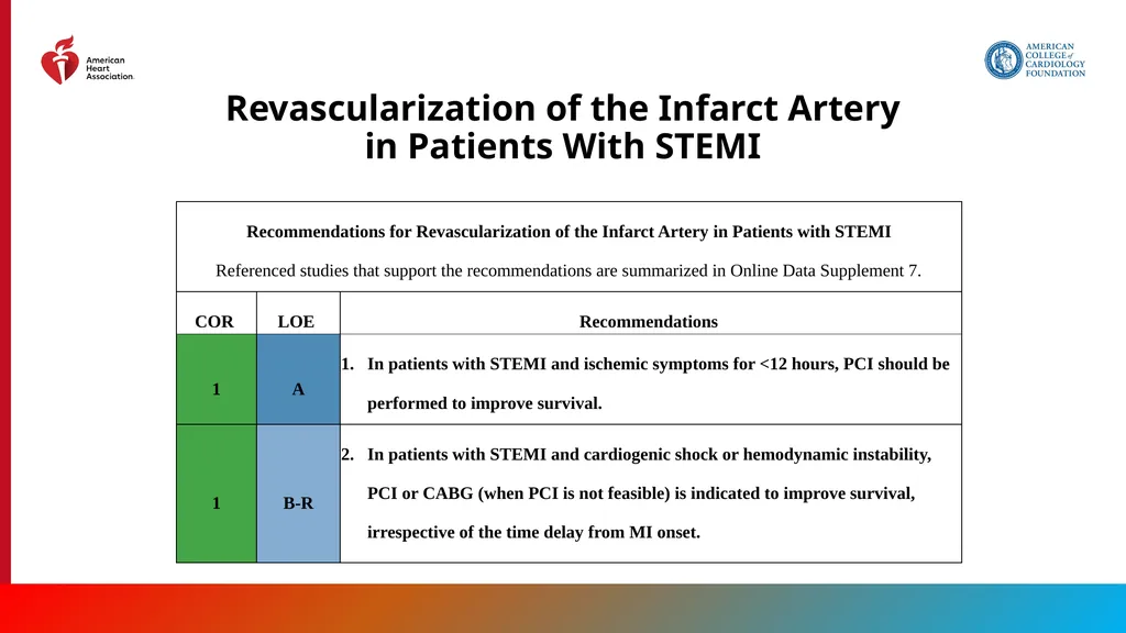
36 Revascularization of the Infarct Artery in Patients With STEMI<br>
37
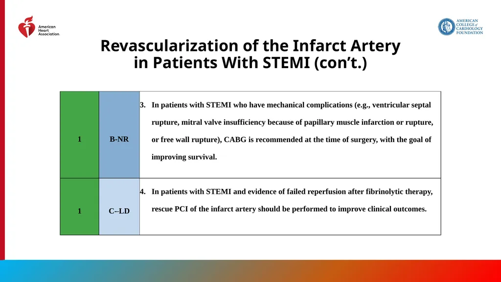
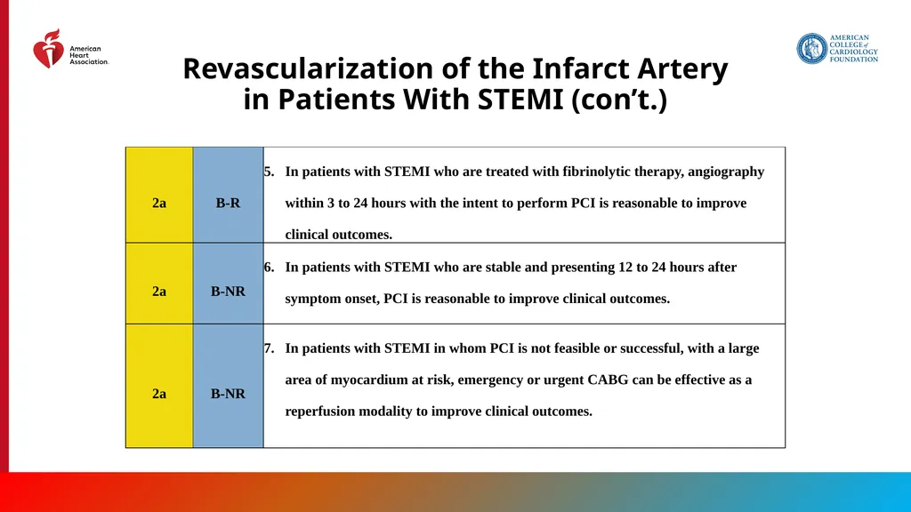
37 Revascularization of the Infarct Artery in Patients With STEMI (con’t.)<br>
38
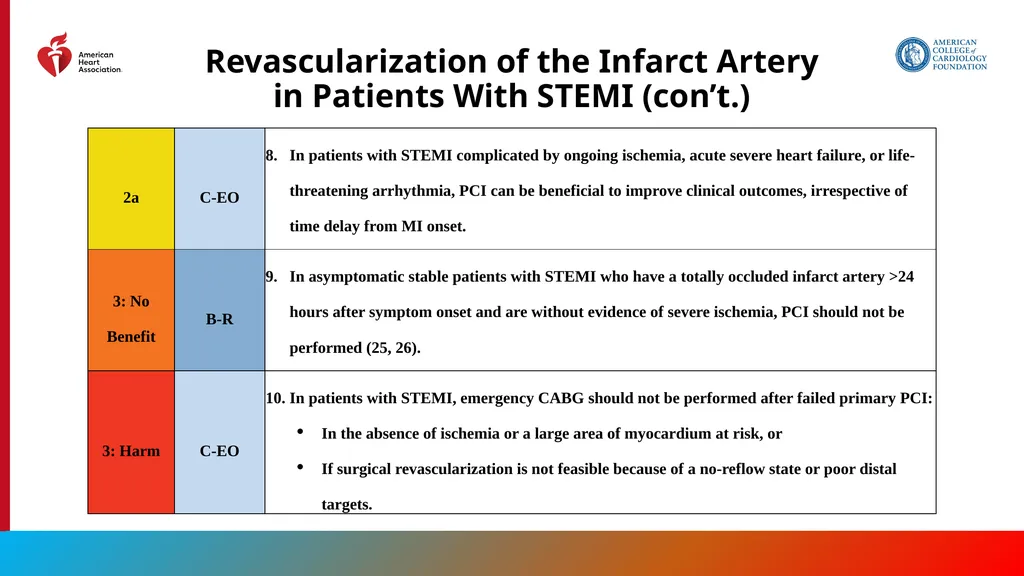
38 Revascularization of the Infarct Artery in Patients With STEMI (con’t.)<br>
39
39 Revascularization of the Infarct Artery in Patients With STEMI (con’t.)<br>
40
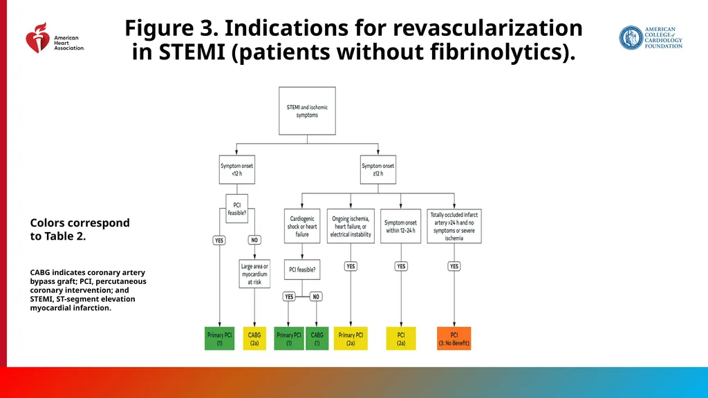
40 Figure 3. Indications for revascularization in STEMI (patients without fibrinolytics). CABG indicates coronary artery bypass graft; PCI, percutaneous coronary intervention; and STEMI, ST-segment elevation myocardial infarction. Colors correspond to Table 2.<br>
41
41 Revascularization of the Non-Infarct Artery in Patients With STEMI<br>
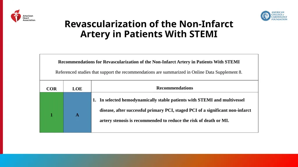
42
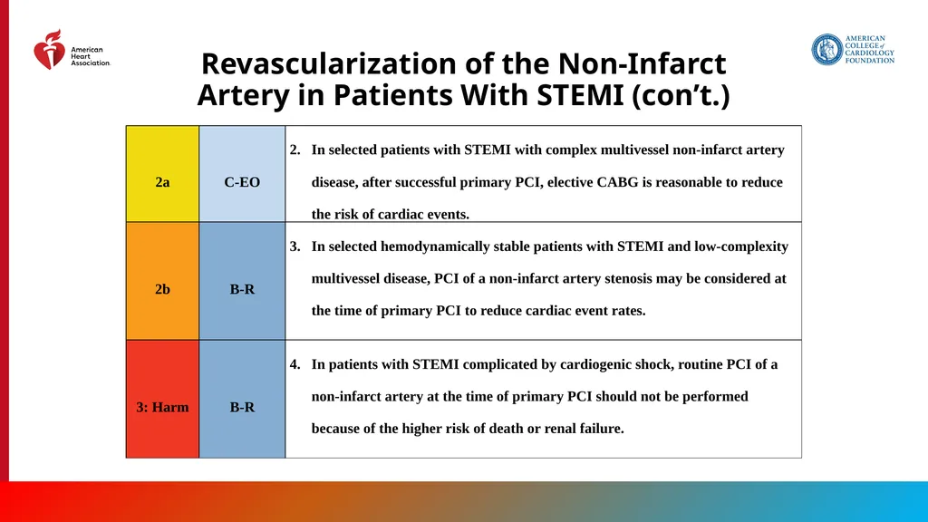
42 Revascularization of the Non-Infarct Artery in Patients With STEMI (con’t.)<br>
43
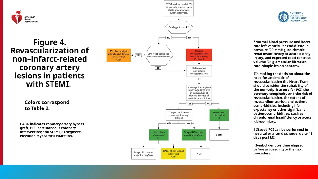
43 Figure 4. Revascularization of non–infarct-related coronary artery lesions in patients with STEMI. CABG indicates coronary artery bypass graft; PCI, percutaneous coronary intervention; and STEMI, ST-segment–elevation myocardial infarction. Colors correspond to Table 2. *Normal blood pressure and heart rate left ventricular end-diastolic pressure ˂20 mmHg, no chronic renal insufficiency or acute kidney injury, and expected total contrast volume ˂3× glomerular filtration rate, simple lesion anatomy.
†In making the decision about the need for and mode of revascularization the Heart Team should consider the suitability of the non-culprit artery for PCI, the coronary complexity and the risk of revascularization, the extent of myocardium at risk, and patient comorbidities, including life expectancy or other significant patient comorbidities, such as chronic renal insufficiency or acute kidney injury.
‡ Staged PCI can be performed in hospital or after discharge, up to 45 days post MI.
Symbol denotes time elapsed before proceeding to the next procedure.<br>
†In making the decision about the need for and mode of revascularization the Heart Team should consider the suitability of the non-culprit artery for PCI, the coronary complexity and the risk of revascularization, the extent of myocardium at risk, and patient comorbidities, including life expectancy or other significant patient comorbidities, such as chronic renal insufficiency or acute kidney injury.
‡ Staged PCI can be performed in hospital or after discharge, up to 45 days post MI.
Symbol denotes time elapsed before proceeding to the next procedure.<br>
44
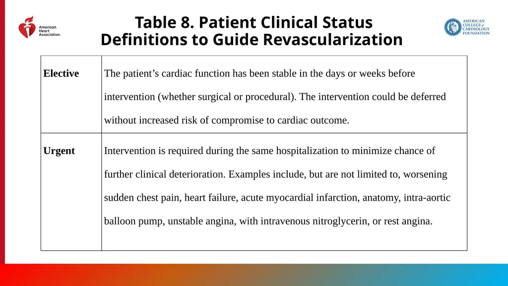
44 Table 8. Patient Clinical Status Definitions to Guide Revascularization<br>
45
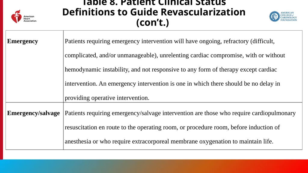
45 Table 8. Patient Clinical Status Definitions to Guide Revascularization (con’t.)<br>
46
46 Revascularization in Non–ST-Segment–Elevation Acute Coronary Syndrome (NSTE-ACS)<br>
47
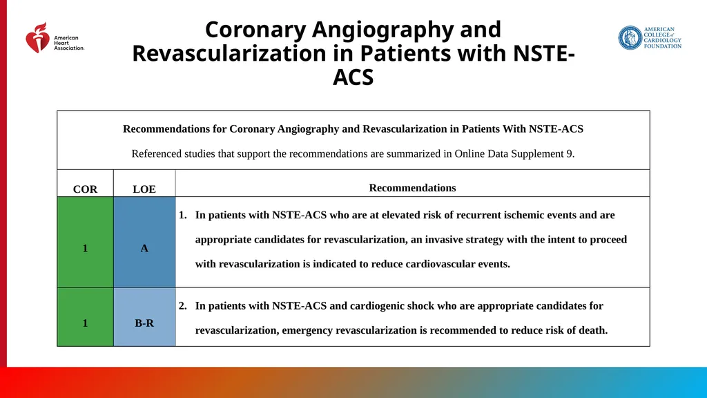
47 Coronary Angiography and Revascularization in Patients with NSTE-ACS<br>
48
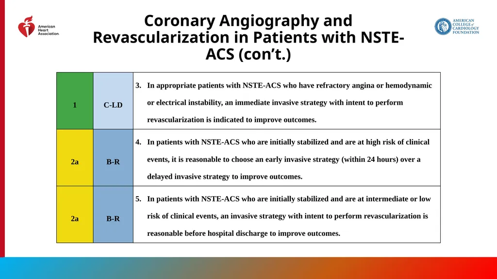
48 Coronary Angiography and Revascularization in Patients with NSTE-ACS (con’t.)<br>
49
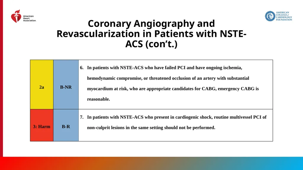
49 Coronary Angiography and Revascularization in Patients with NSTE-ACS (con’t.)<br>
50
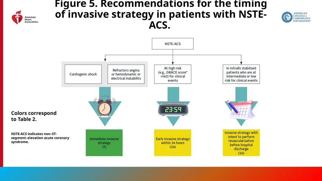
50 Figure 5. Recommendations for the timing of invasive strategy in patients with NSTE-ACS. Colors correspond to Table 2. NSTE-ACS indicates non–ST-segment–elevation acute coronary syndrome.<br>
51
51 Revascularization in SIHD<br>
52
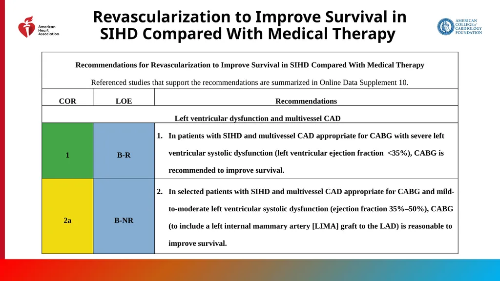
52 Revascularization to Improve Survival in SIHD Compared With Medical Therapy<br>
53
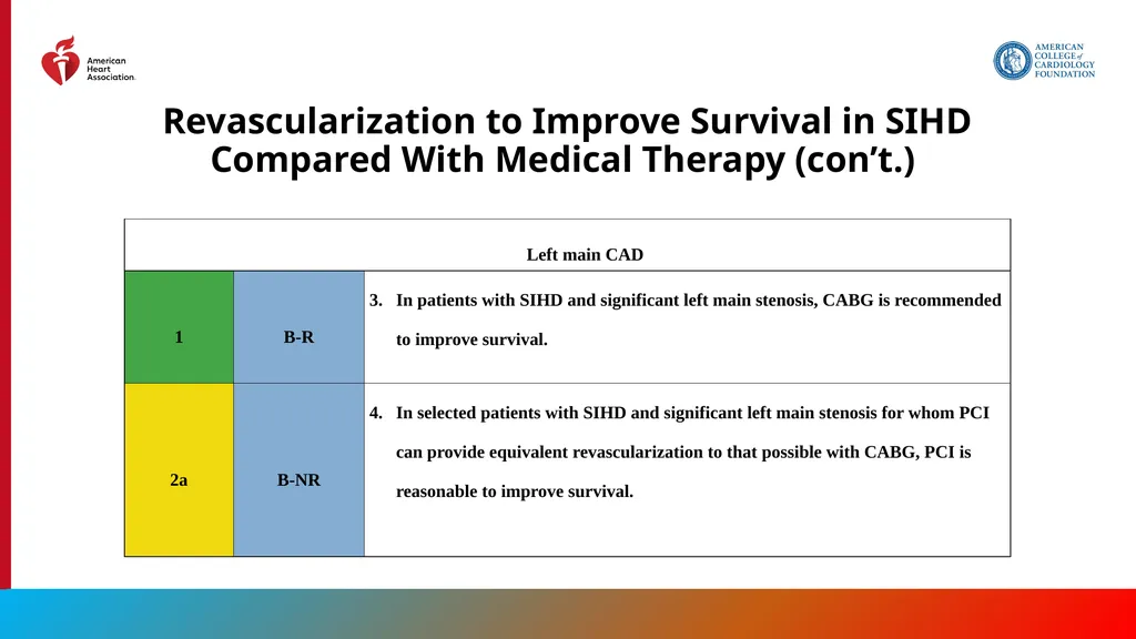
53 Revascularization to Improve Survival in SIHD Compared With Medical Therapy (con’t.)<br>
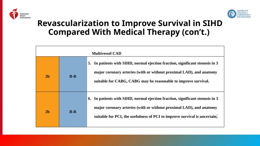
54
54 Revascularization to Improve Survival in SIHD Compared With Medical Therapy (con’t.)<br>
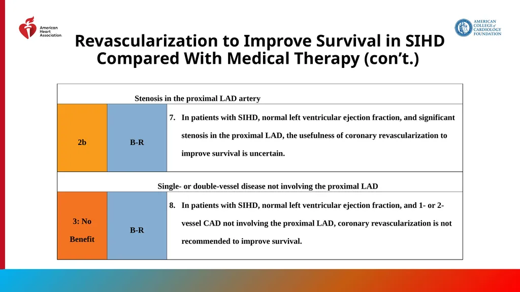
55
55 Revascularization to Improve Survival in SIHD Compared With Medical Therapy (con’t.)<br>
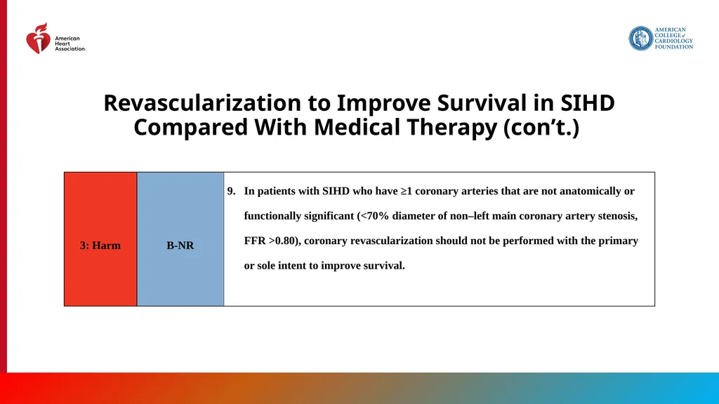
56
56 Revascularization to Improve Survival in SIHD Compared With Medical Therapy (con’t.)<br>
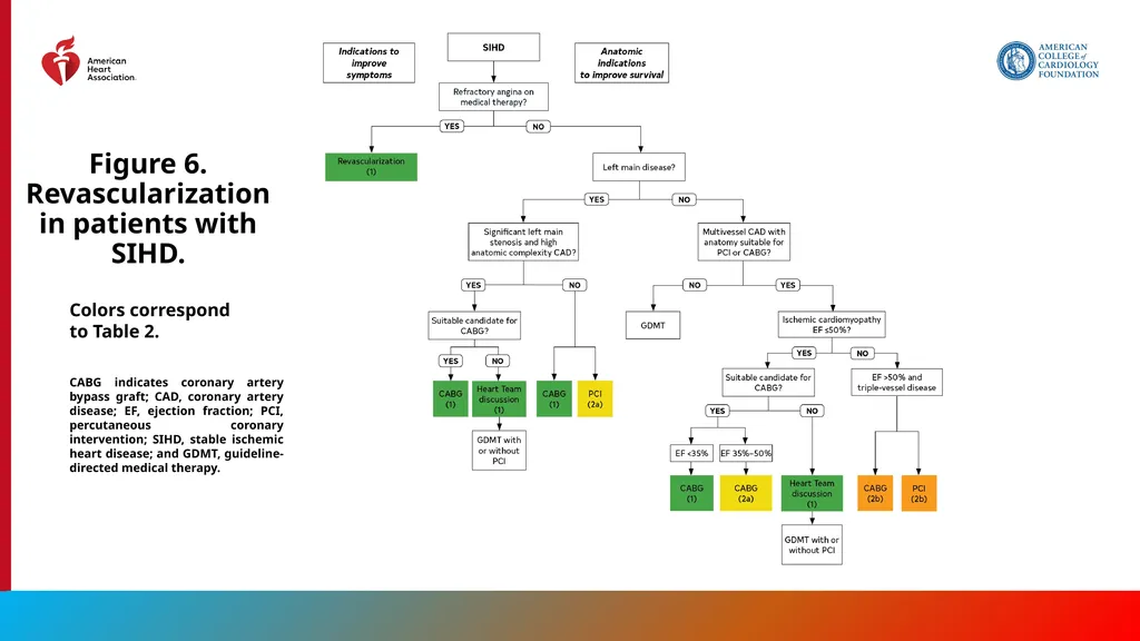
57
57 Figure 6. Revascularization in patients with SIHD. Colors correspond to Table 2. CABG indicates coronary artery bypass graft; CAD, coronary artery disease; EF, ejection fraction; PCI, percutaneous coronary intervention; SIHD, stable ischemic heart disease; and GDMT, guideline-directed medical therapy.<br>
58
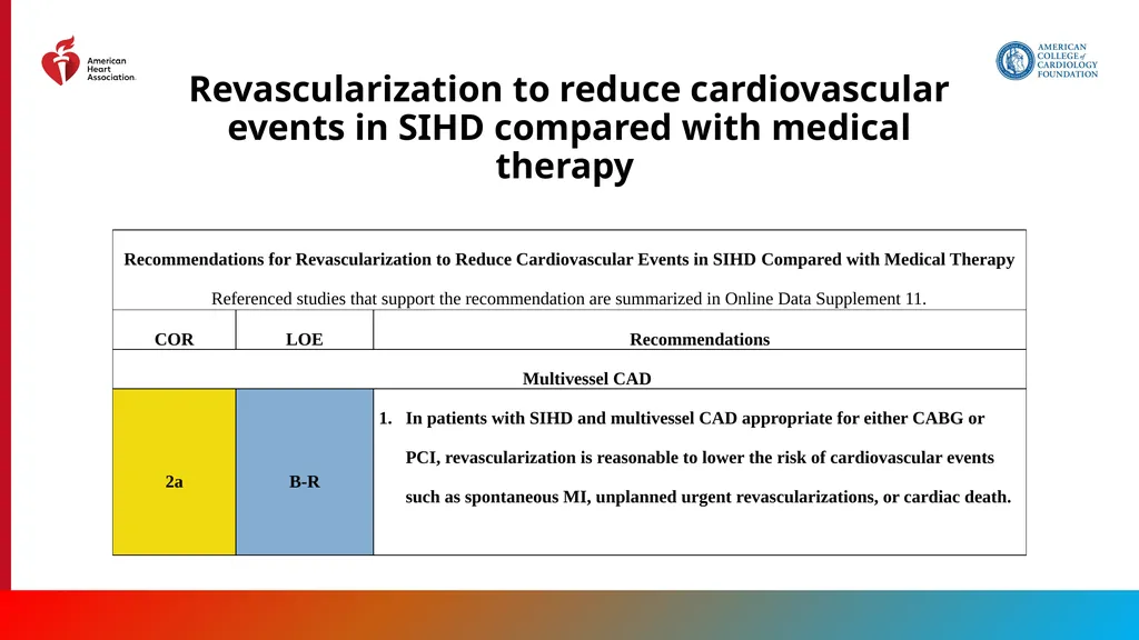
58 Revascularization to reduce cardiovascular events in SIHD compared with medical therapy<br>
59
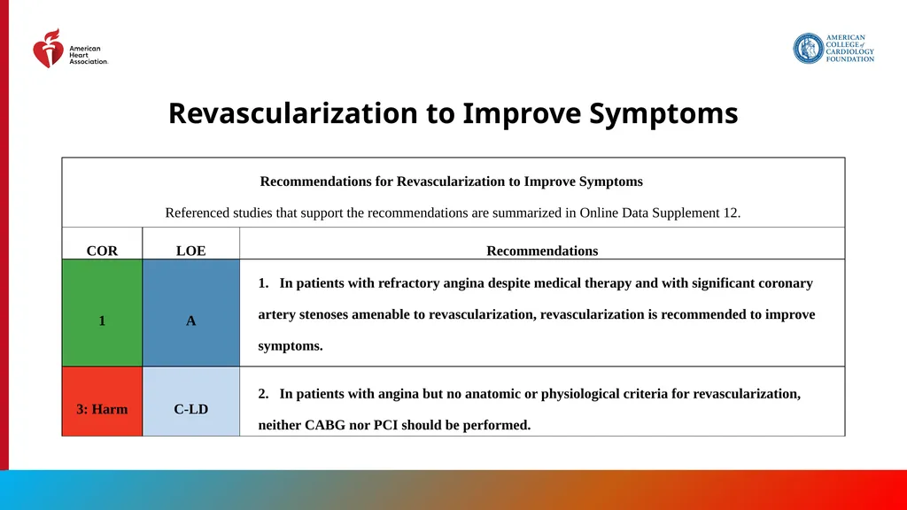
59 Revascularization to Improve Symptoms<br>
60
60 Situations in Which PCI or CABG Would Be Preferred<br>
61
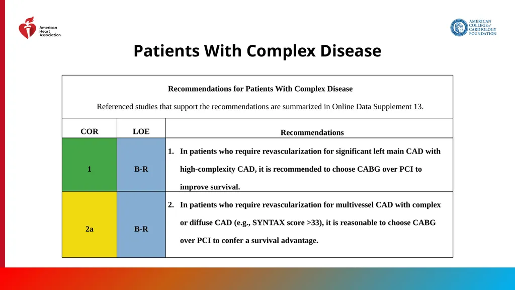
61 Patients With Complex Disease<br>
62
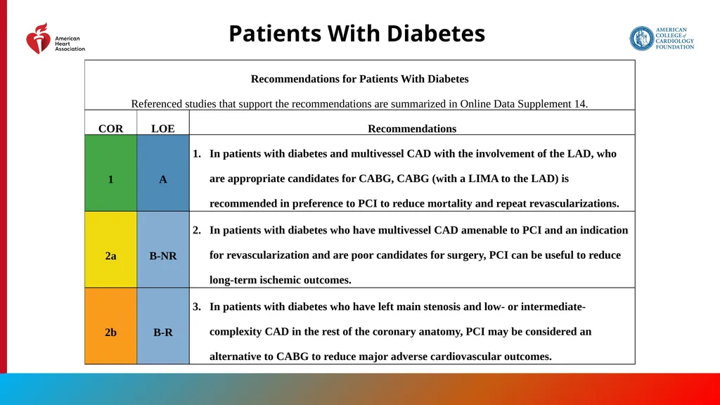
62 Patients With Diabetes<br>
63
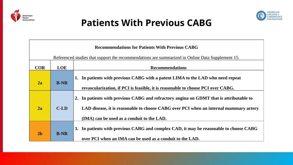
63 Patients With Previous CABG<br>
64
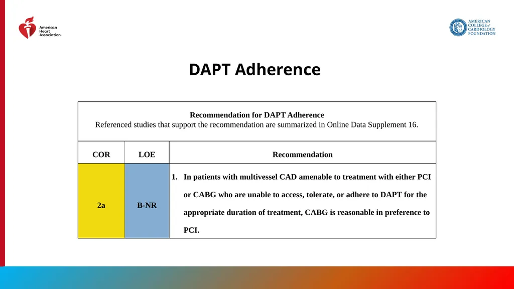
64 DAPT Adherence<br>
65
65 Special Populations and Situations<br>
66
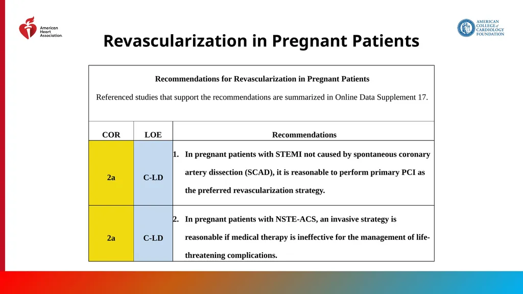
66 Revascularization in Pregnant Patients<br>
67
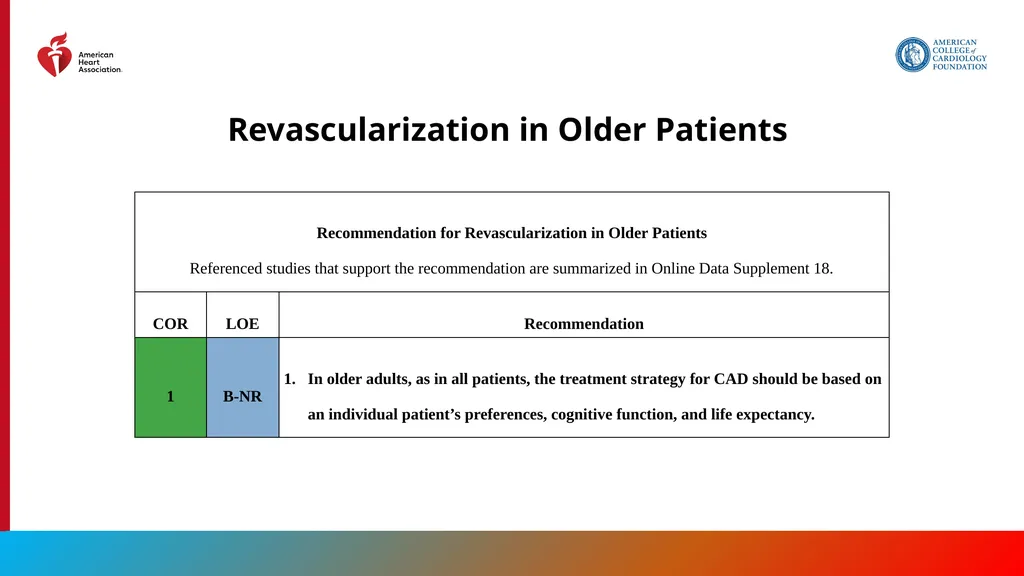
67 Revascularization in Older Patients<br>
68
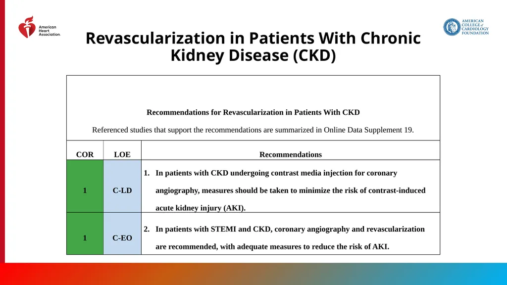
68 Revascularization in Patients With Chronic Kidney Disease (CKD)<br>
69
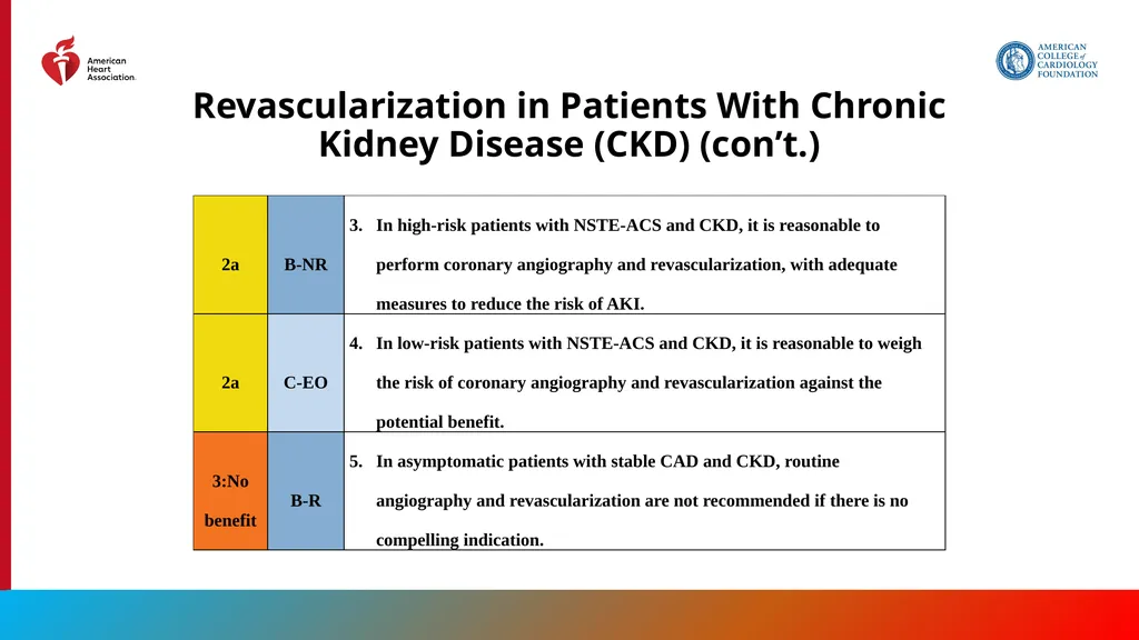
69 Revascularization in Patients With Chronic Kidney Disease (CKD) (con’t.)<br>
70
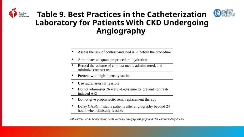
70 Table 9. Best Practices in the Catheterization Laboratory for Patients With CKD Undergoing Angiography AKI indicates acute kidney injury; CABG, coronary artery bypass graft; and CKD, chronic kidney disease.<br>
71
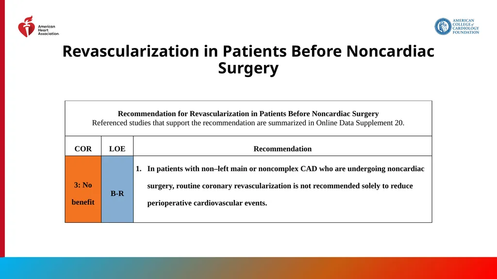
71 Revascularization in Patients Before Noncardiac Surgery<br>
72
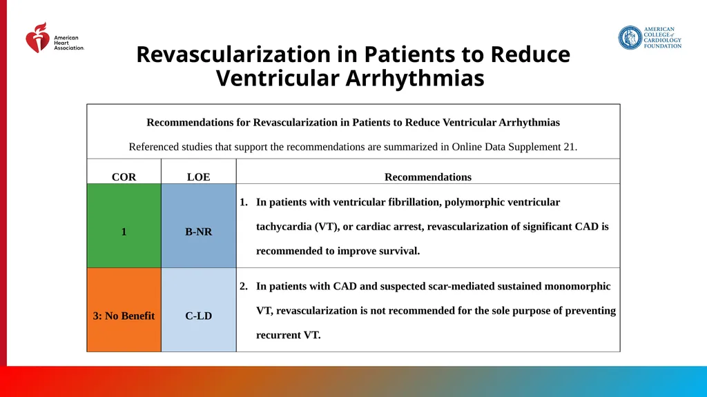
72 Revascularization in Patients to Reduce Ventricular Arrhythmias<br>
73
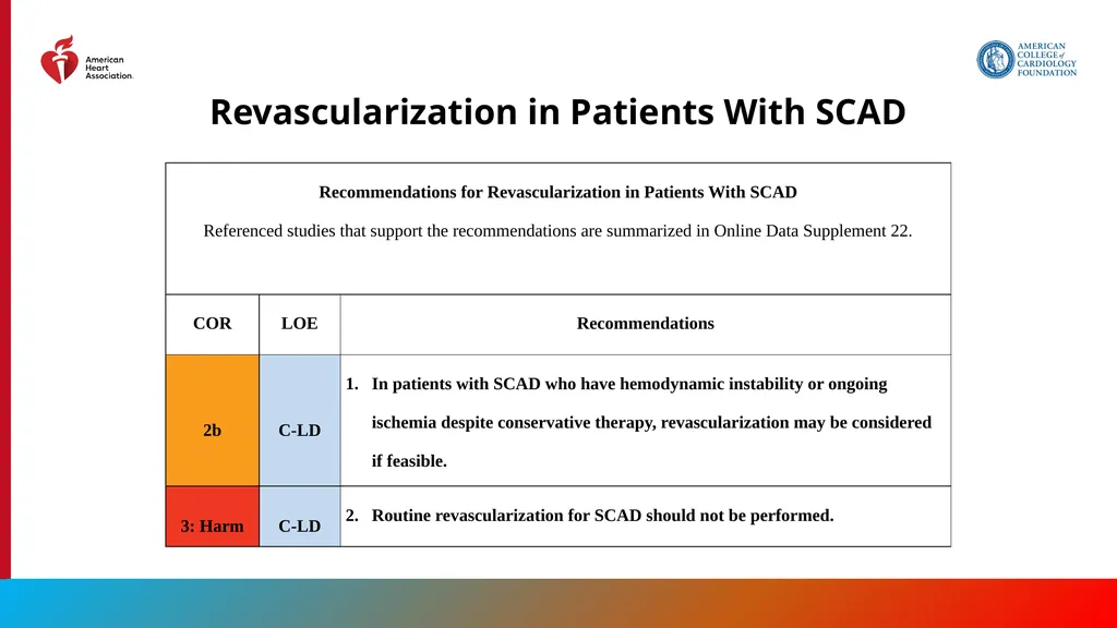
73 Revascularization in Patients With SCAD<br>
74
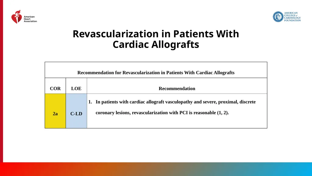
74 Revascularization in Patients With Cardiac Allografts<br>
75
75 General Procedural Issues for PCI<br>
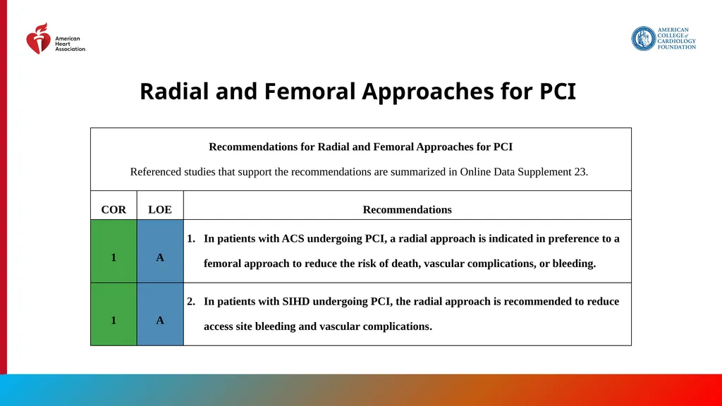
76
76 Radial and Femoral Approaches for PCI<br>
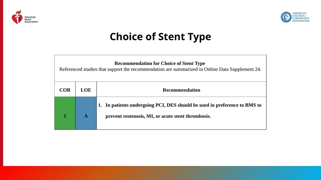
77
77 Choice of Stent Type<br>
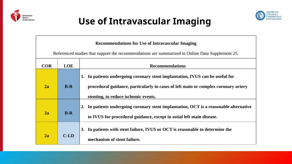
78
78 Use of Intravascular Imaging<br>
79
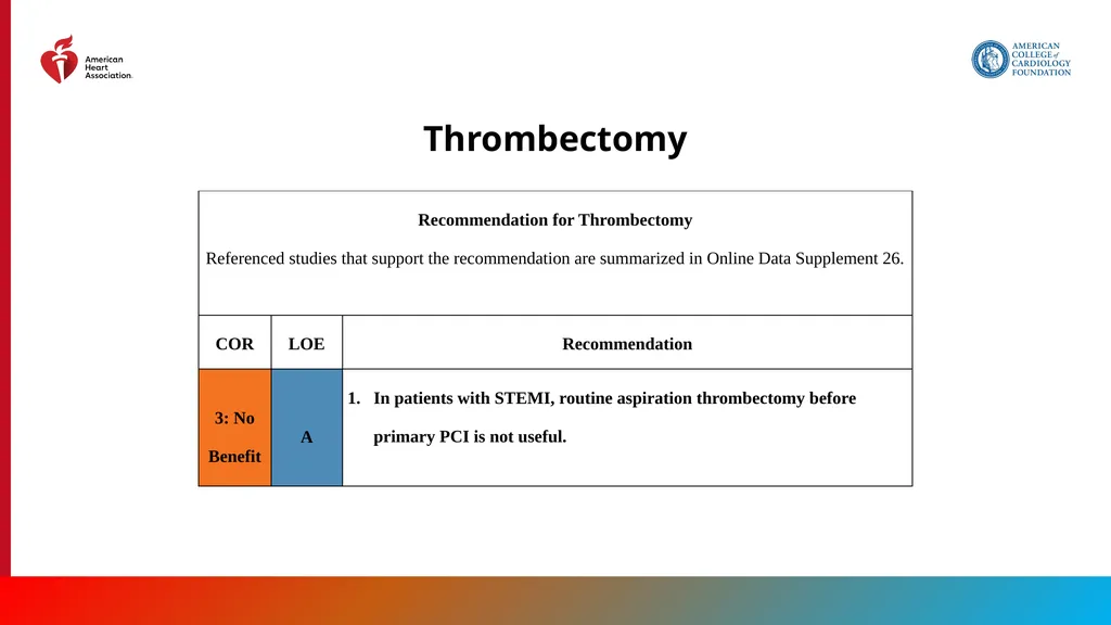
79 Thrombectomy<br>
80
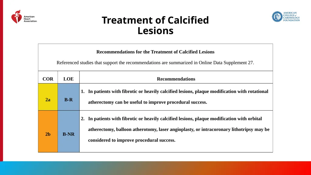
80 Treatment of Calcified Lesions<br>
81
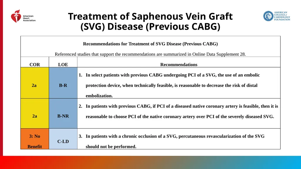
81 Treatment of Saphenous Vein Graft (SVG) Disease (Previous CABG)<br>
82
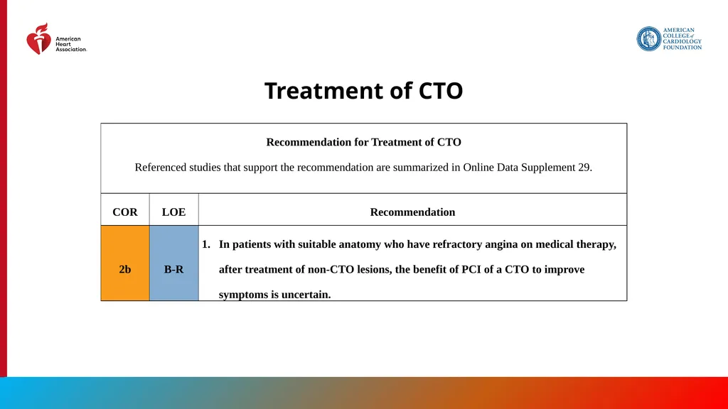
82 Treatment of CTO<br>
83
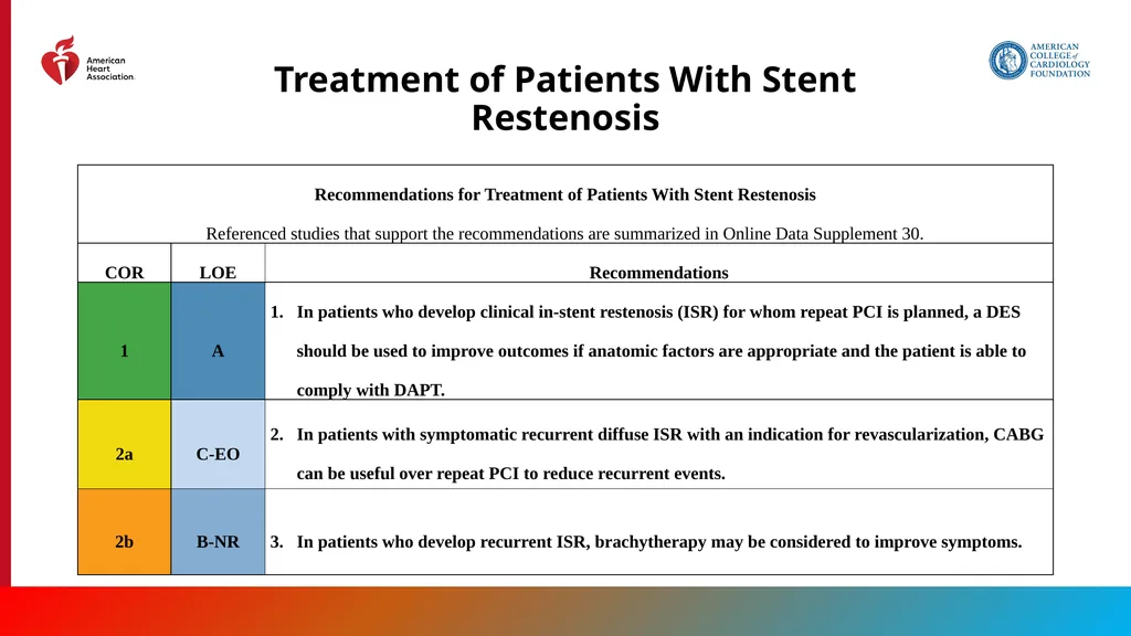
83 Treatment of Patients With Stent Restenosis<br>
84
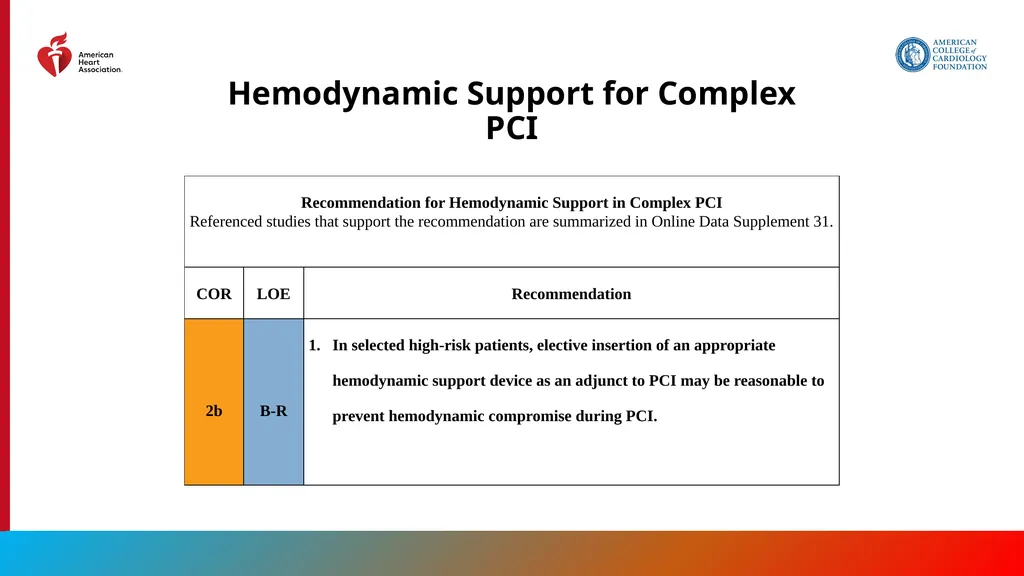
84 Hemodynamic Support for Complex PCI<br>
85
85 Pharmacotherapy in Patients Undergoing PCI<br>
86
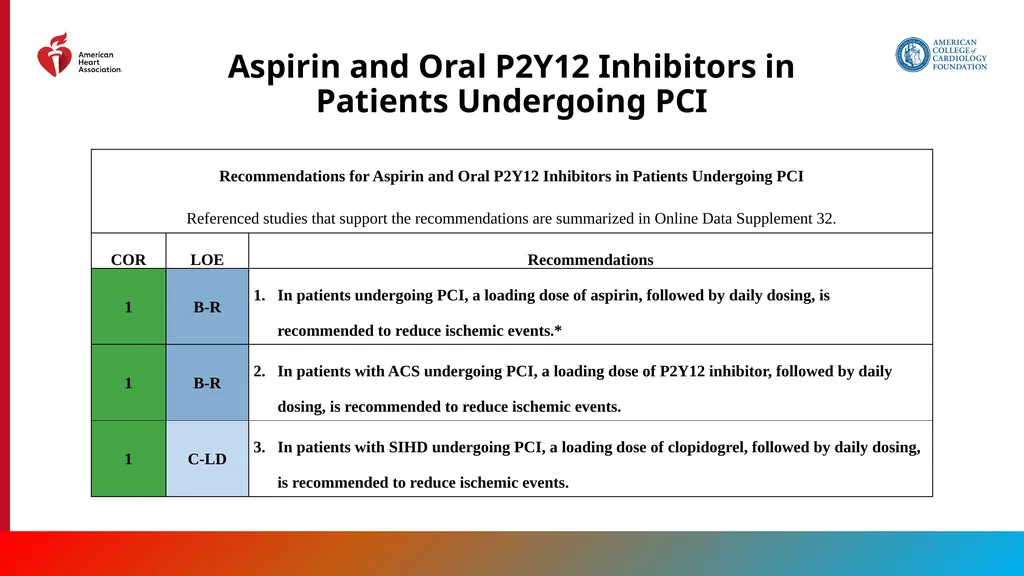
86 Aspirin and Oral P2Y12 Inhibitors in Patients Undergoing PCI<br>
87
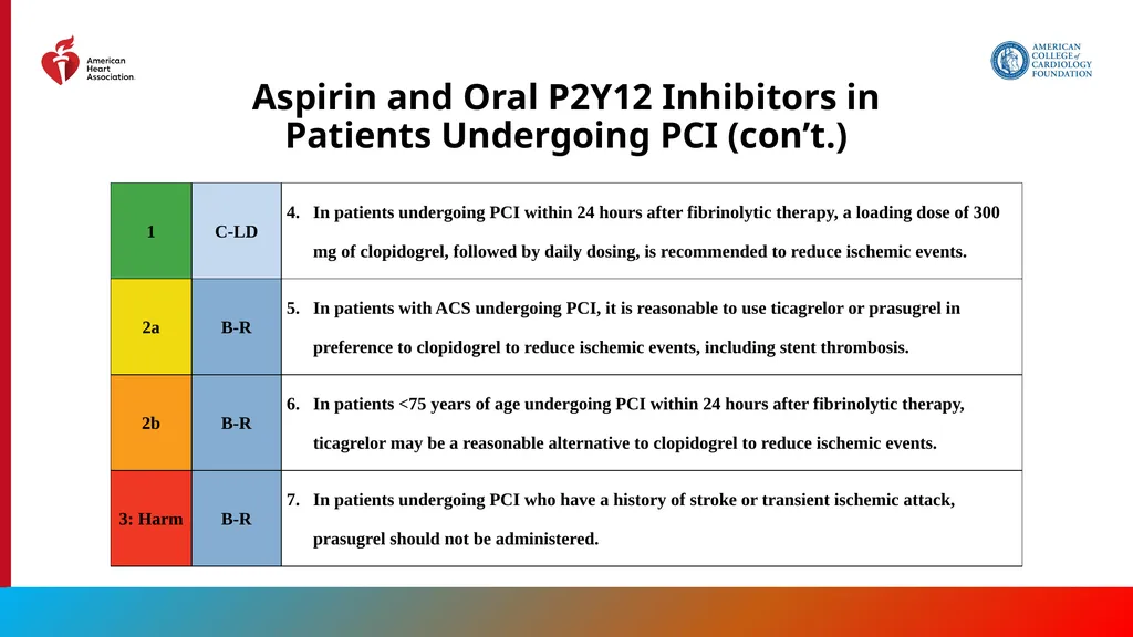
87 Aspirin and Oral P2Y12 Inhibitors in Patients Undergoing PCI (con’t.)<br>
88
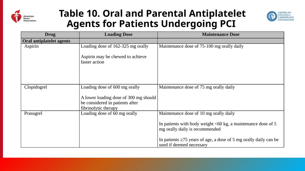
88 Table 10. Oral and Parental Antiplatelet Agents for Patients Undergoing PCI<br>
89
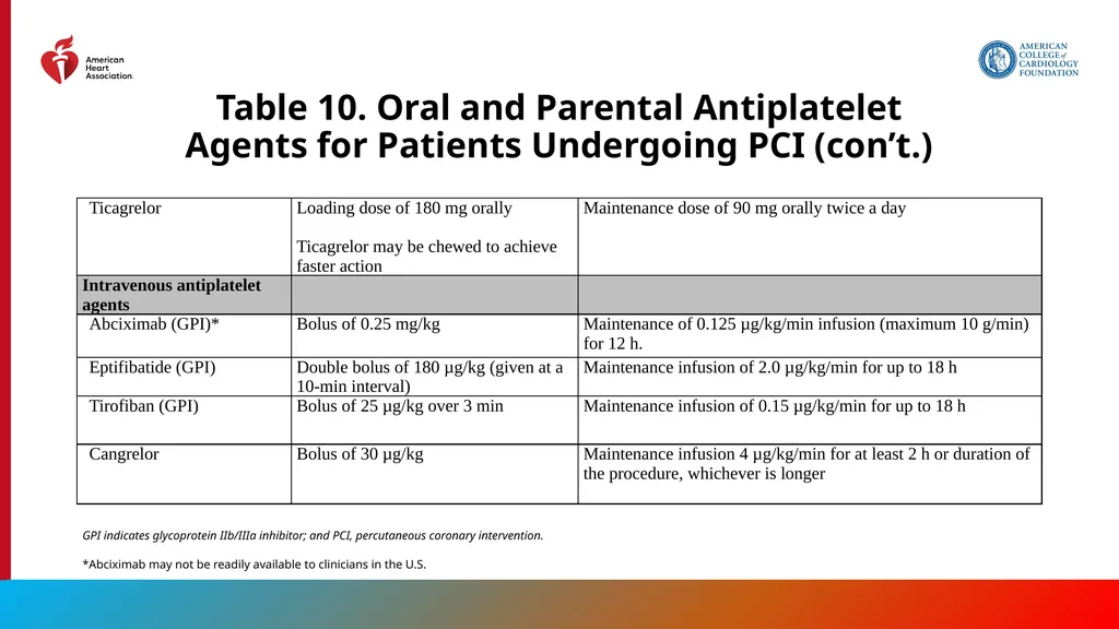
89 Table 10. Oral and Parental Antiplatelet Agents for Patients Undergoing PCI (con’t.) GPI indicates glycoprotein IIb/IIIa inhibitor; and PCI, percutaneous coronary intervention. *Abciximab may not be readily available to clinicians in the U.S.<br>
90
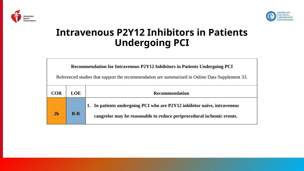
90 Intravenous P2Y12 Inhibitors in Patients Undergoing PCI<br>
91
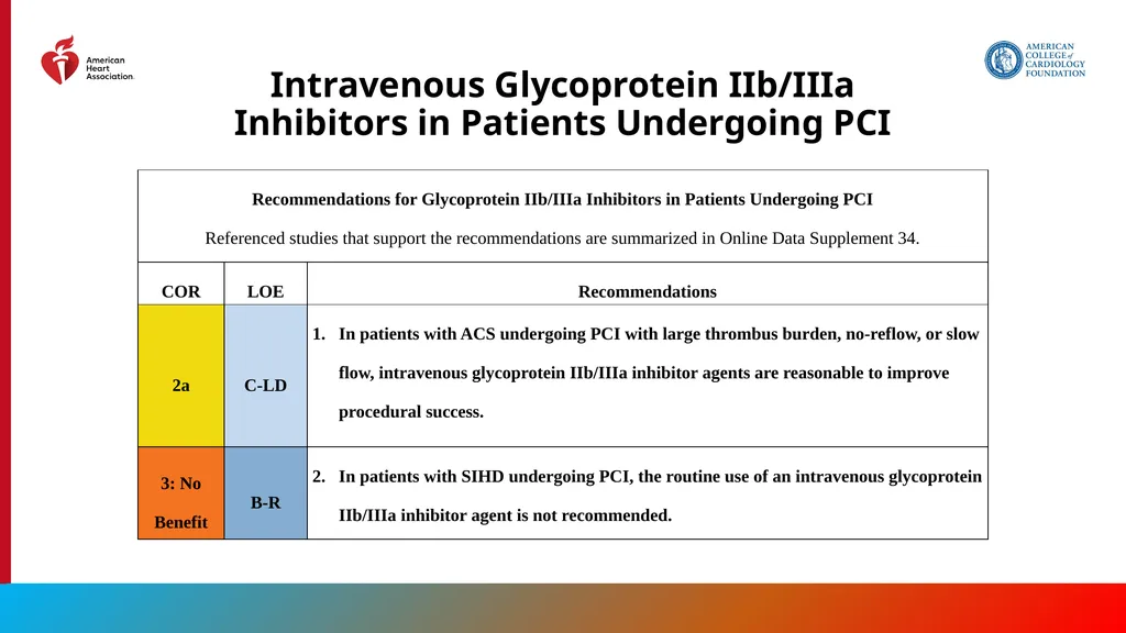
91 Intravenous Glycoprotein IIb/IIIa Inhibitors in Patients Undergoing PCI<br>
92
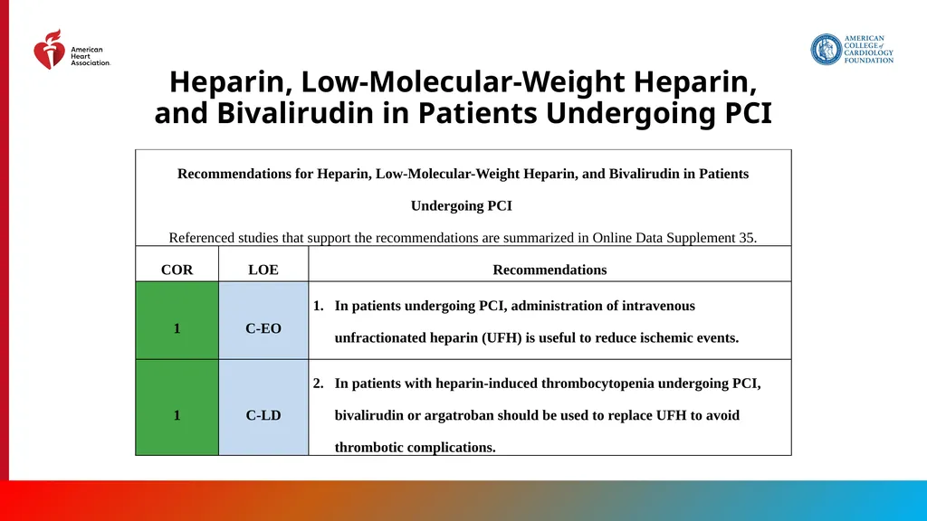
92 Heparin, Low-Molecular-Weight Heparin, and Bivalirudin in Patients Undergoing PCI<br>
93
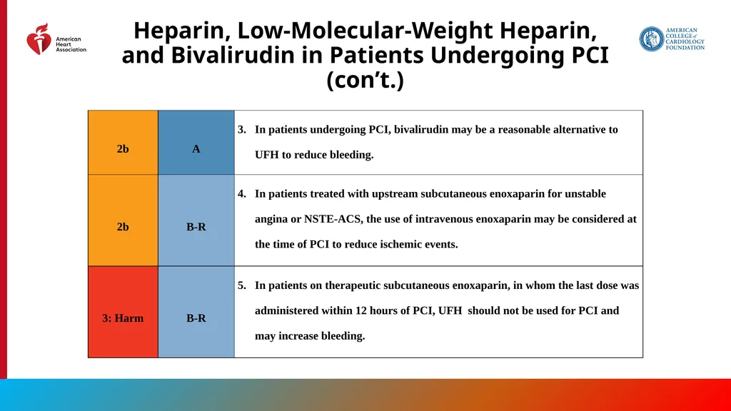
93 Heparin, Low-Molecular-Weight Heparin, and Bivalirudin in Patients Undergoing PCI (con’t.)<br>
94
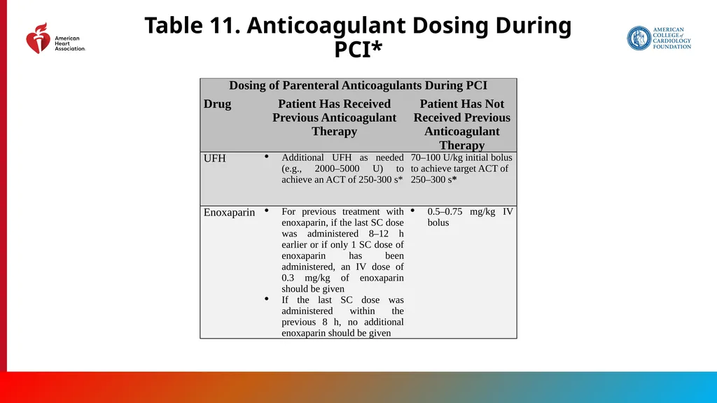
Table 11. Anticoagulant Dosing During PCI* 94<br>
95
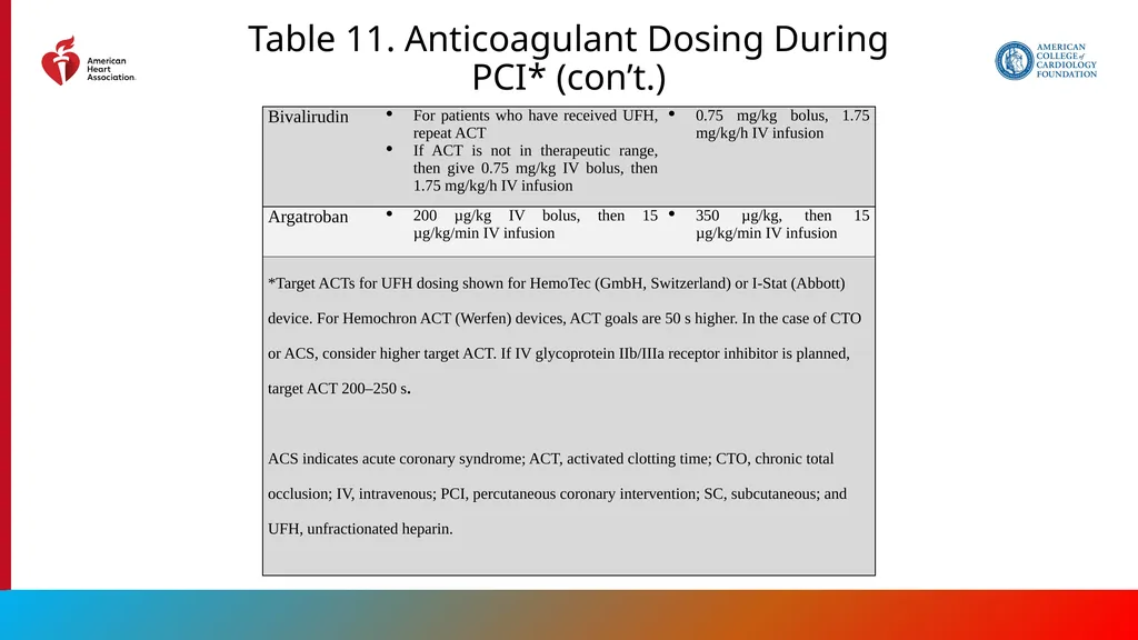
95 Table 11. Anticoagulant Dosing During PCI* (con’t.)<br>
96
96 General Procedural Issues for CABG<br>
97
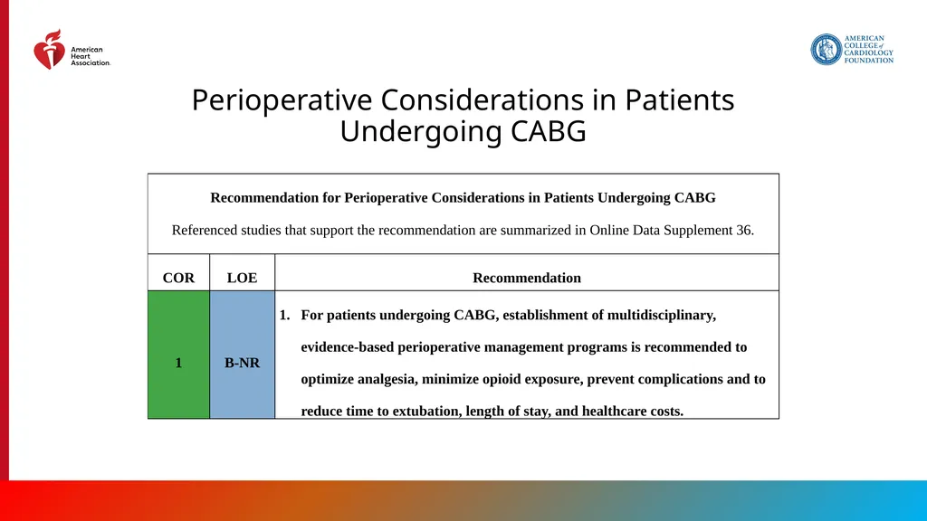
97 Perioperative Considerations in Patients Undergoing CABG<br>
98
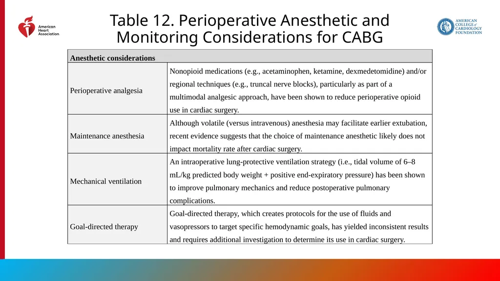
98 Table 12. Perioperative Anesthetic and Monitoring Considerations for CABG<br>
99
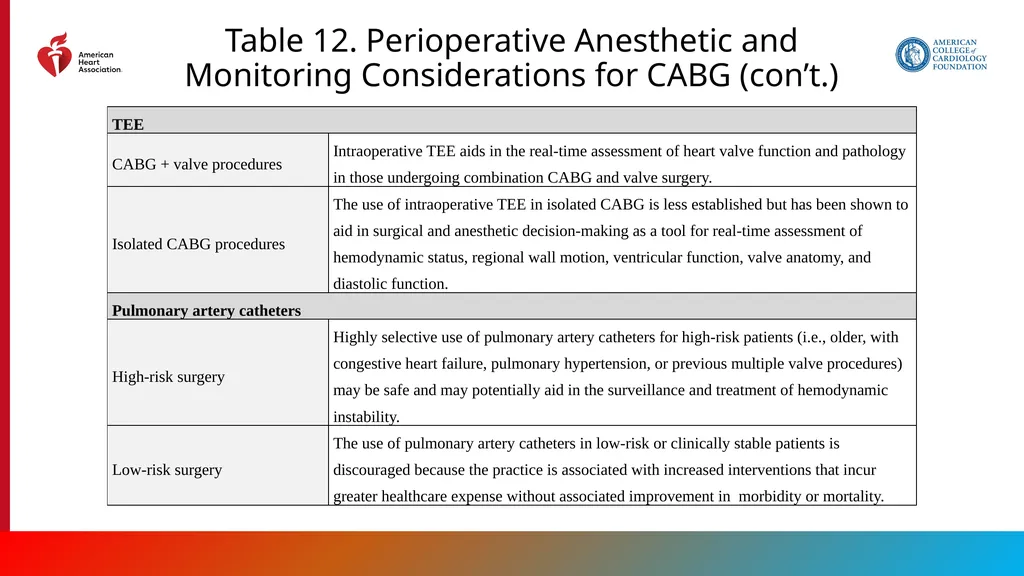
99 Table 12. Perioperative Anesthetic and Monitoring Considerations for CABG (con’t.)<br>
100
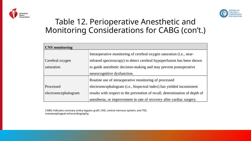
100 Table 12. Perioperative Anesthetic and Monitoring Considerations for CABG (con’t.) CABG indicates coronary artery bypass graft; CNS, central nervous system; and TEE, transesophageal echocardiography.<br>
101
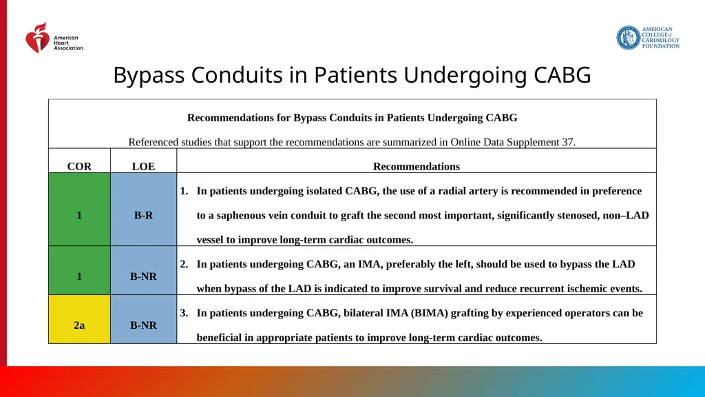
101 Bypass Conduits in Patients Undergoing CABG<br>
102
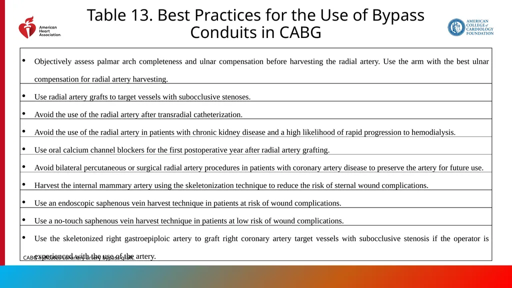
102 Table 13. Best Practices for the Use of Bypass Conduits in CABG CABG indicates coronary artery bypass graft.<br>
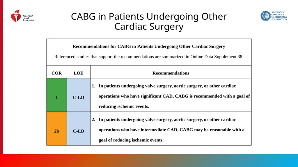
103
103 CABG in Patients Undergoing Other Cardiac Surgery<br>
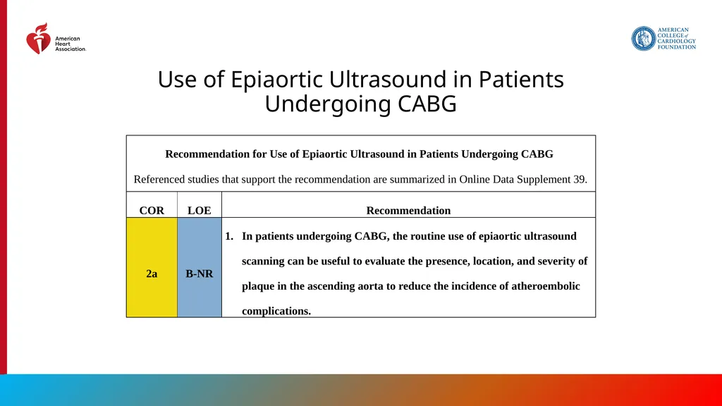
104
104 Use of Epiaortic Ultrasound in Patients Undergoing CABG<br>
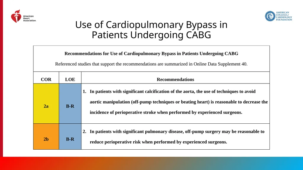
105
105 Use of Cardiopulmonary Bypass in Patients Undergoing CABG<br>
106
106 Pharmacotherapy in Patients Undergoing CABG<br>
107
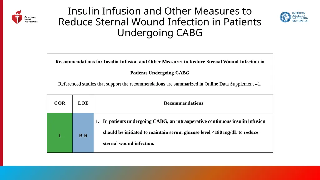
107 Insulin Infusion and Other Measures to Reduce Sternal Wound Infection in Patients Undergoing CABG<br>
108
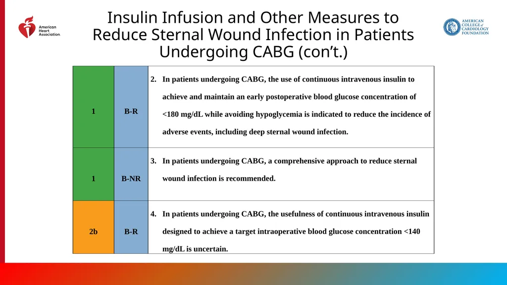
108 Insulin Infusion and Other Measures to Reduce Sternal Wound Infection in Patients Undergoing CABG (con’t.)<br>
109
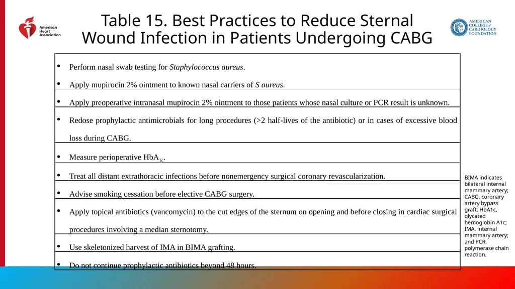
109 Table 15. Best Practices to Reduce Sternal Wound Infection in Patients Undergoing CABG BIMA indicates bilateral internal mammary artery; CABG, coronary artery bypass graft; HbA1c, glycated hemoglobin A1c; IMA, internal mammary artery; and PCR, polymerase chain reaction.<br>
110
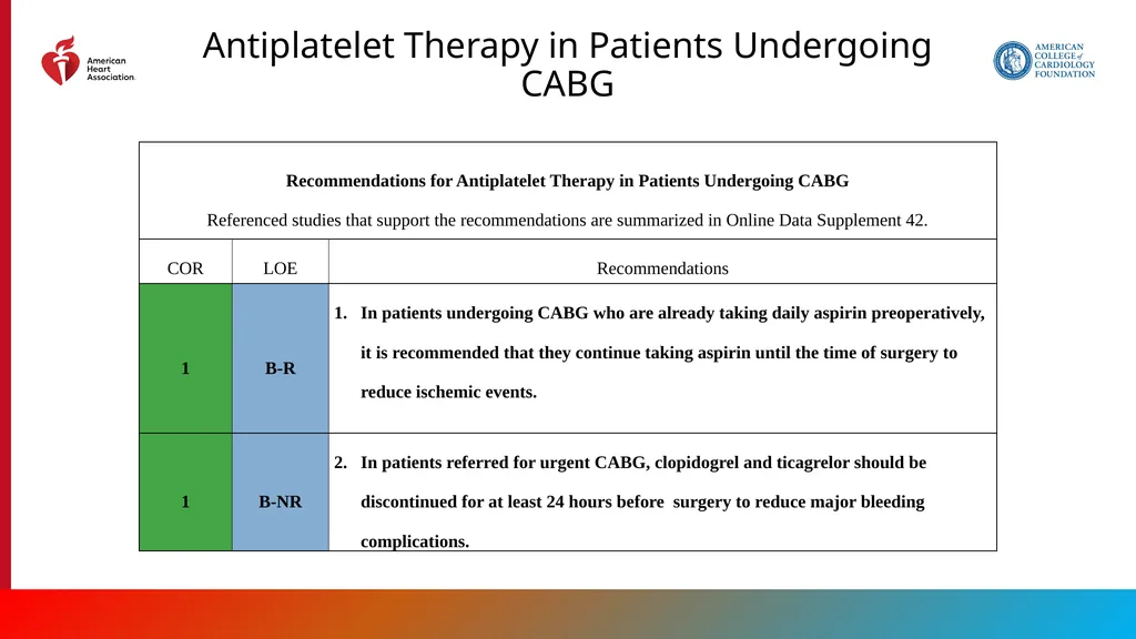
110 Antiplatelet Therapy in Patients Undergoing CABG<br>
111
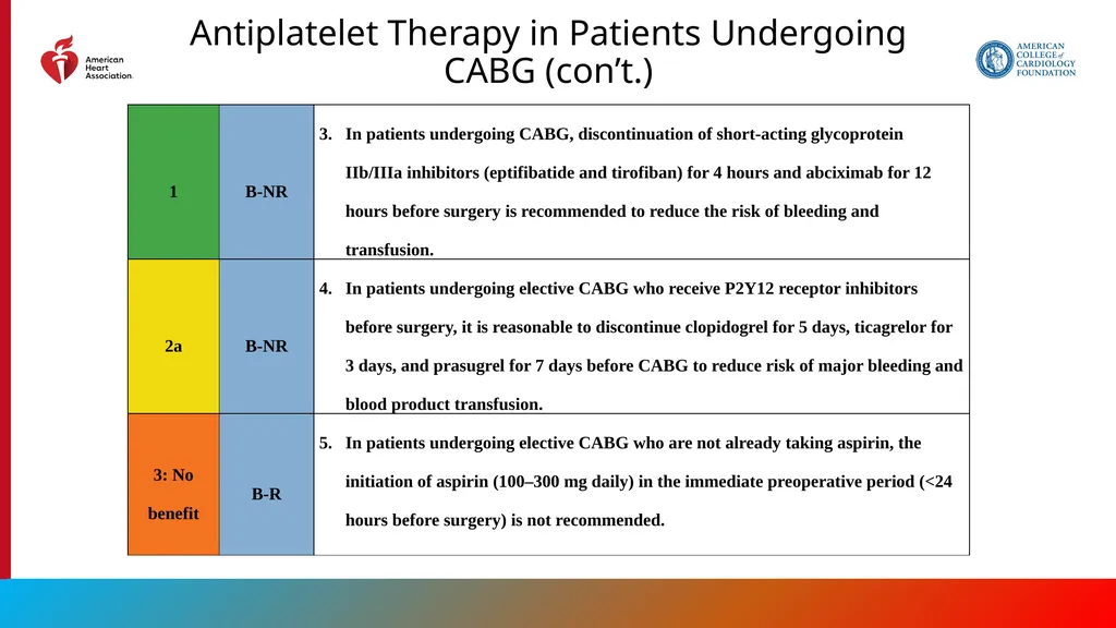
111 Antiplatelet Therapy in Patients Undergoing CABG (con’t.)<br>
112
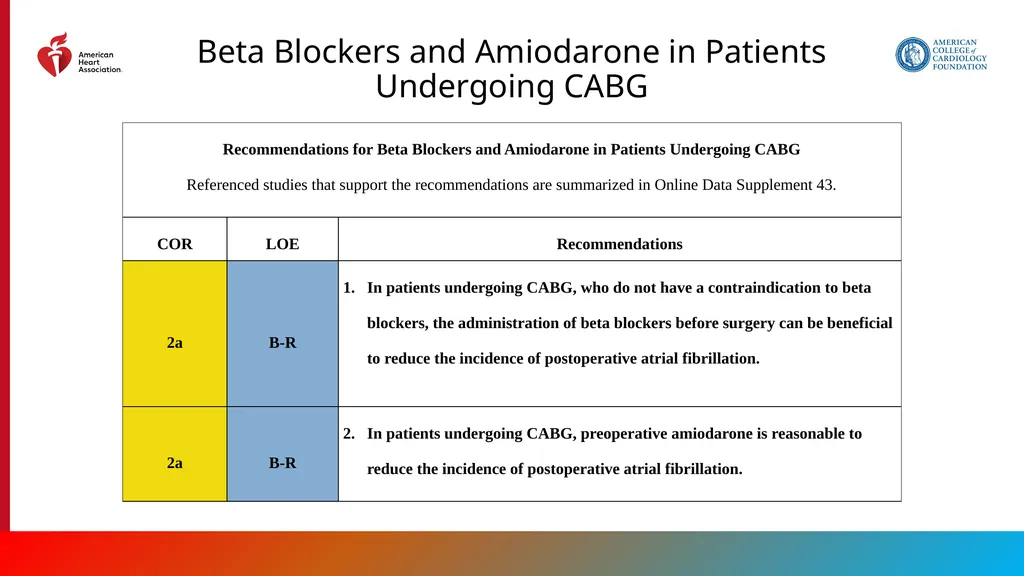
112 Beta Blockers and Amiodarone in Patients Undergoing CABG<br>
113
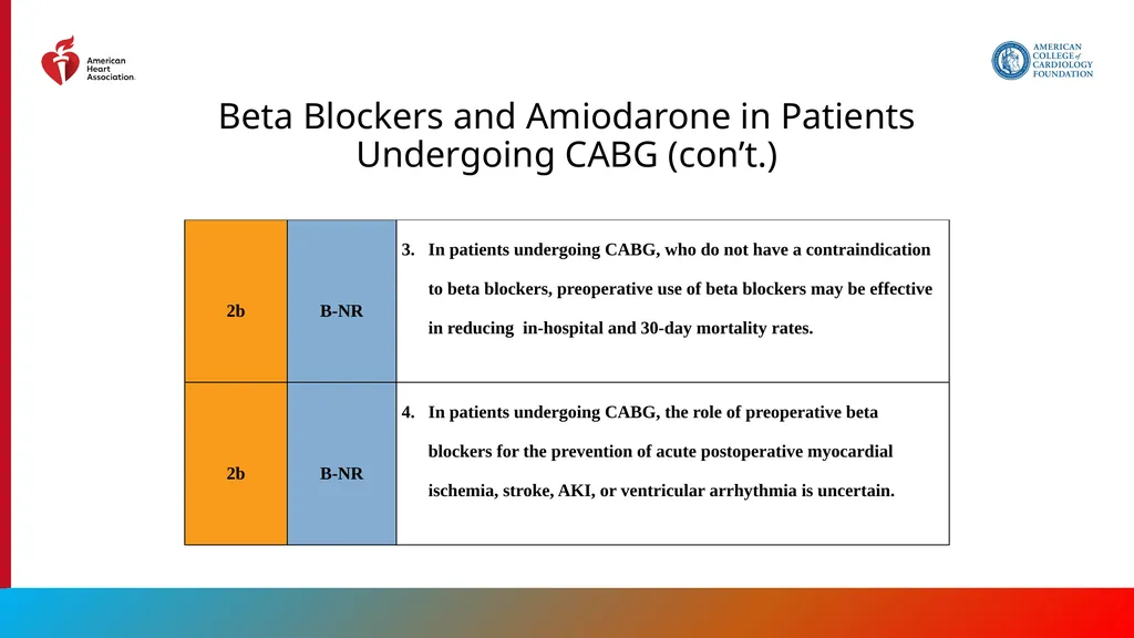
113 Beta Blockers and Amiodarone in Patients Undergoing CABG (con’t.)<br>
114
114 Pharmacotherapy in Patients After Revascularization<br>
115
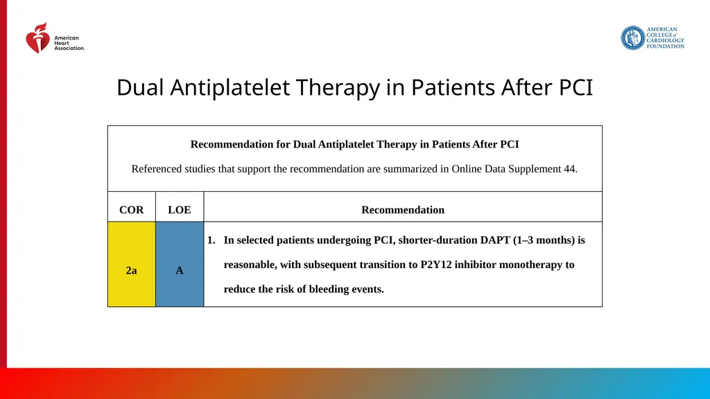
115 Dual Antiplatelet Therapy in Patients After PCI<br>
116
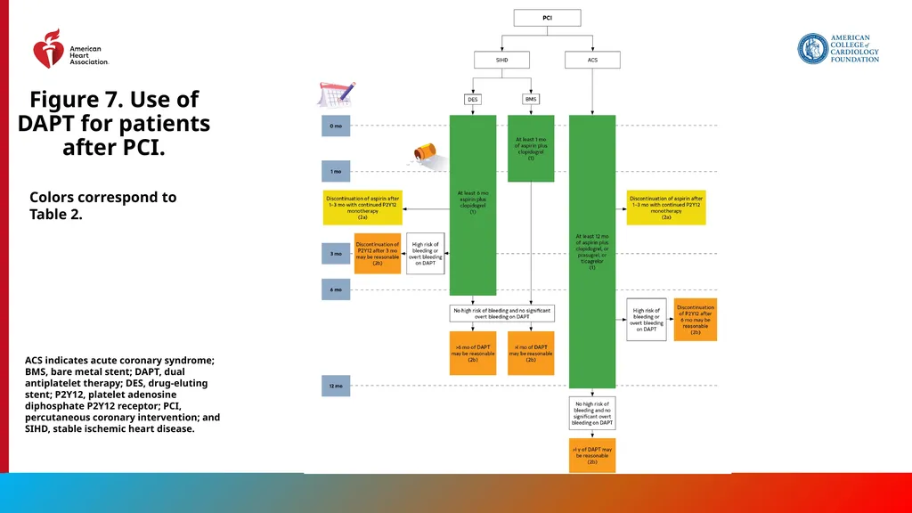
116 Figure 7. Use of DAPT for patients after PCI. Colors correspond to Table 2. ACS indicates acute coronary syndrome; BMS, bare metal stent; DAPT, dual antiplatelet therapy; DES, drug-eluting stent; P2Y12, platelet adenosine diphosphate P2Y12 receptor; PCI, percutaneous coronary intervention; and SIHD, stable ischemic heart disease.<br>
117
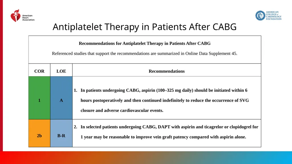
117 Antiplatelet Therapy in Patients After CABG<br>
118
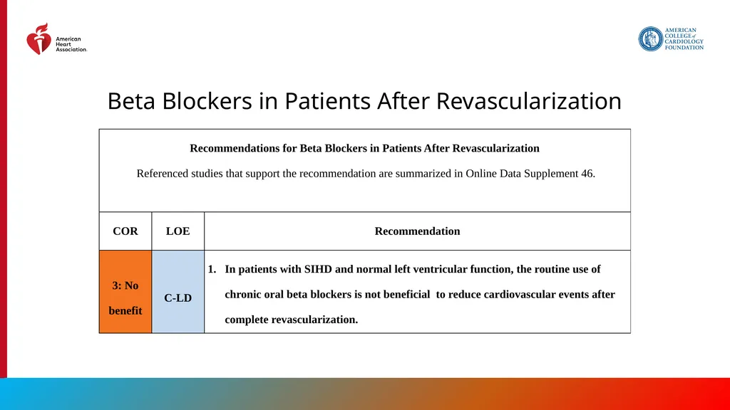
118 Beta Blockers in Patients After Revascularization<br>
119
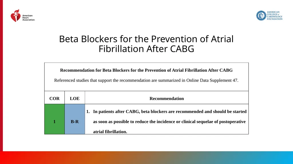
119 Beta Blockers for the Prevention of Atrial Fibrillation After CABG<br>
120
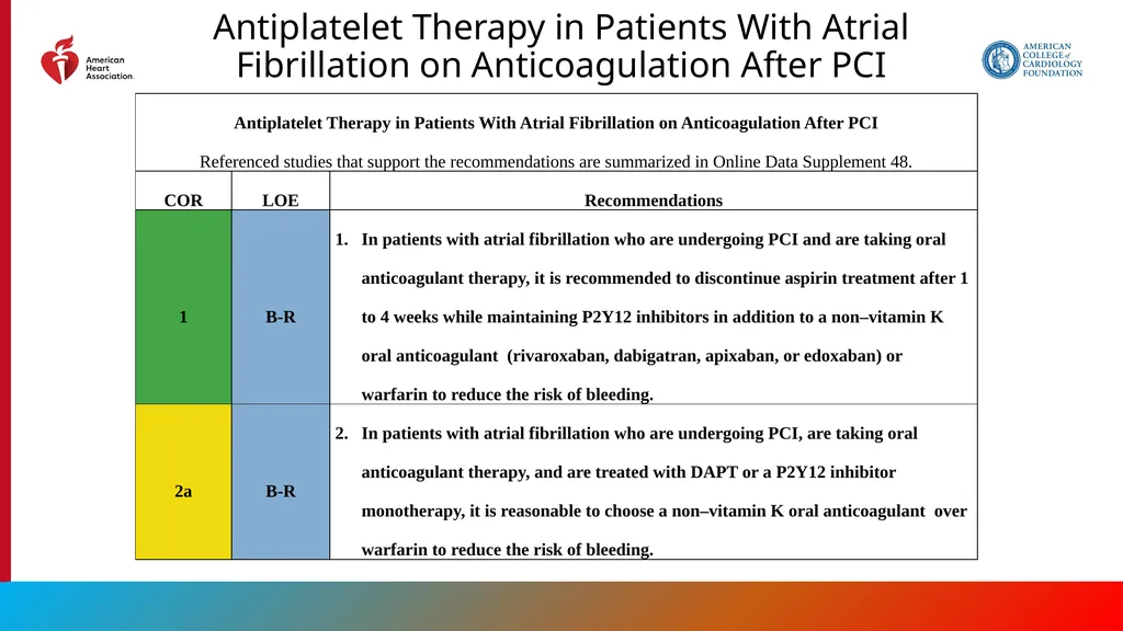
120 Antiplatelet Therapy in Patients With Atrial Fibrillation on Anticoagulation After PCI<br>
121
121 Recommendations for Addressing Psychosocial Factors and Lifestyle Changes After Revascularization<br>
122
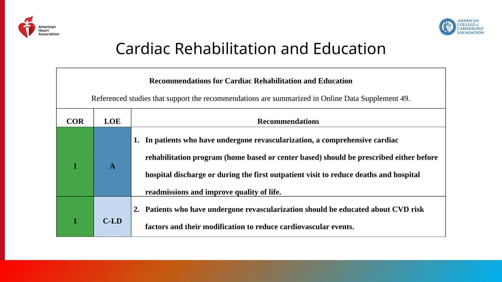
122 Cardiac Rehabilitation and Education<br>
123
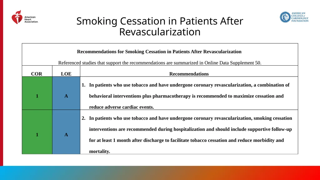
123 Smoking Cessation in Patients After Revascularization<br>
124
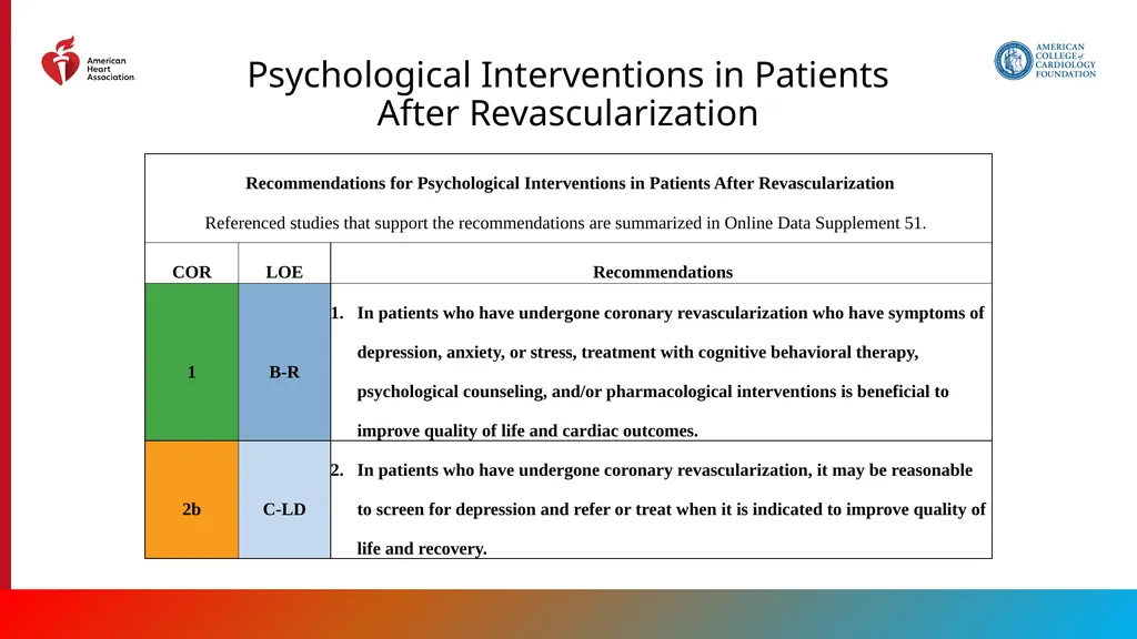
124 Psychological Interventions in Patients After Revascularization<br>
125
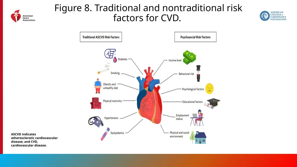
125 Figure 8. Traditional and nontraditional risk factors for CVD. ASCVD indicates atherosclerotic cardiovascular disease; and CVD, cardiovascular disease.<br>
126
126 Revascularization Outcomes<br>
127
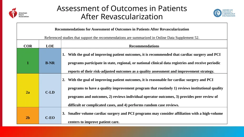
127 Assessment of Outcomes in Patients After Revascularization<br>
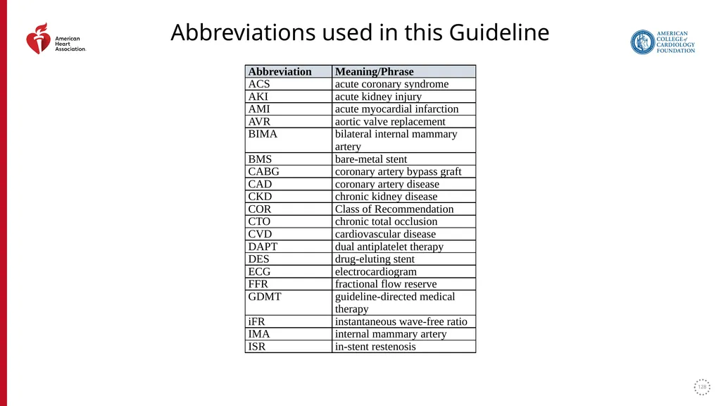
128
128 Abbreviations used in this Guideline<br>
129
Abbreviations used in this Guideline<br>