
ERYTHROCYTE SEDIMENTATION RATE (ESR) Relevance


Description: ERYTHROCYTE SEDIMENTATION RATE (ESR) Relevance ESR: The rate at which the red cells settle or sediment. It is often required by a physician to rule out the presence of organic disease (aid in diagnosis), or to follow the progress of a
Related Topics
Download Presentation
"ERYTHROCYTE SEDIMENTATION RATE (ESR) Relevance" is the property of its rightful owner. Permission is granted to download and print the materials on this website for personal, non-commercial use only, and to display it on your personal computer provided you do not modify the materials and that you retain all copyright notices contained in the materials. By downloading content from our website, you accept the terms of this agreement.
Presentation Transcript
slide1. ERYTHROCYTE SEDIMENTATION RATE (ESR)<br>
slide2. Relevance ESR: The rate at which the red cells settle or sediment.
It is often required by a physician to rule out the presence of organic disease (aid in diagnosis), or to follow the progress of a disease.
ESR is generally done as part of complete blood tests.<br>
slide3. Principle In the circulating blood the red cells remain uniformly suspended in the plasma.
However, when a sample of anticoagulated blood, is allowed to stand in a narrow vertical tube, the RBCs being heavier (denser) than the plasma, settle or sediment gradually towards the bottom of the tube (rouleaux formation).<br>
slide4. Sedimentation of red cells The settling of red cells in a sample of anticoagulated blood occurs in 3 stages:
The first stage, the RBCs pile up (like a stack of coins), and form rouleaux that become heavier during the first 10-15 minutes.<br>
slide5. Sedimentation of red cells The second stage, the rouleau being heavier sink to the bottom. This stage lasts for 40–45 minutes.
The third stage, there is packing of massed bunches of red cells at the bottom of the blood column. This stage lasts for about 10–12 minutes.<br>
slide6. Factors affecting ESR Technical and mechanical factors
(The length of the tube, Diameter of the bore)
Physiological factors
(Age, Sex, High altitude, Pregnancy, Body temperature)
Viscosity of Blood
Nature of Anticoagulant Used<br>
slide7. Clinical significance of ESR As an indicator of bodily reaction to tissue injury and inflammation.
Value of ESR as a prognostic tool.<br>
slide8. Normal values Males : 3–9 mm 1st hour.
Females : 5–12 mm 1st hour.<br>
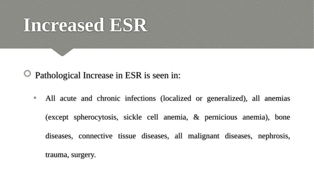
slide9. Increased ESR Pathological Increase in ESR is seen in:
All acute and chronic infections (localized or generalized), all anemias (except spherocytosis, sickle cell anemia, & pernicious anemia), bone diseases, connective tissue diseases, all malignant diseases, nephrosis, trauma, surgery.<br>
slide10. Decreased ESR Pathological decrease in ESR is seen in:
Polycythemia, anemias (spherocytosis, sickle cell anemia, & pernicious anemia), afibrinogenemia, severe allergic reactions.<br>
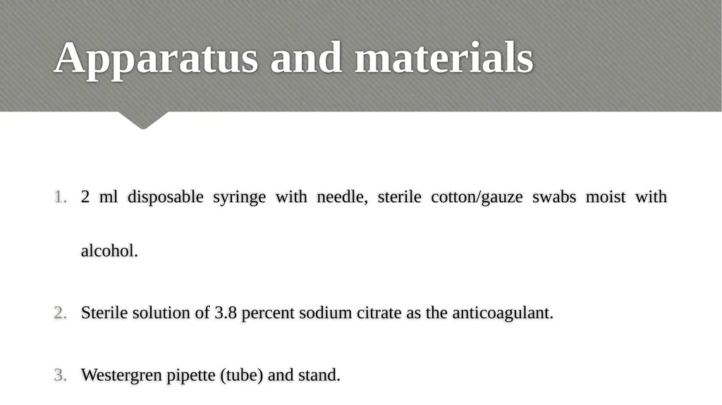
slide11. Apparatus and materials 2 ml disposable syringe with needle, sterile cotton/gauze swabs moist with alcohol.
Sterile solution of 3.8 percent sodium citrate as the anticoagulant.
Westergren pipette (tube) and stand.<br>
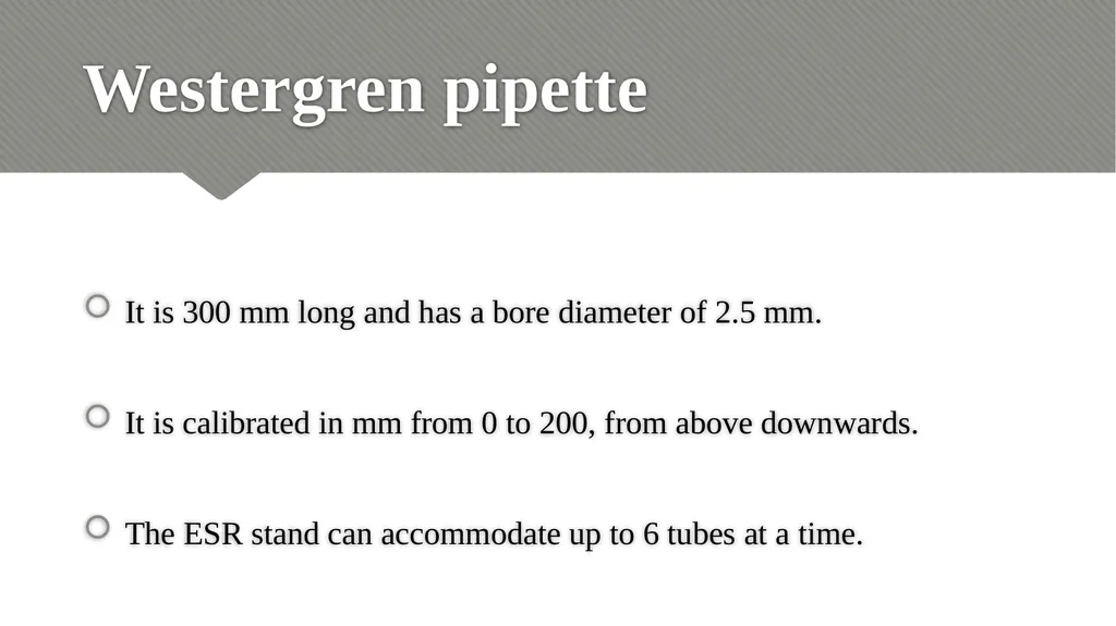
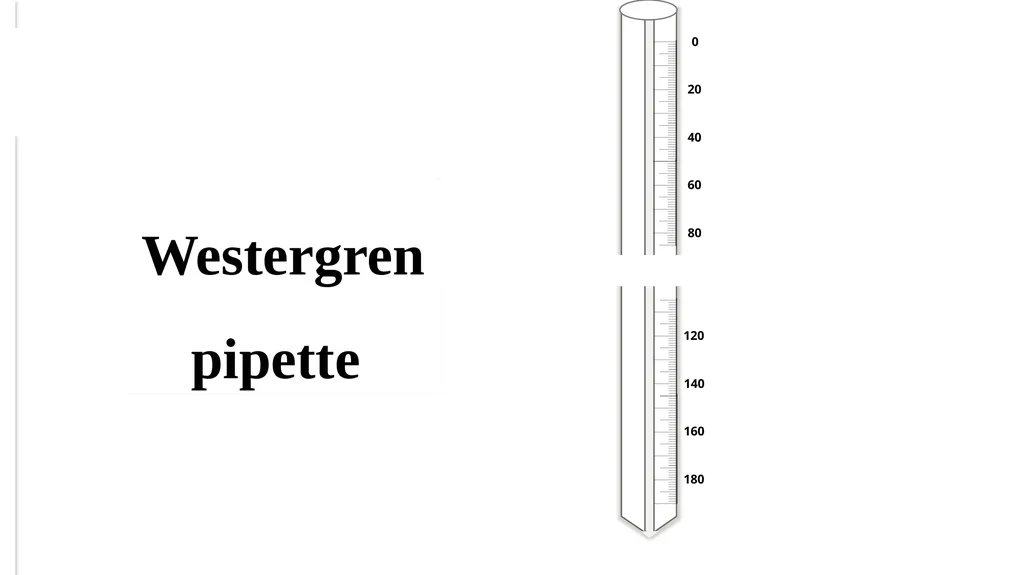
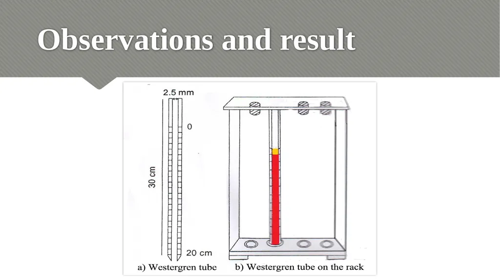
slide12. Westergren pipette It is 300 mm long and has a bore diameter of 2.5 mm.
It is calibrated in mm from 0 to 200, from above downwards.
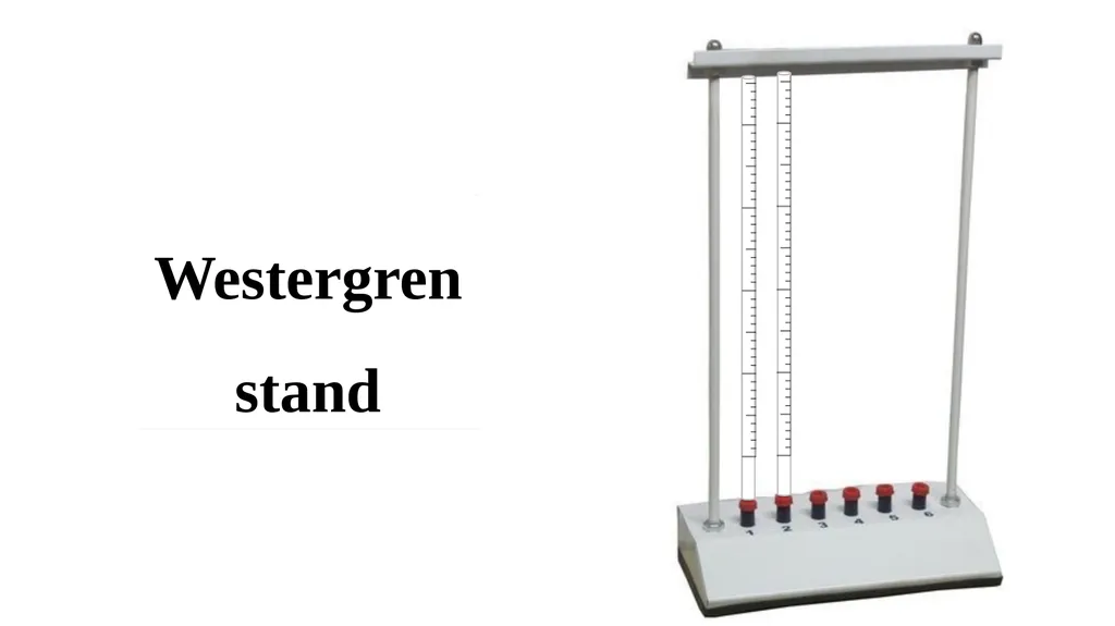
The ESR stand can accommodate up to 6 tubes at a time.<br>
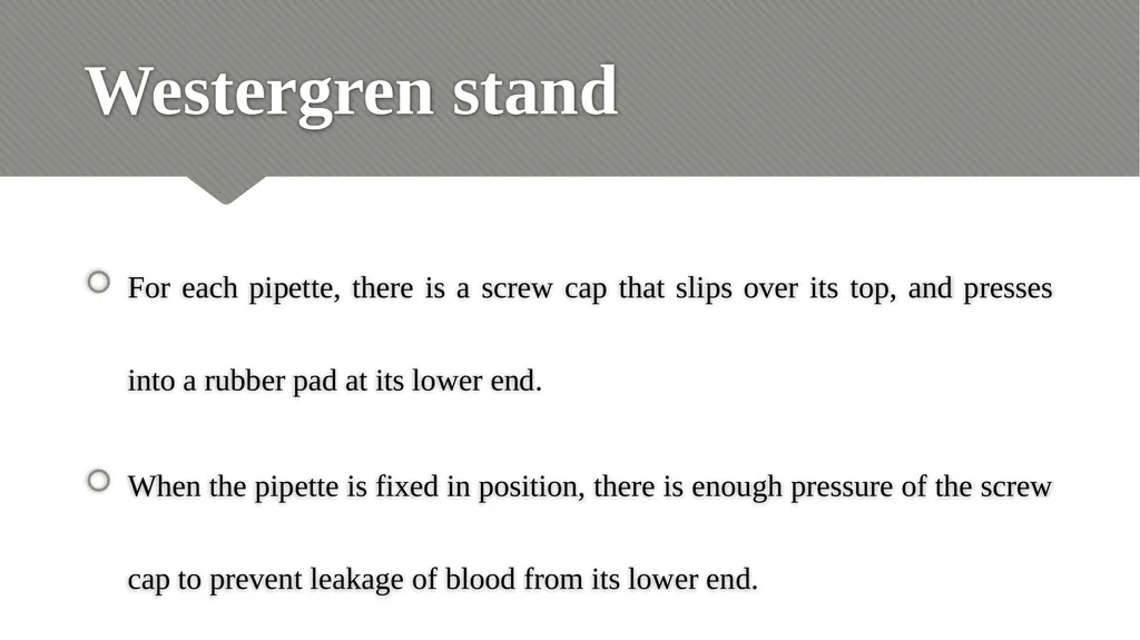
slide13. Westergren stand For each pipette, there is a screw cap that slips over its top, and presses into a rubber pad at its lower end.
When the pipette is fixed in position, there is enough pressure of the screw cap to prevent leakage of blood from its lower end.<br>
slide14. 0 20 40 180 160 140 Westergren
pipette 60 80 120<br>
slide15. Westergren
stand<br>
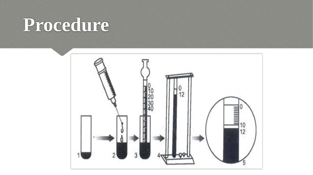
slide16. Procedure Draw 2.0 ml of venous blood and transfer it into a vial containing 0.5 ml of 3.8% sodium citrate solution.
Mix the contents by inverting or swirling the vial.
Do not shake, as it will cause frothing.<br>
slide17. Procedure Fill the Westergren’s pipette with blood-citrate mixture by sucking, after placing the tip of your finger over the top of the pipette to control the flow of blood into and out of it, or with a rubber bulb. Bring the blood column to exact zero mark.<br>
slide18. Procedure<br>
slide19. Procedure Keeping your finger over the pipette, transfer it to the Westergren stand by firmly pressing its lower end into the rubber cushion.
Now slip the upper end of the pipette under the screw cap.
Confirm that there is no leakage of blood and that the pipette will remain vertical.<br>
slide20. Procedure Leave the pipette undisturbed for one hour, at the end of which read the mm of clear plasma above the red cells.
Express your results as: ………………mm 1st hour.<br>
slide21. Observations and result<br>
slide22. Sources of error These include:
Tilting of the tube and high temperature lead to high values.
Low temperature gives false low values.
Hemolysed blood may obscure the sharp line separating red cells and the plasma.<br>
slide23. PRECAUTIONS The test should preferably be done within 2–3 hours of collecting the blood sample, and at a room temperature of 20–35°C.
The hematocrit should be checked and correction factor applied in cases of anemia.<br>
slide24. PRECAUTIONS Clotted or hemolysed blood must be discarded.
The Westergren pipette should not be disturbed from the vertical position for the duration of the test.<br>
slide2. Relevance ESR: The rate at which the red cells settle or sediment.
It is often required by a physician to rule out the presence of organic disease (aid in diagnosis), or to follow the progress of a disease.
ESR is generally done as part of complete blood tests.<br>
slide3. Principle In the circulating blood the red cells remain uniformly suspended in the plasma.
However, when a sample of anticoagulated blood, is allowed to stand in a narrow vertical tube, the RBCs being heavier (denser) than the plasma, settle or sediment gradually towards the bottom of the tube (rouleaux formation).<br>
slide4. Sedimentation of red cells The settling of red cells in a sample of anticoagulated blood occurs in 3 stages:
The first stage, the RBCs pile up (like a stack of coins), and form rouleaux that become heavier during the first 10-15 minutes.<br>
slide5. Sedimentation of red cells The second stage, the rouleau being heavier sink to the bottom. This stage lasts for 40–45 minutes.
The third stage, there is packing of massed bunches of red cells at the bottom of the blood column. This stage lasts for about 10–12 minutes.<br>
slide6. Factors affecting ESR Technical and mechanical factors
(The length of the tube, Diameter of the bore)
Physiological factors
(Age, Sex, High altitude, Pregnancy, Body temperature)
Viscosity of Blood
Nature of Anticoagulant Used<br>
slide7. Clinical significance of ESR As an indicator of bodily reaction to tissue injury and inflammation.
Value of ESR as a prognostic tool.<br>
slide8. Normal values Males : 3–9 mm 1st hour.
Females : 5–12 mm 1st hour.<br>
slide9. Increased ESR Pathological Increase in ESR is seen in:
All acute and chronic infections (localized or generalized), all anemias (except spherocytosis, sickle cell anemia, & pernicious anemia), bone diseases, connective tissue diseases, all malignant diseases, nephrosis, trauma, surgery.<br>
slide10. Decreased ESR Pathological decrease in ESR is seen in:
Polycythemia, anemias (spherocytosis, sickle cell anemia, & pernicious anemia), afibrinogenemia, severe allergic reactions.<br>
slide11. Apparatus and materials 2 ml disposable syringe with needle, sterile cotton/gauze swabs moist with alcohol.
Sterile solution of 3.8 percent sodium citrate as the anticoagulant.
Westergren pipette (tube) and stand.<br>
slide12. Westergren pipette It is 300 mm long and has a bore diameter of 2.5 mm.
It is calibrated in mm from 0 to 200, from above downwards.
The ESR stand can accommodate up to 6 tubes at a time.<br>
slide13. Westergren stand For each pipette, there is a screw cap that slips over its top, and presses into a rubber pad at its lower end.
When the pipette is fixed in position, there is enough pressure of the screw cap to prevent leakage of blood from its lower end.<br>
slide14. 0 20 40 180 160 140 Westergren
pipette 60 80 120<br>
slide15. Westergren
stand<br>
slide16. Procedure Draw 2.0 ml of venous blood and transfer it into a vial containing 0.5 ml of 3.8% sodium citrate solution.
Mix the contents by inverting or swirling the vial.
Do not shake, as it will cause frothing.<br>
slide17. Procedure Fill the Westergren’s pipette with blood-citrate mixture by sucking, after placing the tip of your finger over the top of the pipette to control the flow of blood into and out of it, or with a rubber bulb. Bring the blood column to exact zero mark.<br>
slide18. Procedure<br>
slide19. Procedure Keeping your finger over the pipette, transfer it to the Westergren stand by firmly pressing its lower end into the rubber cushion.
Now slip the upper end of the pipette under the screw cap.
Confirm that there is no leakage of blood and that the pipette will remain vertical.<br>
slide20. Procedure Leave the pipette undisturbed for one hour, at the end of which read the mm of clear plasma above the red cells.
Express your results as: ………………mm 1st hour.<br>
slide21. Observations and result<br>
slide22. Sources of error These include:
Tilting of the tube and high temperature lead to high values.
Low temperature gives false low values.
Hemolysed blood may obscure the sharp line separating red cells and the plasma.<br>
slide23. PRECAUTIONS The test should preferably be done within 2–3 hours of collecting the blood sample, and at a room temperature of 20–35°C.
The hematocrit should be checked and correction factor applied in cases of anemia.<br>
slide24. PRECAUTIONS Clotted or hemolysed blood must be discarded.
The Westergren pipette should not be disturbed from the vertical position for the duration of the test.<br>