04
Autolysis involves two steps:
the release of digestive enzymes normally present in organs like the intestine and liver, and
the breakdown of organic matter by bacteria, or putrefaction.<br>
05
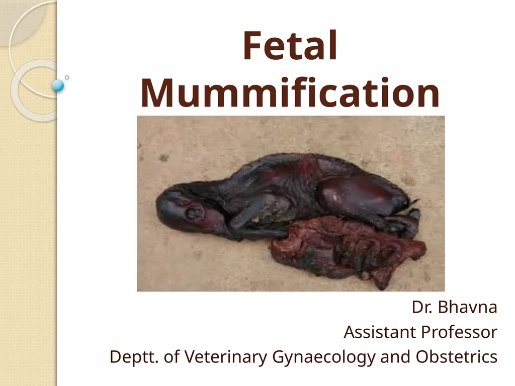
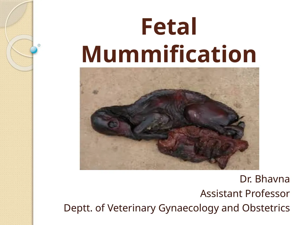
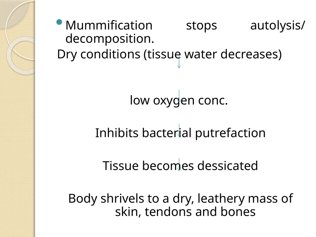
Mummification stops autolysis/ decomposition.
Dry conditions (tissue water decreases)
low oxygen conc.
Inhibits bacterial putrefaction
Tissue becomes dessicated
Body shrivels to a dry, leathery mass of skin, tendons and bones<br>
06
Events that must be present for fetal mummification The fetus must die after the development of bones is complete.
Uterine and fetal fluids must be resorbed relatively rapidly.
There must be no oxygen in the uterus until the mummification process is complete.
There must be no bacteria in the uterus.<br>
07
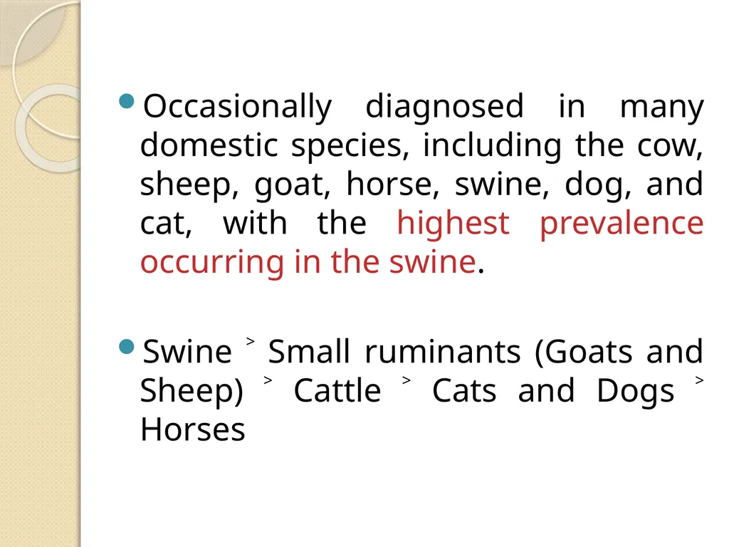
Occasionally diagnosed in many domestic species, including the cow, sheep, goat, horse, swine, dog, and cat, with the highest prevalence occurring in the swine.
Swine ˃ Small ruminants (Goats and Sheep) ˃ Cattle ˃ Cats and Dogs ˃ Horses<br>
08
Two types:
The hematic or chocolate mummification (in cattle)
The papyraceous (in other species)
Produces a dry, stiff fetoplacental unit with no exudate.<br>
09
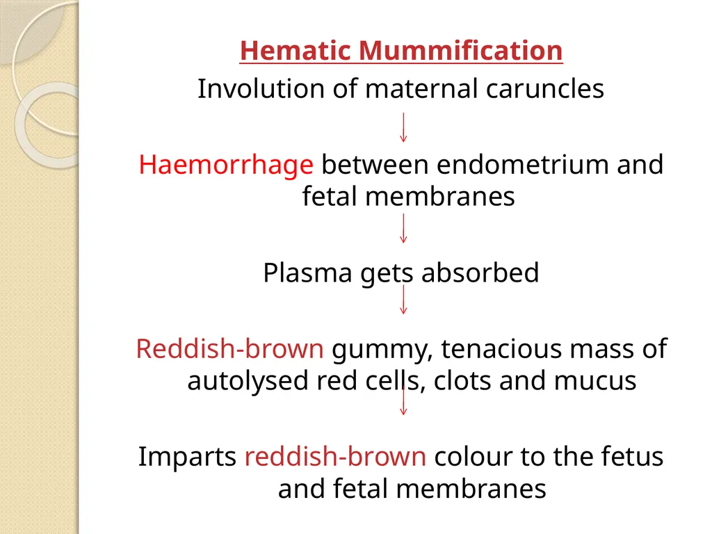
Hematic Mummification
Involution of maternal caruncles
Haemorrhage between endometrium and fetal membranes
Plasma gets absorbed
Reddish-brown gummy, tenacious mass of autolysed red cells, clots and mucus
Imparts reddish-brown colour to the fetus and fetal membranes<br>
10
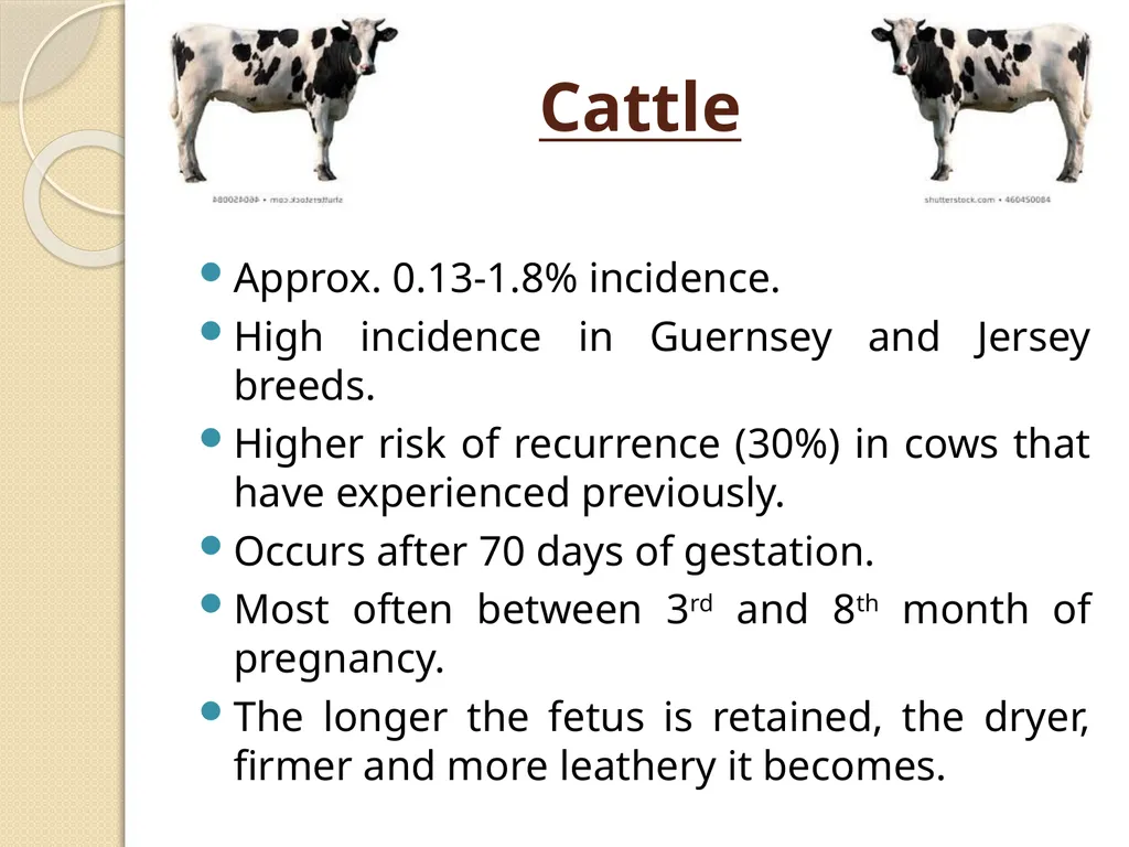
Cattle Approx. 0.13-1.8% incidence.
High incidence in Guernsey and Jersey breeds.
Higher risk of recurrence (30%) in cows that have experienced previously.
Occurs after 70 days of gestation.
Most often between 3rd and 8th month of pregnancy.
The longer the fetus is retained, the dryer, firmer and more leathery it becomes.<br>
11
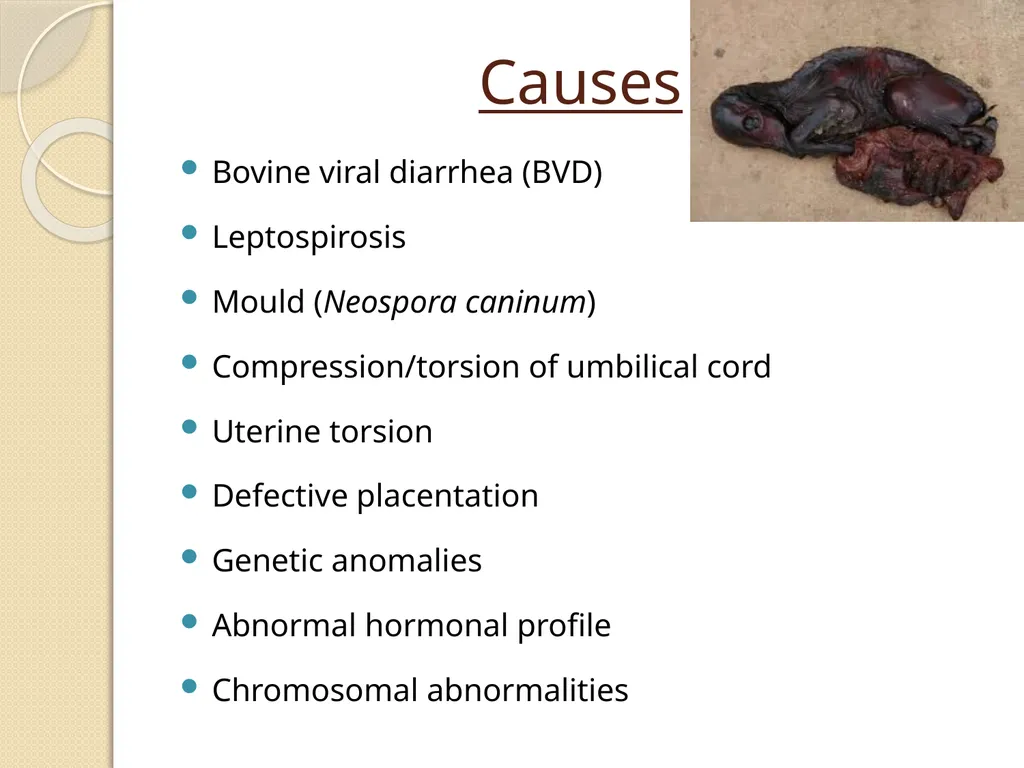
Causes Bovine viral diarrhea (BVD)
Leptospirosis
Mould (Neospora caninum)
Compression/torsion of umbilical cord
Uterine torsion
Defective placentation
Genetic anomalies
Abnormal hormonal profile
Chromosomal abnormalities<br>
12
Clinical findings and Diagnosis Cow’s abdomen to be unusually small for the given stage of pregnancy.
Body changes incident to parturition and calving fail to occur.
Transrectal palpation – compact, firm and immobile mass without placental fluid or placentomes and no fremitus.
USG – absence of heartbeat and fetal fluids.<br>
13
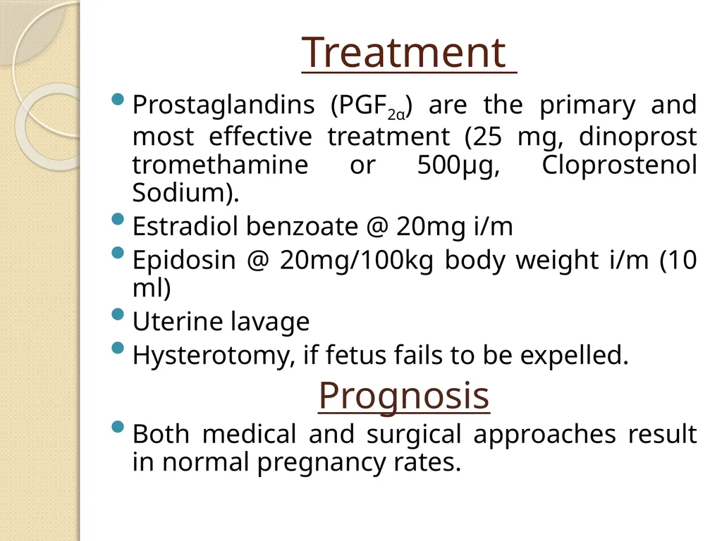
Treatment Prostaglandins (PGF2α) are the primary and most effective treatment (25 mg, dinoprost tromethamine or 500μg, Cloprostenol Sodium).
Estradiol benzoate @ 20mg i/m
Epidosin @ 20mg/100kg body weight i/m (10 ml)
Uterine lavage
Hysterotomy, if fetus fails to be expelled.
Prognosis
Both medical and surgical approaches result in normal pregnancy rates.<br>
14
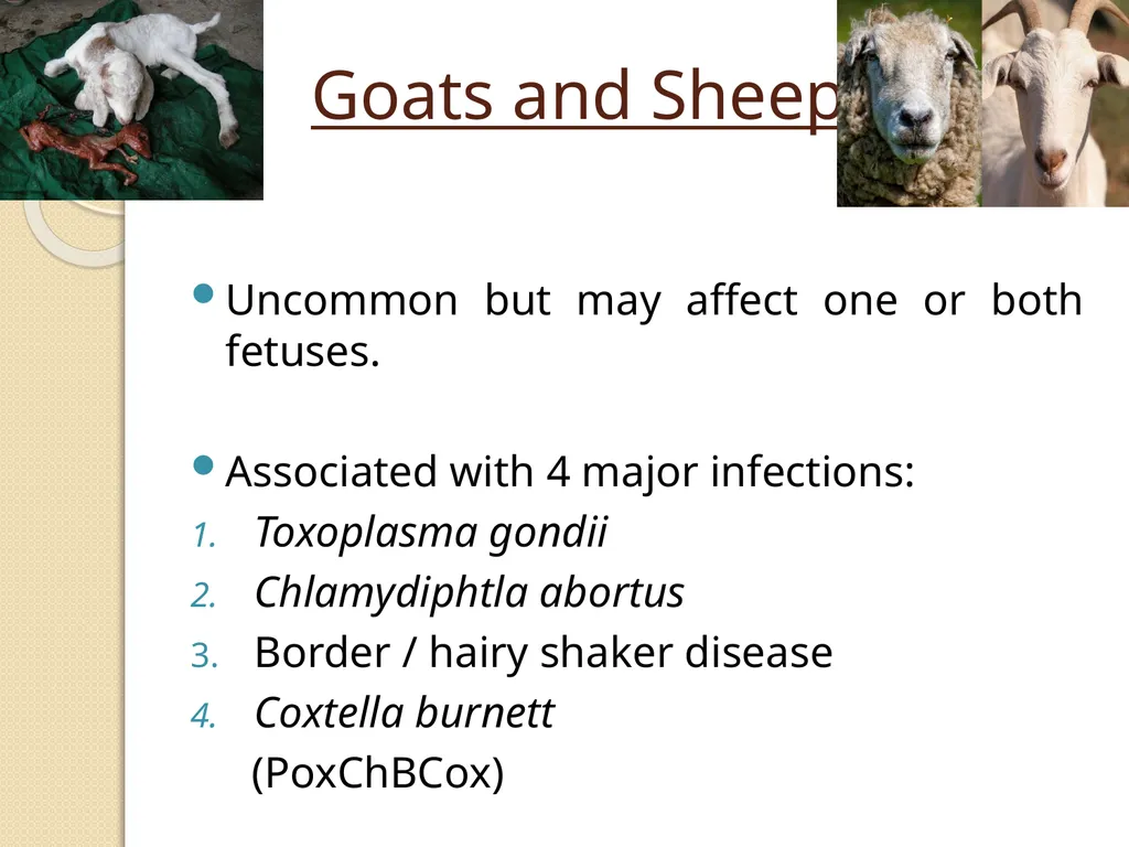
Goats and Sheep Uncommon but may affect one or both fetuses.
Associated with 4 major infections:
Toxoplasma gondii
Chlamydiphtla abortus
Border / hairy shaker disease
Coxtella burnett
(PoxChBCox)<br>
15
Energy and protein deficiencies, particularly on days 90 – 120 of gestation.
Mummified fetuses are spontaneously aborted.<br>
16
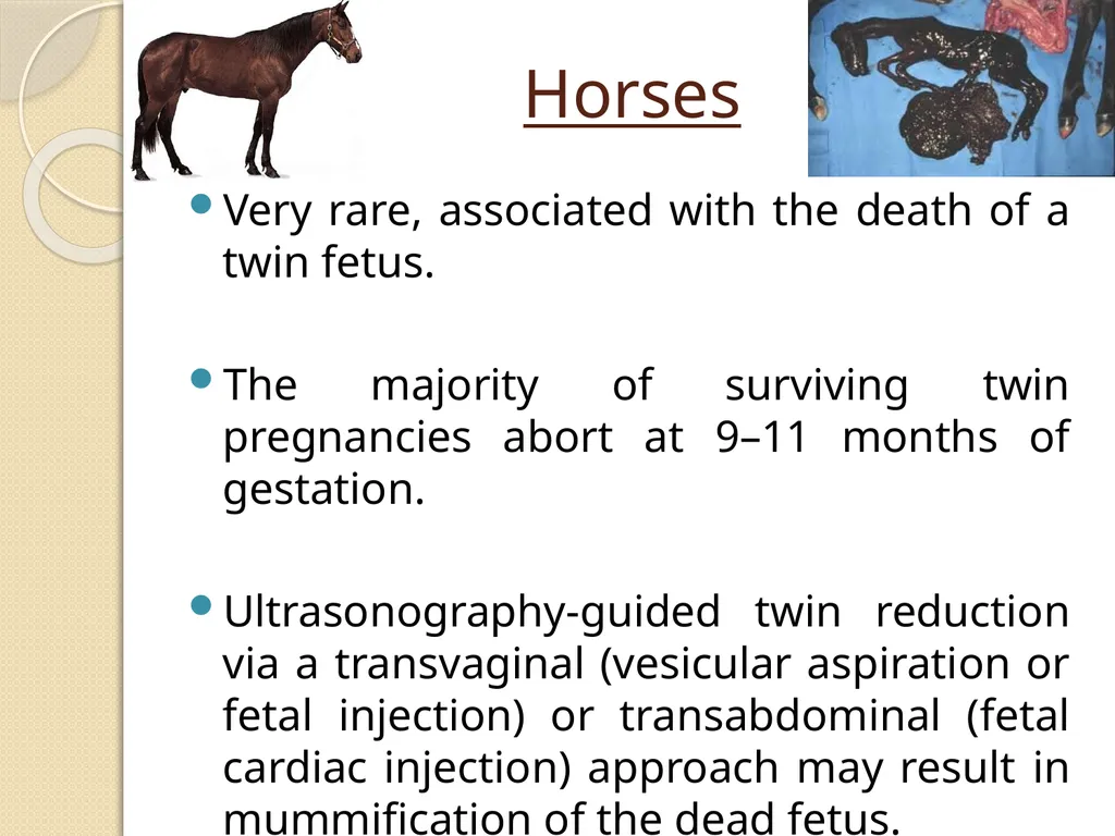
Horses Very rare, associated with the death of a twin fetus.
The majority of surviving twin pregnancies abort at 9–11 months of gestation.
Ultrasonography-guided twin reduction via a transvaginal (vesicular aspiration or fetal injection) or transabdominal (fetal cardiac injection) approach may result in mummification of the dead fetus.<br>
17
Clinical findings Mummified fetuses are found unexpectedly during a normal pregnancy, dystocia during foaling or prolonged pregnancy.
Diagnosis
Uncomplicated
Transrectal palpation and ultrasonography show a hard and bony structure without fluid in the uterine lumen.<br>
18
Treatment Where elevated progesterone is associated with a CL, PGF2α (25 mg dinoprost tromethamine IM) is indicated.
In the absence of a CL, 17β-estradiol (5 mg IM) or PGE1 (200 μg Misoprostol) administered locally on or in the cervix 24 hours before oxytocin is administered.
Caesarean section, if large in size.
Uterine lavage to remove debris is indicated.<br>
19
Prevention Close monitoring to determine time of ovulation and so enable the diagnosis of double ovulation and confirm a twin pregnancy early (on day 14) is recommended.
Allows for early twin reduction in order to optimize success, and minimizes the risk of fetal mummification, reduces the risk of twins, and optimizes the future fertility of the mare.<br>
20
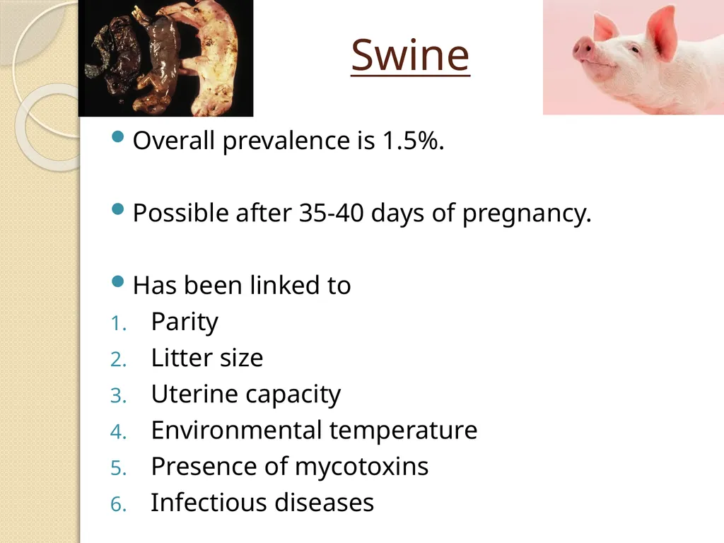
Swine Overall prevalence is 1.5%.
Possible after 35-40 days of pregnancy.
Has been linked to
Parity
Litter size
Uterine capacity
Environmental temperature
Presence of mycotoxins
Infectious diseases<br>
21
Presence of one mummy in an otherwise normal litter may indicate physiological death, whereas the presence of multiple mummified fetuses indicates an infectious cause.<br>
22
Infectious agents potentially associated with fetal mummification in swine :
1. Porcine parvovirus (PPV)
2. Aujeszky’s disease / pseudorabies virus (AD/PRV)
3. Encephalomyocarditis virus (EMCV)
4. Erysipelas (bacteria)
5. Japanese encephalitis virus (JEV)
6. Porcine circovirus 2 (PCV2)
7. Porcine reproductive and respiratory syndrome virus (PRRSV)
8. Swine fever virus (SFV; African and classical)
9. Swine influenza virus (SIV)
10. Teschovirus<br>
23
Clinical findings Infection during early gestation (˂40 days) causes absorption of the infected fetuses, and the dam returns to estrus in a regular (18–24 days after heat) or irregular (25–38 days after heat) manner or fails to farrow.
Infection between 40 and 70 days of pregnancy, causes infection to fetuses, resulting in the mummification of fetuses of different sizes at birth, alongside the healthy piglets.<br>
24
Diagnosis The number of mummified fetuses relative to litter size and age at which death occurs is an indication of the potential etiologic agent.
Insufficient space or a large litter - mummified fetuses associated with a normal litter size.
Infectious agent - the litter size will be normal, but more mummified fetuses and less live piglets will be observed.<br>
25
Prevention Proper care/comfort
Optimal nutrition
Effective stress management
Rigorous sanitary protocols
Vaccination<br>
26
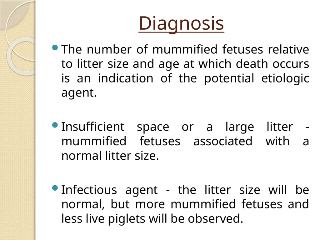
Dogs Most common cause is canine herpesvirus (CHV).
Difficult to diagnose esp. if bitch consumes or hides the fetus.
Proper vaccination may prevent.<br>
27
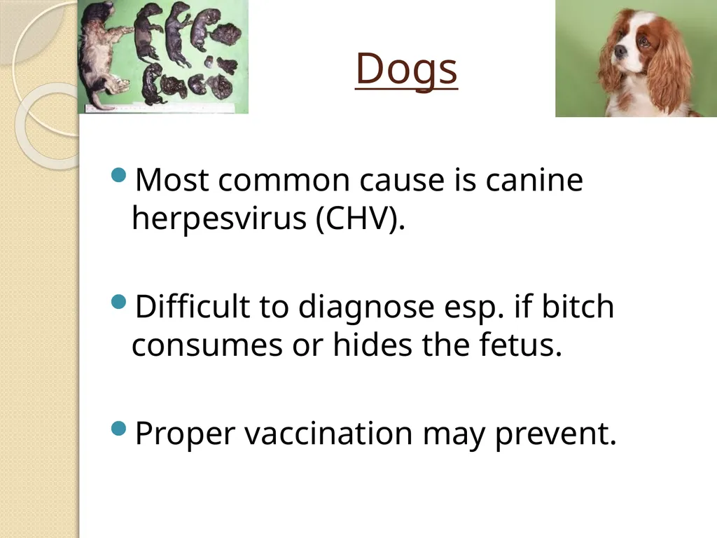
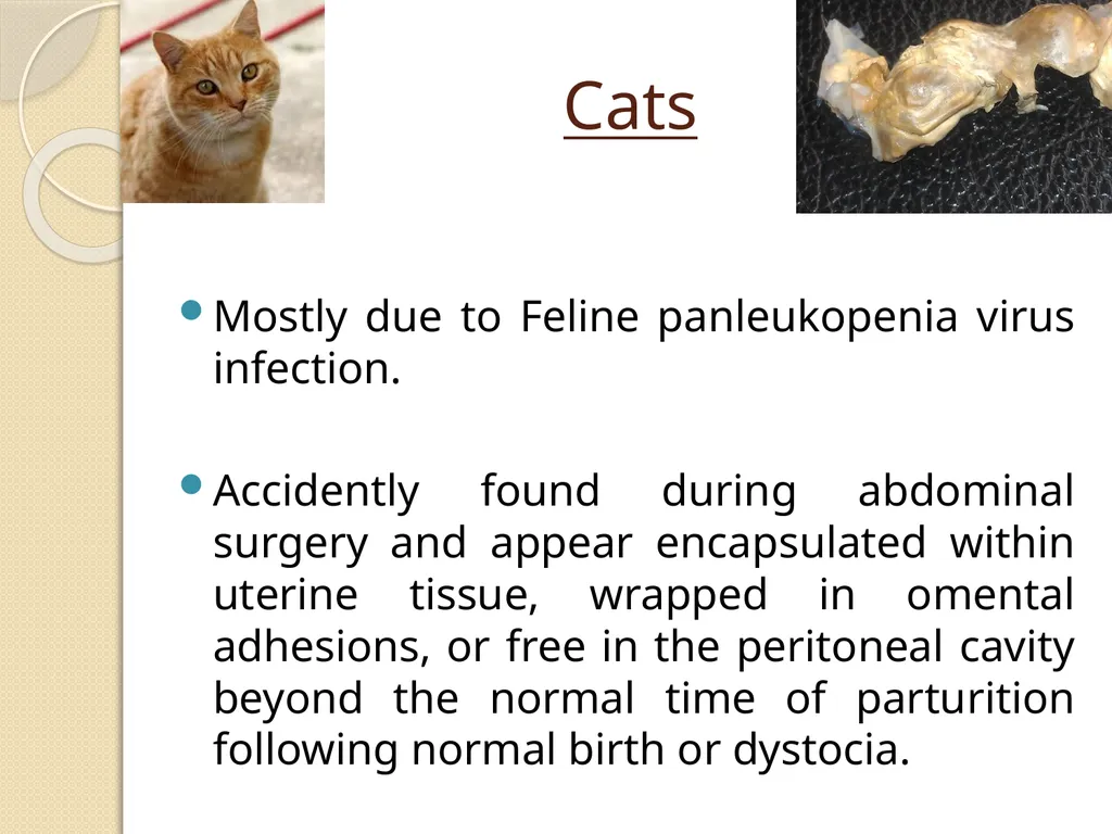
Cats Mostly due to Feline panleukopenia virus infection.
Accidently found during abdominal surgery and appear encapsulated within uterine tissue, wrapped in omental adhesions, or free in the peritoneal cavity beyond the normal time of parturition following normal birth or dystocia.<br>