
HeFSSA Heart Failure Management Meeting 2025



Description: HeFSSA Heart Failure Management Meeting 2025 Professor Eric Klug Faculty of Health Sciences, University of Witwatersrand, Netcare Sunninghill Hospital Diagnosis of HFpEF Making a firm diagnosis of chronic heart failure with preserved
Related Topics
Download Presentation
"HeFSSA Heart Failure Management Meeting 2025" is the property of its rightful owner. Permission is granted to download and print the materials on this website for personal, non-commercial use only, and to display it on your personal computer provided you do not modify the materials and that you retain all copyright notices contained in the materials. By downloading content from our website, you accept the terms of this agreement.
Presentation Transcript
slide1. HeFSSA Heart Failure Management Meeting 2025 Professor Eric Klug
Faculty of Health Sciences, University of Witwatersrand, Netcare Sunninghill Hospital Diagnosis of HFpEF<br>
slide2. Making a firm diagnosis of chronic heart failure with preserved ejection fraction (HFpEF) remains a challenge The challenge is to increase not only our accuracy, but also number of correct diagnoses of HFpEF! We need useful clinical criteria<br>
slide3. HFpEF (“huff puff”), a common clinical syndrome that is increasing in prevalence with the aging population, is associated with an alarmingly high morbidity and mortality Cambridge Idioms Dictionary defines “huff and puff” as follows: “to complain noisily about something but not be able to do anything about it”.
This is changing in 2025 – trials are now positive for new agents.
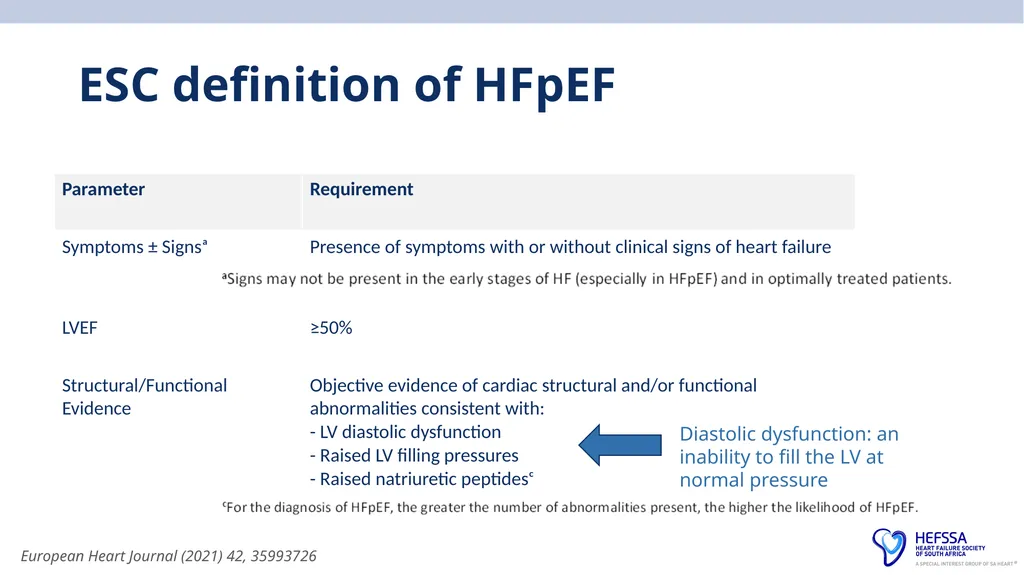
EF > 50% remains a defining feature in HFpEF
Despite heterogeneous contribution of putative pathomechanisms, HFpEF patients do have common patterns of clinical presentation, such as impaired exercise tolerance or physical limitations in activities of daily living<br>
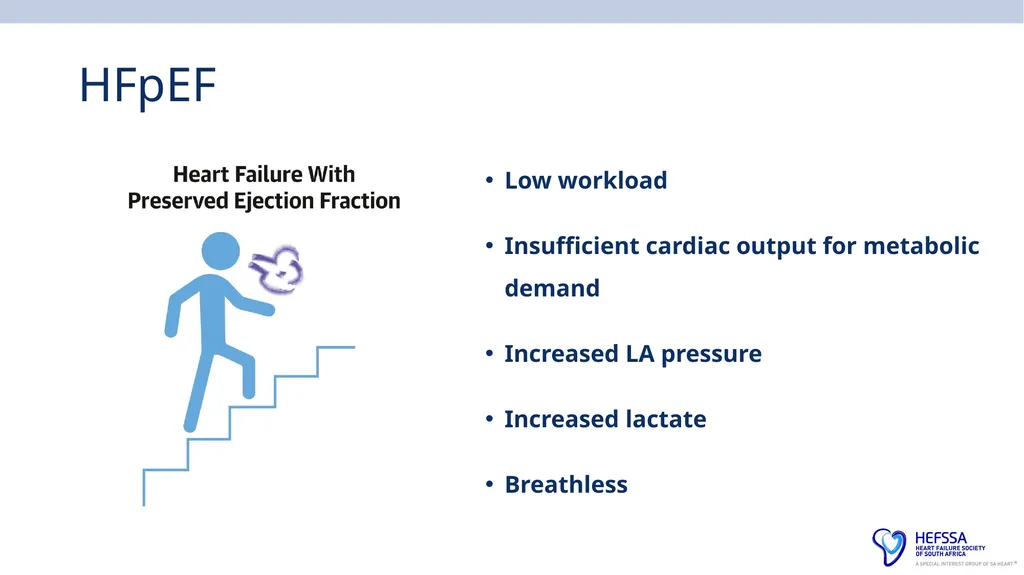
slide4. HFpEF Low workload
Insufficient cardiac output for metabolic demand
Increased LA pressure
Increased lactate
Breathless<br>
slide5. Why diagnose HFpEF Treat risk factors and symptoms
EMPEROR-Preserved, DELIVER, STEP-HFpEF, SUMMIT, FINEARTS-HF
– now we can treat also to improve outcomes
Distinguish from HFmrEF – which may benefit from HFrEF drugs
Diagnose more patients – help more patients<br>
slide6. Point 1: Why is it difficult to diagnose HFpEF? They may sound like and look like any heart failure patient
May be indistinguishable on history from a HFrEF
They also may masquerade , hiding their diagnosis behind other conditions
They are begging for you to recognise themselves for what they are
By correctly labelling them, you immediately trigger the correct algorithms
Common diagnoses such as anaemia, chronic kidney disease, atrial fibrillation, CAD, valvular heart disease, pulmonary hypertension, and lung disease can all mimic HFpEF<br>
slide7. Usual approach HFpEF is usually diagnosed after excluding non-cardiac causes of dyspnoea that can mimic HF, and alternate established diseases with left ventricular EF >50% causing HF symptoms (e.g., constrictive pericarditis, infiltrative cardiomyopathies, isolated right-sided HF, valvular heart disease, non–group 2 pulmonary hypertension)<br>
slide8. Point 2: No clear defining clinical feature Studies have shown no statistically significant difference in the prevalence of signs and symptoms between patients with HFpEF and HFrEF
Patients with either condition often present with dyspnoea on exertion, impaired exercise tolerance, paroxysmal nocturnal dyspnoea or orthopnoea
Each may have similar signs of HF, such as jugular venous distension, rales, S3, S4, hepatomegaly, and oedema
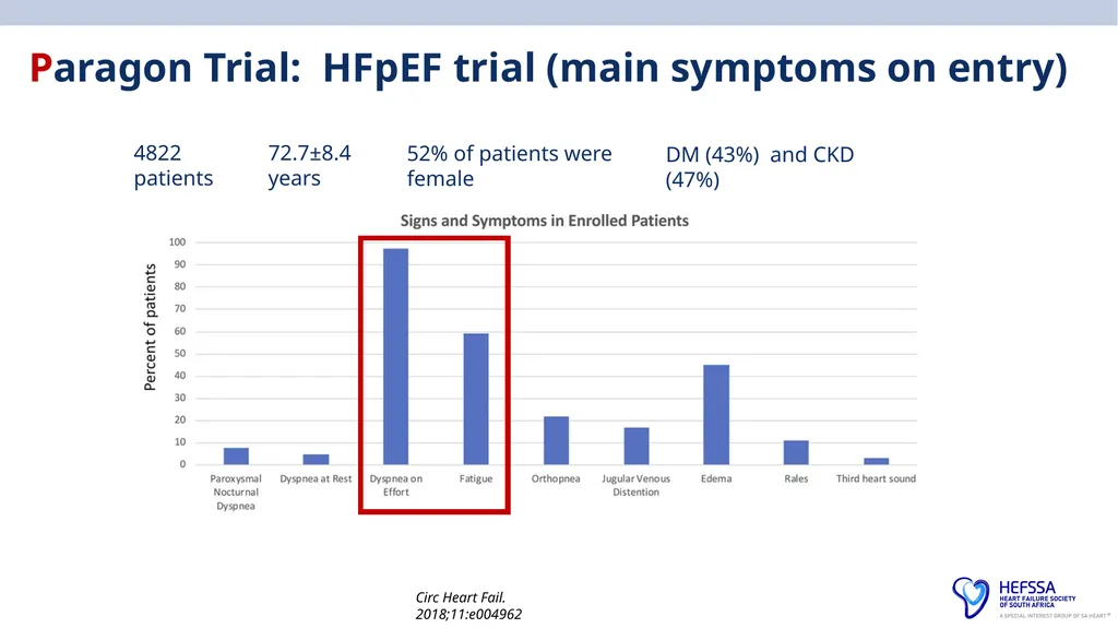
Patients with early stages of HFpEF may present with exertional dyspnoea and/or fatigue in the absence of signs of overt volume overload on physical examination<br>
slide9. ESC definition of HFpEF European Heart Journal (2021) 42, 35993726 Diastolic dysfunction: an inability to fill the LV at normal pressure<br>
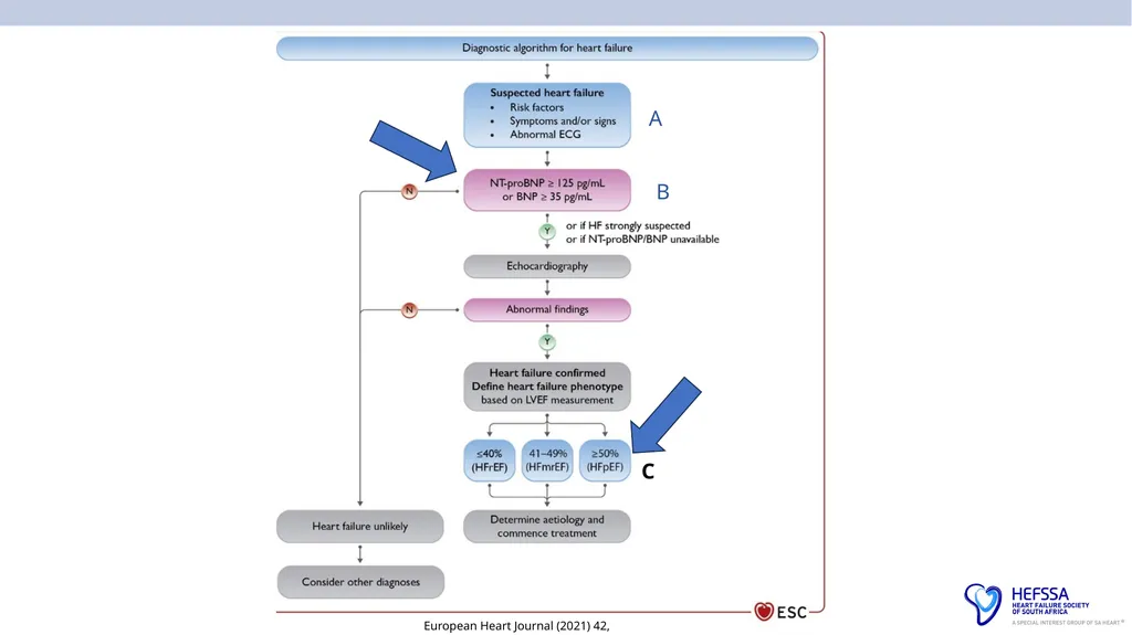
slide10. European Heart Journal (2021) 42, 35993726 A B C<br>
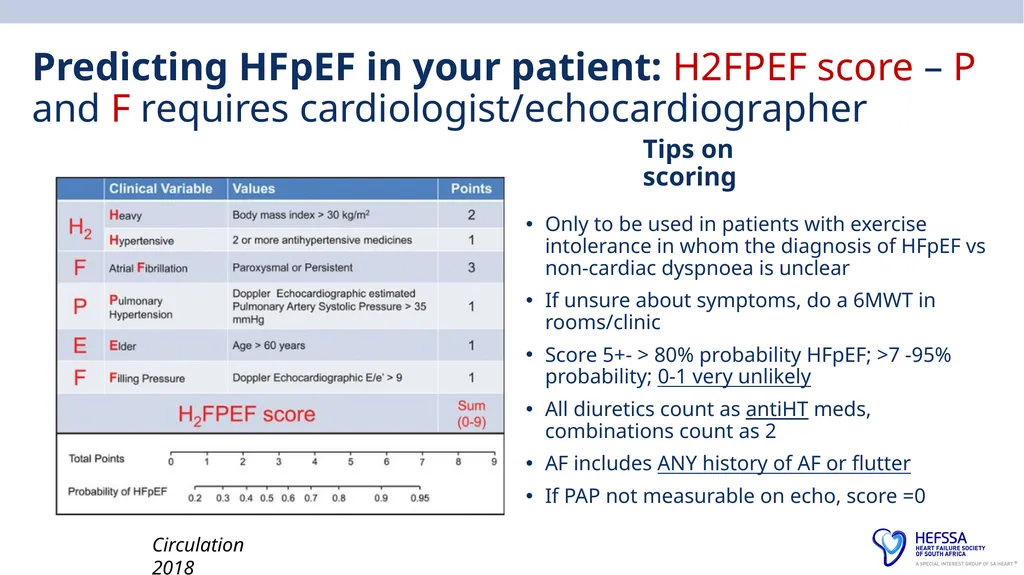
slide11. Predicting HFpEF in your patient: H2FPEF score – P and F requires cardiologist/echocardiographer Tips on scoring Only to be used in patients with exercise intolerance in whom the diagnosis of HFpEF vs non-cardiac dyspnoea is unclear
If unsure about symptoms, do a 6MWT in rooms/clinic
Score 5+- > 80% probability HFpEF; >7 -95% probability; 0-1 very unlikely
All diuretics count as antiHT meds, combinations count as 2
AF includes ANY history of AF or flutter
If PAP not measurable on echo, score =0 Circulation 2018<br>
slide12. Paragon Trial: HFpEF trial (main symptoms on entry) Circ Heart Fail. 2018;11:e004962 4822 patients 72.7±8.4 years 52% of patients were female DM (43%) and CKD (47%)<br>
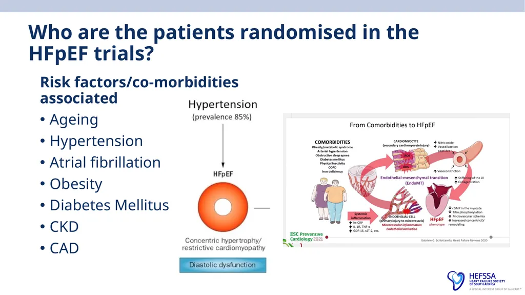
slide13. Who are the patients randomised in the HFpEF trials? Risk factors/co-morbidities associated
Ageing
Hypertension
Atrial fibrillation
Obesity
Diabetes Mellitus
CKD
CAD<br>
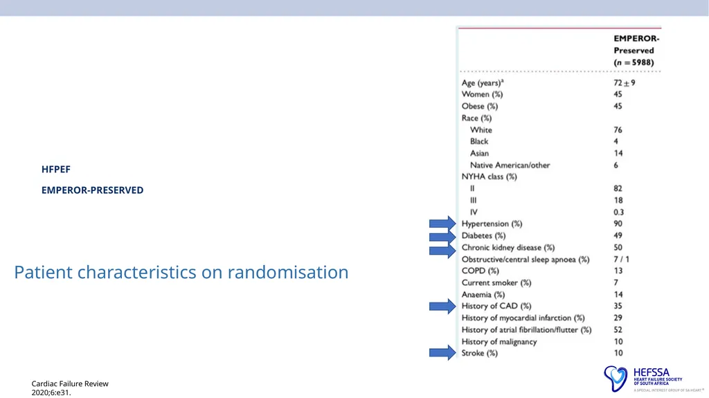
slide14. Cardiac Failure Review 2020;6:e31. HFPEF EMPEROR-PRESERVED Patient characteristics on randomisation<br>
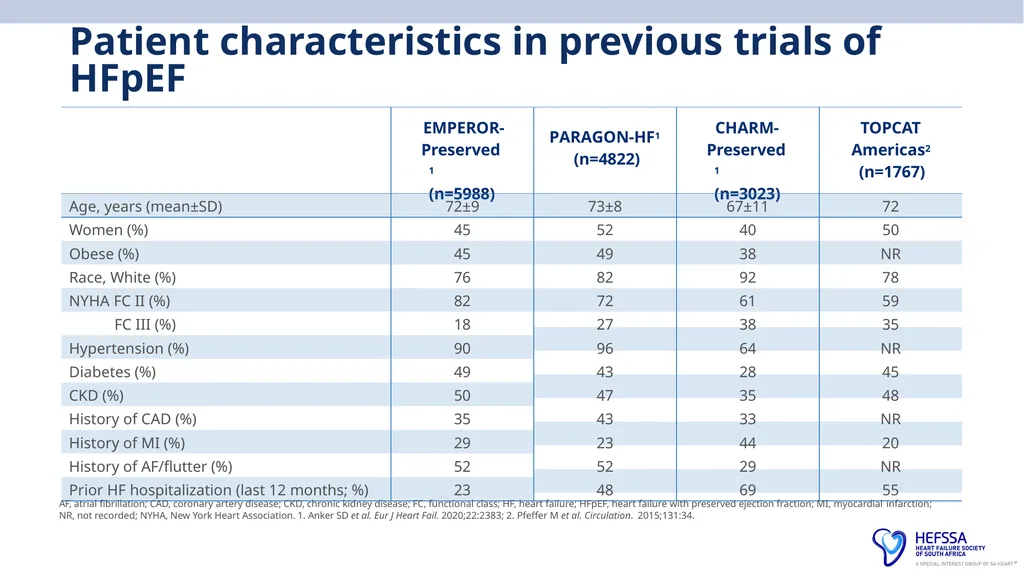
slide15. Patient characteristics in previous trials of HFpEF NR, not recorded; NYHA, New York Heart Association. 1. Anker SD et al. Eur J Heart Fail. 2020;22:2383; 2. Pfeffer M et al. Circulation. 2015;131:34. AF, atrial fibrillation; CAD, coronary artery disease; CKD, chronic kidney disease; FC, functional class; HF, heart failure; HFpEF, heart failure with preserved ejection fraction; MI, myocardial infarction;<br>
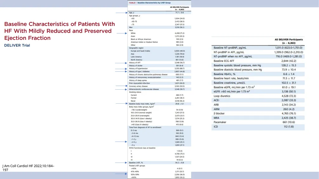
slide16. J Am Coll Cardiol HF 2022;10:184–197<br>
slide17. Africa: INTERCHF: Africa (Mozambique, Nigeria, South Africa, Sudan, and Uganda)<br>
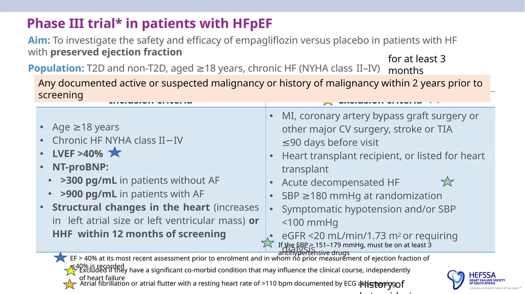
slide18. Phase III trial* in patients with HFpEF
Aim: To investigate the safety and efficacy of empagliflozin versus placebo in patients with HF with preserved ejection fraction
Population: T2D and non-T2D, aged ≥18 years, chronic HF (NYHA class II–IV) for at least 3 months EF > 40% at its most recent assessment prior to enrolment and in whom no prior measurement of ejection fraction of ≤40% is recorded Excluded if they have a significant co-morbid condition that may influence the clinical course, independently of heart failure Atrial fibrillation or atrial flutter with a resting heart rate of >110 bpm documented by ECG at screening If the SBP > 151–179 mmHg, must be on at least 3 antihypertensive drugs History of ketoacidosis Any documented active or suspected malignancy or history of malignancy within 2 years prior to screening<br>
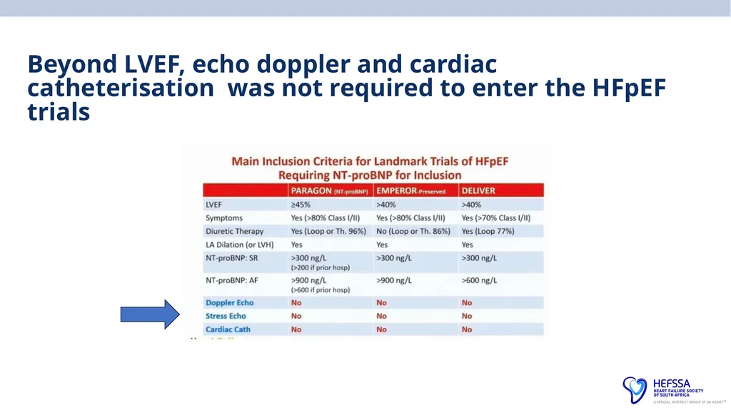
slide19. Beyond LVEF, echo doppler and cardiac catheterisation was not required to enter the HFpEF trials<br>
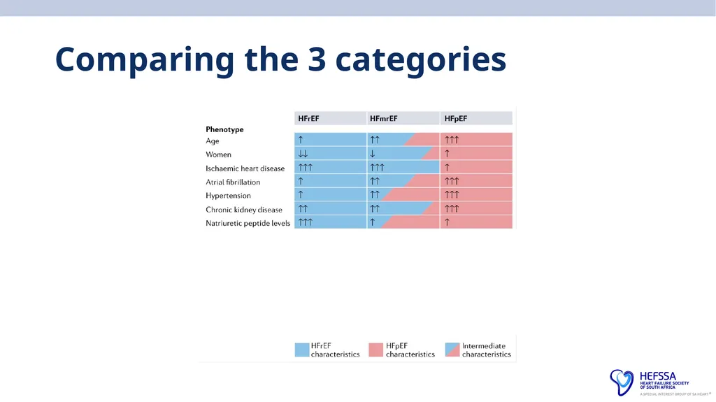
slide20. Comparing the 3 categories<br>
slide22. Point 3: Imaging plays a vital role in evaluation of HFpEF Echocardiography is considered as the single most useful diagnostic test in the evaluation of the patients with HF due to its availability and ability to provide information about cardiac anatomy, valvular structures, wall thickness, and filling pressures
All guidelines require the simultaneous and obligatory presence of signs and/or symptoms of HF, evidence of normal LVEF, and evidence of diastolic dysfunction
In early HFpEF, resting echocardiography may demonstrate only mild (grade I) diastolic dysfunction and normal or indeterminate LV filling pressures
CXR can be normal<br>
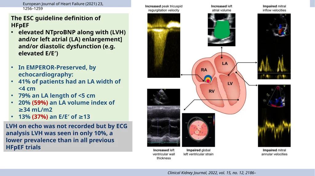
slide23. Clinical Kidney Journal, 2022, vol. 15, no. 12, 2186–2199 The ESC guideline definition of
HFpEF
elevated NTproBNP along with (LVH) and/or left atrial (LA) enlargement] and/or diastolic dysfunction (e.g. elevated E/E′)
In EMPEROR-Preserved, by echocardiography:
41% of patients had an LA width of <4 cm
79% an LA length of <5 cm
20% (59%) an LA volume index of ≥34 mL/m2
13% (37%) an E/E′ of ≥13 European Journal of Heart Failure (2021) 23, 1256–1259 LVH on echo was not recorded but by ECG analysis LVH was seen in only 10%, a lower prevalence than in all previous HFpEF trials<br>
slide24. Point 4: Elevated Pulmonary Artery Systolic Pressure on Echo with Normal LVEF – Consider HFpEF Frequency of elevated pulmonary artery systolic pressure (PASP) among patients with HFpEF is 83%
Predominantly due to pulmonary venous hypertension secondary to passive congestion of the pulmonary vasculature
In patients with normal LVEF, elevated PASP is suggestive of HFpEF until proven otherwise
In a patient with signs and symptoms of HF or possible HF (such as unexplained dyspnoea), elevated PASP in the presence of a normal LVEF should prompt consideration of HFpEF, especially if other causes of elevated PASP (as detailed above) have been excluded<br>
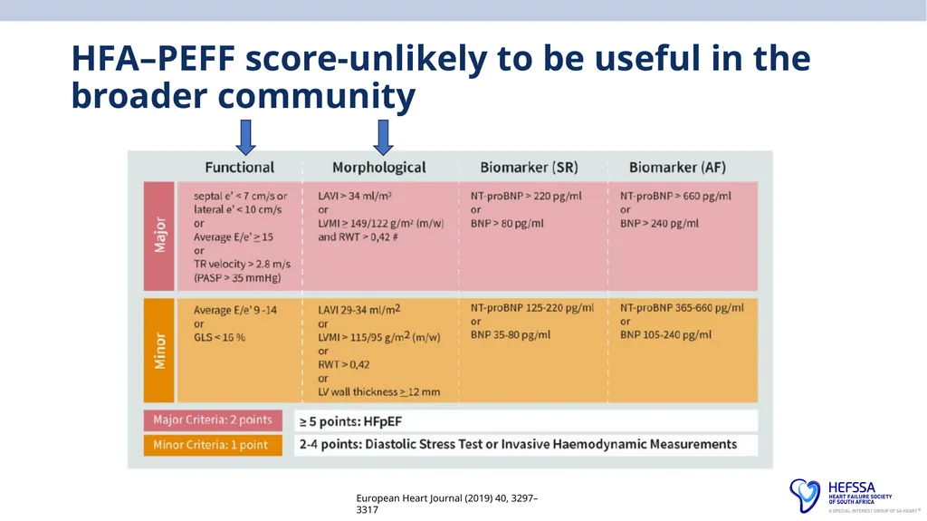
slide25. HFA–PEFF score-unlikely to be useful in the broader community European Heart Journal (2019) 40, 3297–3317<br>
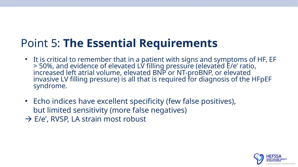
slide26. Point 5: The Essential Requirements It is critical to remember that in a patient with signs and symptoms of HF, EF > 50%, and evidence of elevated LV filling pressure (elevated E/e’ ratio, increased left atrial volume, elevated BNP or NT-proBNP, or elevated invasive LV filling pressure) is all that is required for diagnosis of the HFpEF syndrome. Echo indices have excellent specificity (few false positives), but limited sensitivity (more false negatives)
E/e’, RVSP, LA strain most robust<br>
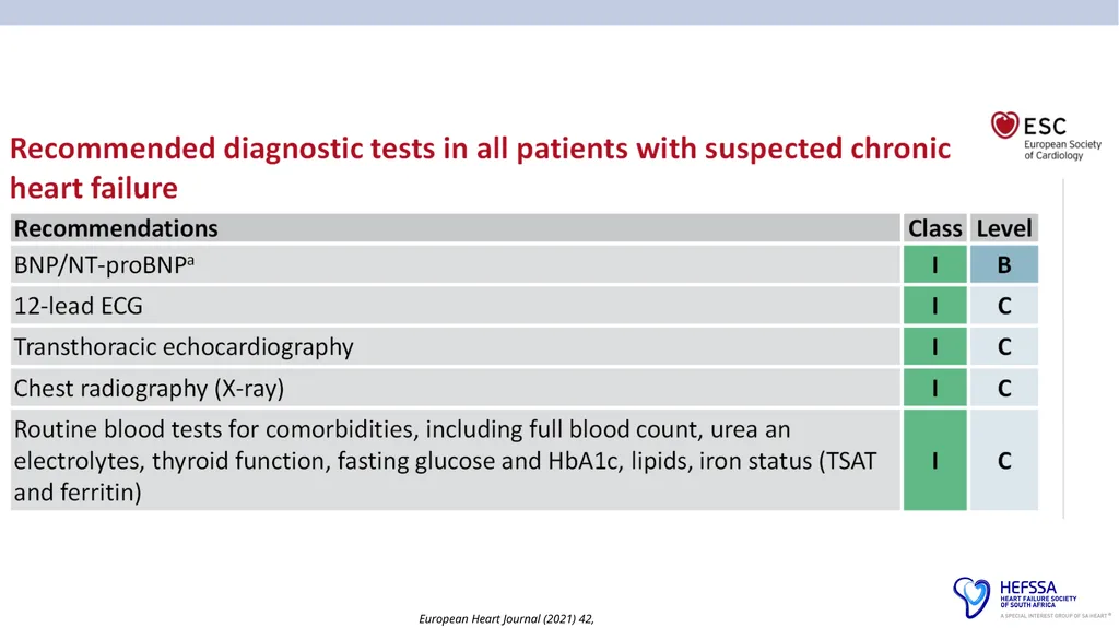
slide27. European Heart Journal (2021) 42, 35993726<br>
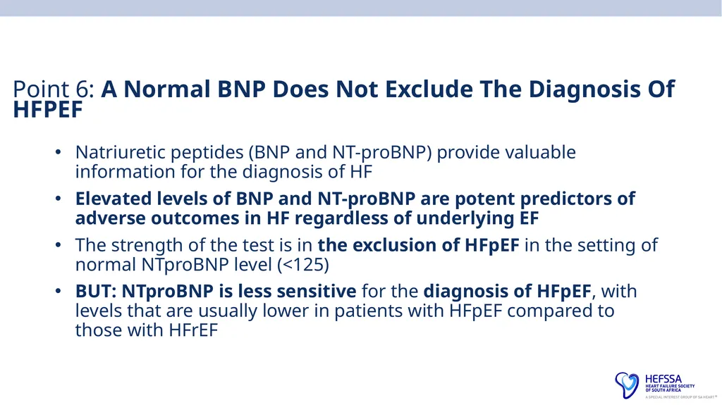
slide28. Point 6: A Normal BNP Does Not Exclude The Diagnosis Of HFPEF Natriuretic peptides (BNP and NT-proBNP) provide valuable information for the diagnosis of HF
Elevated levels of BNP and NT-proBNP are potent predictors of adverse outcomes in HF regardless of underlying EF
The strength of the test is in the exclusion of HFpEF in the setting of normal NTproBNP level (<125)
BUT: NTproBNP is less sensitive for the diagnosis of HFpEF, with levels that are usually lower in patients with HFpEF compared to those with HFrEF<br>
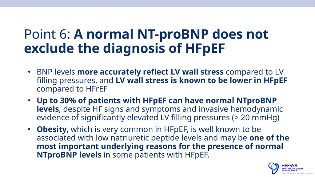
slide29. Point 6: A normal NT-proBNP does not exclude the diagnosis of HFpEF BNP levels more accurately reflect LV wall stress compared to LV filling pressures, and LV wall stress is known to be lower in HFpEF compared to HFrEF
Up to 30% of patients with HFpEF can have normal NTproBNP levels, despite HF signs and symptoms and invasive hemodynamic evidence of significantly elevated LV filling pressures (> 20 mmHg)
Obesity, which is very common in HFpEF, is well known to be associated with low natriuretic peptide levels and may be one of the most important underlying reasons for the presence of normal NTproBNP levels in some patients with HFpEF.<br>
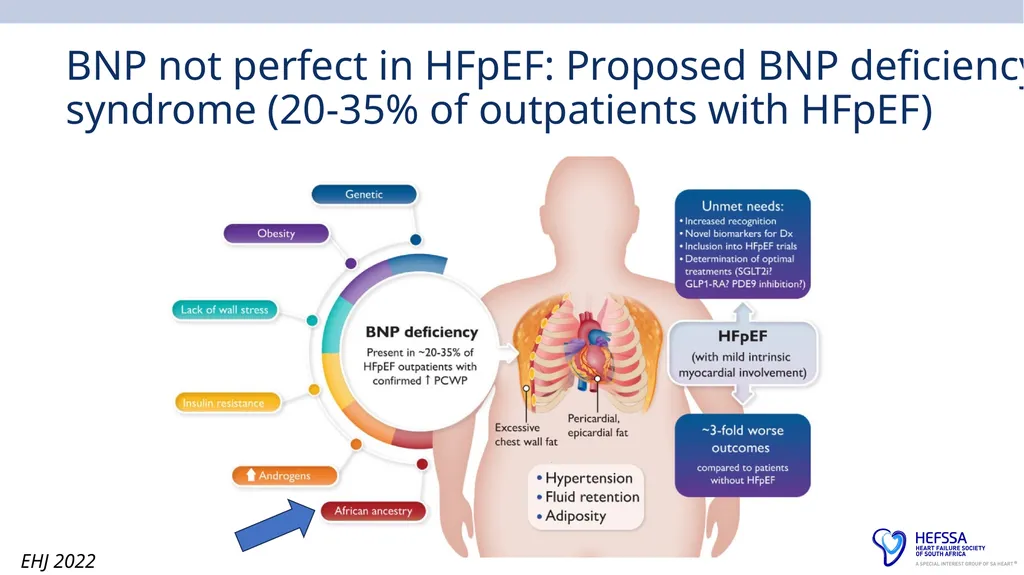
slide30. BNP not perfect in HFpEF: Proposed BNP deficiency syndrome (20-35% of outpatients with HFpEF) EHJ 2022<br>
slide31. Vigilance and persistence in diagnosis Use echocardiography and/or invasive hemodynamic testing to identify objective signs of elevated cardiac filling pressures.
In patients with unexplained dyspnoea or exercise intolerance where HFpEF is suspected:
Begin with exercise echocardiography, measuring E/e’ ratio and PASP at peak stress, alongside cardiopulmonary exercise testing.
If diagnosis remains uncertain, perform exercise cardiac catheterization (using straight leg raise or supine stationary bike) for further evaluation.
This comprehensive approach is seldom applied outside specialised centers, reflecting underdiagnosis and insufficient diagnostic rigor.<br>
slide32. Point 7: Look for coronary artery disease in all patients with HFpEF Epidemiologic and observational studies have documented a CAD prevalence of approximately 50% in HFpEF
Systematic identification of CAD is an important part of management of HFpEF
Consider CT Coronary angiography (if not in AF) in most HFpEF patients, or DCA<br>
slide33. In conclusion: What is HFpEF? Signs and symptoms of HF
Preserved LVEF (>50%)
Objective evidence of elevated LV filling pressures at rest or during exercise – BNP/LA enlarged or dysfunction/PCWP >15 (rest) or >25 exercise
Evidence of a cardiac-predominant problem
Exclude – infiltrative, genetic, valvar, toxin, pericardial, ischaemic aetiology
Diastolic dysfunction on echo not required for diagnosis
When in doubt do a right heart catheterisation<br>
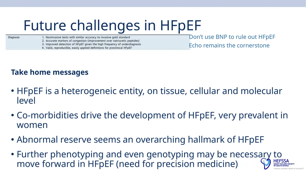
slide34. Future challenges in HFpEF Take home messages HFpEF is a heterogeneic entity, on tissue, cellular and molecular level
Co-morbidities drive the development of HFpEF, very prevalent in women
Abnormal reserve seems an overarching hallmark of HFpEF
Further phenotyping and even genotyping may be necessary to move forward in HFpEF (need for precision medicine) Don’t use BNP to rule out HFpEF Echo remains the cornerstone<br>
slide35. Thank you! Any questions?<br>
Faculty of Health Sciences, University of Witwatersrand, Netcare Sunninghill Hospital Diagnosis of HFpEF<br>
slide2. Making a firm diagnosis of chronic heart failure with preserved ejection fraction (HFpEF) remains a challenge The challenge is to increase not only our accuracy, but also number of correct diagnoses of HFpEF! We need useful clinical criteria<br>
slide3. HFpEF (“huff puff”), a common clinical syndrome that is increasing in prevalence with the aging population, is associated with an alarmingly high morbidity and mortality Cambridge Idioms Dictionary defines “huff and puff” as follows: “to complain noisily about something but not be able to do anything about it”.
This is changing in 2025 – trials are now positive for new agents.
EF > 50% remains a defining feature in HFpEF
Despite heterogeneous contribution of putative pathomechanisms, HFpEF patients do have common patterns of clinical presentation, such as impaired exercise tolerance or physical limitations in activities of daily living<br>
slide4. HFpEF Low workload
Insufficient cardiac output for metabolic demand
Increased LA pressure
Increased lactate
Breathless<br>
slide5. Why diagnose HFpEF Treat risk factors and symptoms
EMPEROR-Preserved, DELIVER, STEP-HFpEF, SUMMIT, FINEARTS-HF
– now we can treat also to improve outcomes
Distinguish from HFmrEF – which may benefit from HFrEF drugs
Diagnose more patients – help more patients<br>
slide6. Point 1: Why is it difficult to diagnose HFpEF? They may sound like and look like any heart failure patient
May be indistinguishable on history from a HFrEF
They also may masquerade , hiding their diagnosis behind other conditions
They are begging for you to recognise themselves for what they are
By correctly labelling them, you immediately trigger the correct algorithms
Common diagnoses such as anaemia, chronic kidney disease, atrial fibrillation, CAD, valvular heart disease, pulmonary hypertension, and lung disease can all mimic HFpEF<br>
slide7. Usual approach HFpEF is usually diagnosed after excluding non-cardiac causes of dyspnoea that can mimic HF, and alternate established diseases with left ventricular EF >50% causing HF symptoms (e.g., constrictive pericarditis, infiltrative cardiomyopathies, isolated right-sided HF, valvular heart disease, non–group 2 pulmonary hypertension)<br>
slide8. Point 2: No clear defining clinical feature Studies have shown no statistically significant difference in the prevalence of signs and symptoms between patients with HFpEF and HFrEF
Patients with either condition often present with dyspnoea on exertion, impaired exercise tolerance, paroxysmal nocturnal dyspnoea or orthopnoea
Each may have similar signs of HF, such as jugular venous distension, rales, S3, S4, hepatomegaly, and oedema
Patients with early stages of HFpEF may present with exertional dyspnoea and/or fatigue in the absence of signs of overt volume overload on physical examination<br>
slide9. ESC definition of HFpEF European Heart Journal (2021) 42, 35993726 Diastolic dysfunction: an inability to fill the LV at normal pressure<br>
slide10. European Heart Journal (2021) 42, 35993726 A B C<br>
slide11. Predicting HFpEF in your patient: H2FPEF score – P and F requires cardiologist/echocardiographer Tips on scoring Only to be used in patients with exercise intolerance in whom the diagnosis of HFpEF vs non-cardiac dyspnoea is unclear
If unsure about symptoms, do a 6MWT in rooms/clinic
Score 5+- > 80% probability HFpEF; >7 -95% probability; 0-1 very unlikely
All diuretics count as antiHT meds, combinations count as 2
AF includes ANY history of AF or flutter
If PAP not measurable on echo, score =0 Circulation 2018<br>
slide12. Paragon Trial: HFpEF trial (main symptoms on entry) Circ Heart Fail. 2018;11:e004962 4822 patients 72.7±8.4 years 52% of patients were female DM (43%) and CKD (47%)<br>
slide13. Who are the patients randomised in the HFpEF trials? Risk factors/co-morbidities associated
Ageing
Hypertension
Atrial fibrillation
Obesity
Diabetes Mellitus
CKD
CAD<br>
slide14. Cardiac Failure Review 2020;6:e31. HFPEF EMPEROR-PRESERVED Patient characteristics on randomisation<br>
slide15. Patient characteristics in previous trials of HFpEF NR, not recorded; NYHA, New York Heart Association. 1. Anker SD et al. Eur J Heart Fail. 2020;22:2383; 2. Pfeffer M et al. Circulation. 2015;131:34. AF, atrial fibrillation; CAD, coronary artery disease; CKD, chronic kidney disease; FC, functional class; HF, heart failure; HFpEF, heart failure with preserved ejection fraction; MI, myocardial infarction;<br>
slide16. J Am Coll Cardiol HF 2022;10:184–197<br>
slide17. Africa: INTERCHF: Africa (Mozambique, Nigeria, South Africa, Sudan, and Uganda)<br>
slide18. Phase III trial* in patients with HFpEF
Aim: To investigate the safety and efficacy of empagliflozin versus placebo in patients with HF with preserved ejection fraction
Population: T2D and non-T2D, aged ≥18 years, chronic HF (NYHA class II–IV) for at least 3 months EF > 40% at its most recent assessment prior to enrolment and in whom no prior measurement of ejection fraction of ≤40% is recorded Excluded if they have a significant co-morbid condition that may influence the clinical course, independently of heart failure Atrial fibrillation or atrial flutter with a resting heart rate of >110 bpm documented by ECG at screening If the SBP > 151–179 mmHg, must be on at least 3 antihypertensive drugs History of ketoacidosis Any documented active or suspected malignancy or history of malignancy within 2 years prior to screening<br>
slide19. Beyond LVEF, echo doppler and cardiac catheterisation was not required to enter the HFpEF trials<br>
slide20. Comparing the 3 categories<br>
slide22. Point 3: Imaging plays a vital role in evaluation of HFpEF Echocardiography is considered as the single most useful diagnostic test in the evaluation of the patients with HF due to its availability and ability to provide information about cardiac anatomy, valvular structures, wall thickness, and filling pressures
All guidelines require the simultaneous and obligatory presence of signs and/or symptoms of HF, evidence of normal LVEF, and evidence of diastolic dysfunction
In early HFpEF, resting echocardiography may demonstrate only mild (grade I) diastolic dysfunction and normal or indeterminate LV filling pressures
CXR can be normal<br>
slide23. Clinical Kidney Journal, 2022, vol. 15, no. 12, 2186–2199 The ESC guideline definition of
HFpEF
elevated NTproBNP along with (LVH) and/or left atrial (LA) enlargement] and/or diastolic dysfunction (e.g. elevated E/E′)
In EMPEROR-Preserved, by echocardiography:
41% of patients had an LA width of <4 cm
79% an LA length of <5 cm
20% (59%) an LA volume index of ≥34 mL/m2
13% (37%) an E/E′ of ≥13 European Journal of Heart Failure (2021) 23, 1256–1259 LVH on echo was not recorded but by ECG analysis LVH was seen in only 10%, a lower prevalence than in all previous HFpEF trials<br>
slide24. Point 4: Elevated Pulmonary Artery Systolic Pressure on Echo with Normal LVEF – Consider HFpEF Frequency of elevated pulmonary artery systolic pressure (PASP) among patients with HFpEF is 83%
Predominantly due to pulmonary venous hypertension secondary to passive congestion of the pulmonary vasculature
In patients with normal LVEF, elevated PASP is suggestive of HFpEF until proven otherwise
In a patient with signs and symptoms of HF or possible HF (such as unexplained dyspnoea), elevated PASP in the presence of a normal LVEF should prompt consideration of HFpEF, especially if other causes of elevated PASP (as detailed above) have been excluded<br>
slide25. HFA–PEFF score-unlikely to be useful in the broader community European Heart Journal (2019) 40, 3297–3317<br>
slide26. Point 5: The Essential Requirements It is critical to remember that in a patient with signs and symptoms of HF, EF > 50%, and evidence of elevated LV filling pressure (elevated E/e’ ratio, increased left atrial volume, elevated BNP or NT-proBNP, or elevated invasive LV filling pressure) is all that is required for diagnosis of the HFpEF syndrome. Echo indices have excellent specificity (few false positives), but limited sensitivity (more false negatives)
E/e’, RVSP, LA strain most robust<br>
slide27. European Heart Journal (2021) 42, 35993726<br>
slide28. Point 6: A Normal BNP Does Not Exclude The Diagnosis Of HFPEF Natriuretic peptides (BNP and NT-proBNP) provide valuable information for the diagnosis of HF
Elevated levels of BNP and NT-proBNP are potent predictors of adverse outcomes in HF regardless of underlying EF
The strength of the test is in the exclusion of HFpEF in the setting of normal NTproBNP level (<125)
BUT: NTproBNP is less sensitive for the diagnosis of HFpEF, with levels that are usually lower in patients with HFpEF compared to those with HFrEF<br>
slide29. Point 6: A normal NT-proBNP does not exclude the diagnosis of HFpEF BNP levels more accurately reflect LV wall stress compared to LV filling pressures, and LV wall stress is known to be lower in HFpEF compared to HFrEF
Up to 30% of patients with HFpEF can have normal NTproBNP levels, despite HF signs and symptoms and invasive hemodynamic evidence of significantly elevated LV filling pressures (> 20 mmHg)
Obesity, which is very common in HFpEF, is well known to be associated with low natriuretic peptide levels and may be one of the most important underlying reasons for the presence of normal NTproBNP levels in some patients with HFpEF.<br>
slide30. BNP not perfect in HFpEF: Proposed BNP deficiency syndrome (20-35% of outpatients with HFpEF) EHJ 2022<br>
slide31. Vigilance and persistence in diagnosis Use echocardiography and/or invasive hemodynamic testing to identify objective signs of elevated cardiac filling pressures.
In patients with unexplained dyspnoea or exercise intolerance where HFpEF is suspected:
Begin with exercise echocardiography, measuring E/e’ ratio and PASP at peak stress, alongside cardiopulmonary exercise testing.
If diagnosis remains uncertain, perform exercise cardiac catheterization (using straight leg raise or supine stationary bike) for further evaluation.
This comprehensive approach is seldom applied outside specialised centers, reflecting underdiagnosis and insufficient diagnostic rigor.<br>
slide32. Point 7: Look for coronary artery disease in all patients with HFpEF Epidemiologic and observational studies have documented a CAD prevalence of approximately 50% in HFpEF
Systematic identification of CAD is an important part of management of HFpEF
Consider CT Coronary angiography (if not in AF) in most HFpEF patients, or DCA<br>
slide33. In conclusion: What is HFpEF? Signs and symptoms of HF
Preserved LVEF (>50%)
Objective evidence of elevated LV filling pressures at rest or during exercise – BNP/LA enlarged or dysfunction/PCWP >15 (rest) or >25 exercise
Evidence of a cardiac-predominant problem
Exclude – infiltrative, genetic, valvar, toxin, pericardial, ischaemic aetiology
Diastolic dysfunction on echo not required for diagnosis
When in doubt do a right heart catheterisation<br>
slide34. Future challenges in HFpEF Take home messages HFpEF is a heterogeneic entity, on tissue, cellular and molecular level
Co-morbidities drive the development of HFpEF, very prevalent in women
Abnormal reserve seems an overarching hallmark of HFpEF
Further phenotyping and even genotyping may be necessary to move forward in HFpEF (need for precision medicine) Don’t use BNP to rule out HFpEF Echo remains the cornerstone<br>
slide35. Thank you! Any questions?<br>