
Impotentia Cocundi and Impotentia Generendi,








Description: Impotentia Cocundi and Impotentia Generendi, Testicular Hypoplasia and Degeneration: Causes and Affect on Semen and Fertility DR ANKESH KUMAR Asstt. Prof. Department of Veterinary Clinical Complex UNIT VII VGO 604 CAUSES OF MALE INFERTILITY
Related Topics
Download Presentation
"Impotentia Cocundi and Impotentia Generendi," is the property of its rightful owner. Permission is granted to download and print the materials on this website for personal, non-commercial use only, and to display it on your personal computer provided you do not modify the materials and that you retain all copyright notices contained in the materials. By downloading content from our website, you accept the terms of this agreement.
Presentation Transcript
slide1. Impotentia Cocundi and Impotentia Generendi, Testicular Hypoplasia and Degeneration: Causes and Affect on Semen and Fertility DR ANKESH KUMAR
Asstt. Prof.
Department of Veterinary Clinical Complex UNIT VII VGO 604<br>
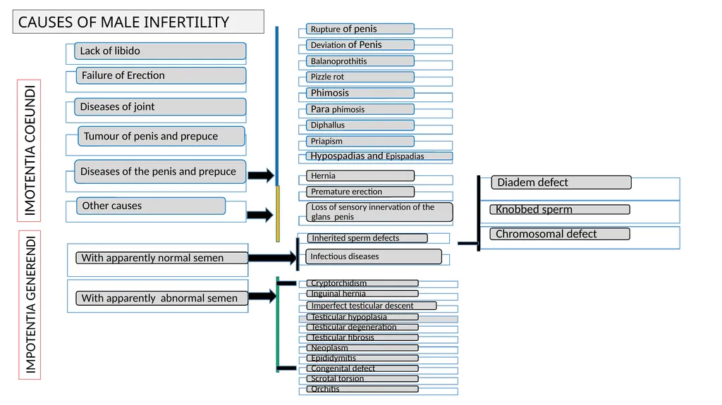
slide2. CAUSES OF MALE INFERTILITY IMOTENTIA COEUNDI IMPOTENTIA GENERENDI<br>
slide3. Testicular Hypoplasia: This is a congenital failure in the development of the Spermatogenic epithelium. It is found in two form
Hereditary and non hereditary
Hereditary: Recessive gene
Non-genetic: Extra X chromosome Klinefelter syndrom
Unilateral is more common than bilateral hypoplasia. In bull left side hypoplasia is more than the right
Symptoms
The scrotum of the affected male is smaller than normal male
The spermatic chord is shorter
Increase in proximal protoplasmic droplet
Sexual desire is not affected
Low concentration of semen
Low mortality
High incidence of proximal protoplasmic droplet
Presence of giant cell and medusa cell in semen
Bilateral
Clear watery
Few and no sperm
Diagnosis
Size of testes- small and flabby
Epididymis – small and hard
Libido-normal
Prognosis- poor
Treatment – affected bull should not be used for breeding purpose<br>
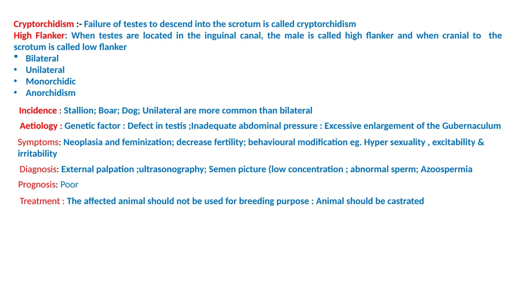
slide4. Cryptorchidism :- Failure of testes to descend into the scrotum is called cryptorchidism
High Flanker: When testes are located in the inguinal canal, the male is called high flanker and when cranial to the scrotum is called low flanker
Bilateral
Unilateral
Monorchidic
Anorchidism Incidence : Stallion; Boar; Dog; Unilateral are more common than bilateral Aetiology : Genetic factor : Defect in testis ;Inadequate abdominal pressure : Excessive enlargement of the Gubernaculum Symptoms: Neoplasia and feminization; decrease fertility; behavioural modification eg. Hyper sexuality , excitability & irritability Diagnosis: External palpation ;ultrasonography; Semen picture (low concentration ; abnormal sperm; Azoospermia Prognosis: Poor Treatment : The affected animal should not be used for breeding purpose : Animal should be castrated<br>
slide5. Testicular Degeneration: The partial or complete failure of epithelium of seminiferous tubules to proceed with spermatogenesis. Acquire disorder and is most common cause of infertility in male domestic animals. It may happen very but reaeration is very slow fast Aetiology : Endless : Commons are hyperthermia; Extreme cold; Advance age; Stress; Irradiation; Autoimmune; hormonal imbalance ; Nutritional Disorder
Incidence: Bilateral is very common
Symptoms: Smaller and softer than normal; libido is not affected; Adhesions between testis and scrotum are usually found; fibrosis and calcification also occur in late stage; Testes becomes hard and shrunken and irregular Semen picture: Depend on degree and involvement testicular tissue
Increasing in the incidence of loos head
Immature spermatozoa
Ejaculate volume is nit affected
Motility falls
Abnormality rise Diagnosis: Based on
history
Careful examination of scrotum
Semen picture
Ultrasonography Prognosis- Fair to good Treatment: Correction of cause ; supplementation of nutrition; Supplementation of vit A; Sexual rest; Keep animal in cool<br>
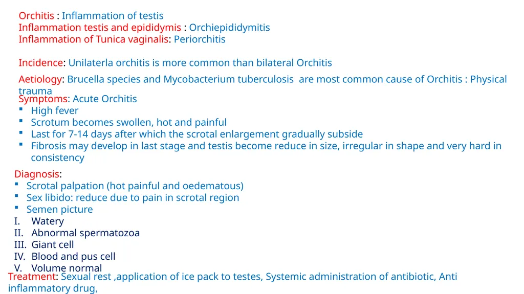
slide6. Orchitis : Inflammation of testis
Inflammation testis and epididymis : Orchiepididymitis
Inflammation of Tunica vaginalis: Periorchitis
Incidence: Unilaterla orchitis is more common than bilateral Orchitis Aetiology: Brucella species and Mycobacterium tuberculosis are most common cause of Orchitis : Physical trauma Symptoms: Acute Orchitis
High fever
Scrotum becomes swollen, hot and painful
Last for 7-14 days after which the scrotal enlargement gradually subside
Fibrosis may develop in last stage and testis become reduce in size, irregular in shape and very hard in consistency Diagnosis:
Scrotal palpation (hot painful and oedematous)
Sex libido: reduce due to pain in scrotal region
Semen picture
Watery
Abnormal spermatozoa
Giant cell
Blood and pus cell
Volume normal Treatment: Sexual rest ,application of ice pack to testes, Systemic administration of antibiotic, Anti inflammatory drug,<br>
slide7. Disease of Accessory Sex Glands Seminal Vasculitis- it is common in bulls, stallion and boar
Aetiology- Bacteria, Virus,
Clinical signs – Most of the affected animal do not show sign of vasculitis, abdominal pain abscess in infected gland, unilateral
Semen picture - Decrease in motility, elevate pH level, Low fructose concentration, brownish colour of semen
Diagnosis- confirmed by per rectal examination ch/by enlarged, tens and painful during the acute phase or lobular fibrous
Prognosis- fair to poor
Treatment- Bull older than 9 years of age seldom recover from vesiculitis. Younger bull recovered within 6 month. Medical treatment of this disease condition is based on long term antibiotic therapy. Amputation of affected gland or removal is only hope for treatment Disease of Prostate Gland Prostatic disease is rare in all animals except dog. In which prostatitis and hyperplasia are common. This may be due to continuous expansion of the gland throughout life that enable development of prostatic hyperplasia in this species.
Prostatitis- Inflammation of the prostrate is common in dog. This is very uncommon in castrated dogs than intact dogs. Prostatitis and Prostatitis hyperplasia often occur together
Aetiology-_Brucella canis , Proteus, Streptococcus
Clinical Sign-Fever, High pulse rate, constipation, arch back, Anorexia, Vomiting, Leucocytosis , weakness, lethargy, Blood in urine,
Treatment- Parental administration of broad range antibiotic for longer period are indicative. If the cause is Brucella then prognosis is poor. In intact male treatment is very difficult. Castration is choice as it prevent the reoccurrences<br>
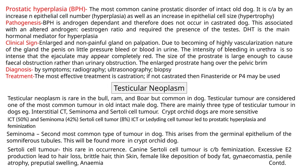
slide8. Prostatic hyperplasia (BPH)- The most common canine prostatic disorder of intact old dog. It is c/a by an increase n epithelial cell number (hyperplasia) as well as an increase in epithelial cell size (hypertrophy)
Pathogenesis-BPH is androgen dependant and therefore does not occur in castrated dog. This associated with an altered androgen: oestrogen ratio and required the presence of the testes. DHT is the main hormonal mediator for hyperplasia
Clinical Sign-Enlarged and non-painful gland on palpation. Due to becoming of highly vascularization nature of the gland the penis on little pressure bleed or blood in urine. The intensity of bleeding in urethra is so intense that the ejaculate may appear completely red. The size of the prostrate is large enough to cause faecal obstruction rather than urinary obstruction. The enlarged prostrate hang over the pelvic brim
Diagnosis- by symptoms; radiography; ultrasonography; biopsy
Treatment-The most effective treatment is castration; if not castrated then Finasteride or P4 may be used Testicular Neoplasm Testicular neoplasm is rare in the bull, ram, and Boar but common in dog. Testicular tumour are considered one of the most common tumour in old intact male dog. There are mainly three type of testicular tumour in dogs eg. Interstitial CT, Seminoma and Sertoli cell tumour. Crypt orchid dogs are more sensitive ICT (50%) and Seminoma (42%) Sertoli cell tumor (8%) ICT or Ledyding cell tumour led to prostatic hyperplasia and feminization Seminoma – Second most common type of tumour in dog. This arises from the germinal epithelium of the somniferous tubules. This will be found more in crypt orchid dog. Sertoli cell tumour- this rare in occurrence. Canine Sertoli cell tumour is c/b feminization. Excessive E2 production lead to hair loss, brittle hair, thin Skin, female like deposition of body fat, gynaecomastia, penile atrophy, preputial swelling, Anaemia Contd.<br>
slide9. Diagnosis- By symptoms, CBC count, radiography, ultrasonography Treatment- Surgical castration
Prognosis- poor<br>
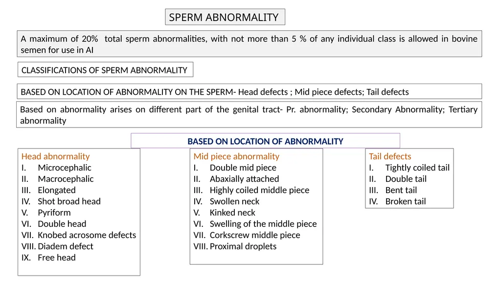
slide10. SPERM ABNORMALITY A maximum of 20% total sperm abnormalities, with not more than 5 % of any individual class is allowed in bovine semen for use in AI CLASSIFICATIONS OF SPERM ABNORMALITY BASED ON LOCATION OF ABNORMALITY ON THE SPERM- Head defects ; Mid piece defects; Tail defects Based on abnormality arises on different part of the genital tract- Pr. abnormality; Secondary Abnormality; Tertiary abnormality Head abnormality
Microcephalic
Macrocephalic
Elongated
Shot broad head
Pyriform
Double head
Knobed acrosome defects
Diadem defect
Free head Mid piece abnormality
Double mid piece
Abaxially attached
Highly coiled middle piece
Swollen neck
Kinked neck
Swelling of the middle piece
Corkscrew middle piece
Proximal droplets Tail defects
Tightly coiled tail
Double tail
Bent tail
Broken tail BASED ON LOCATION OF ABNORMALITY<br>
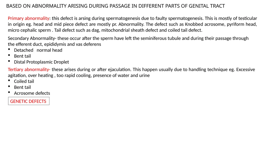
slide11. BASED ON ABNORMALITY ARISING DURING PASSAGE IN DIFFERENT PARTS OF GENITAL TRACT Primary abnormality: this defect is arsing during spermatogenesis due to faulty spermatogenesis. This is mostly of testicular in origin eg. head and mid piece defect are mostly pr. Abnormality. The defect such as Knobbed acrosome, pyriform head, micro cephalic sperm . Tail defect such as dag, mitochondrial sheath defect and coiled tail defect. Secondary Abnormality- these occur after the sperm have left the seminiferous tubule and during their passage through the efferent duct, epididymis and vas deferens
Detached normal head
Bent tail
Distal Protoplasmic Droplet Tertiary abnormality- these arises during or after ejaculation. This happen usually due to handling technique eg. Excessive agitation, over heating , too rapid cooling, presence of water and urine
Coiled tail
Bent tail
Acrosome defects GENETIC DEFECTS<br>
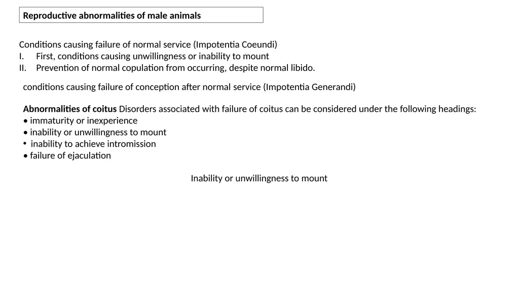
slide12. Reproductive abnormalities of male animals Conditions causing failure of normal service (Impotentia Coeundi)
First, conditions causing unwillingness or inability to mount
Prevention of normal copulation from occurring, despite normal libido. conditions causing failure of conception after normal service (Impotentia Generandi) Abnormalities of coitus Disorders associated with failure of coitus can be considered under the following headings:
• immaturity or inexperience
• inability or unwillingness to mount
inability to achieve intromission
• failure of ejaculation Inability or unwillingness to mount<br>
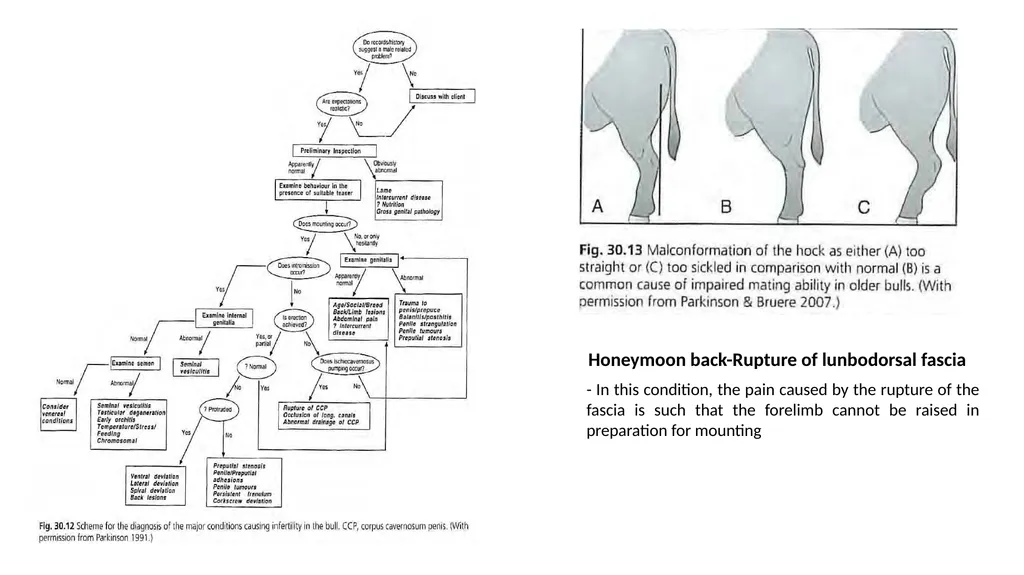
slide13. Honeymoon back-Rupture of lunbodorsal fascia - In this condition, the pain caused by the rupture of the fascia is such that the forelimb cannot be raised in preparation for mounting<br>
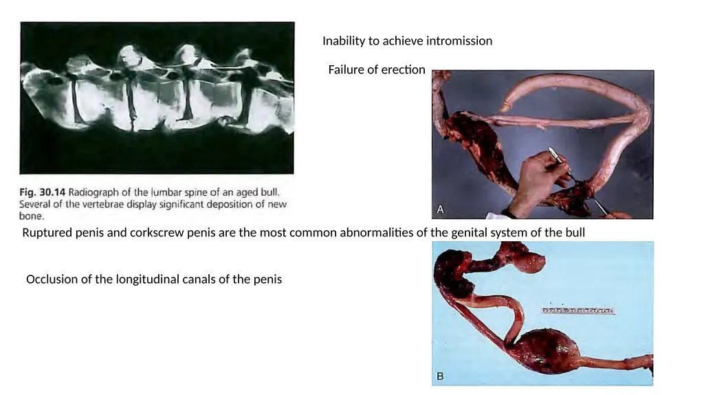
slide14. Inability to achieve intromission Failure of erection Ruptured penis and corkscrew penis are the most common abnormalities of the genital system of the bull Occlusion of the longitudinal canals of the penis<br>
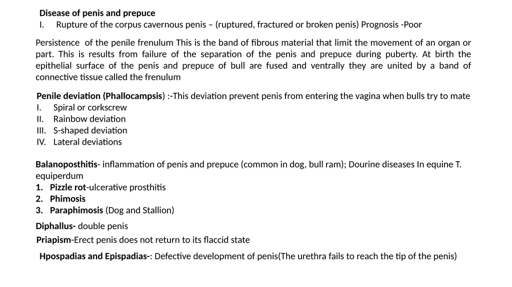
slide15. Disease of penis and prepuce
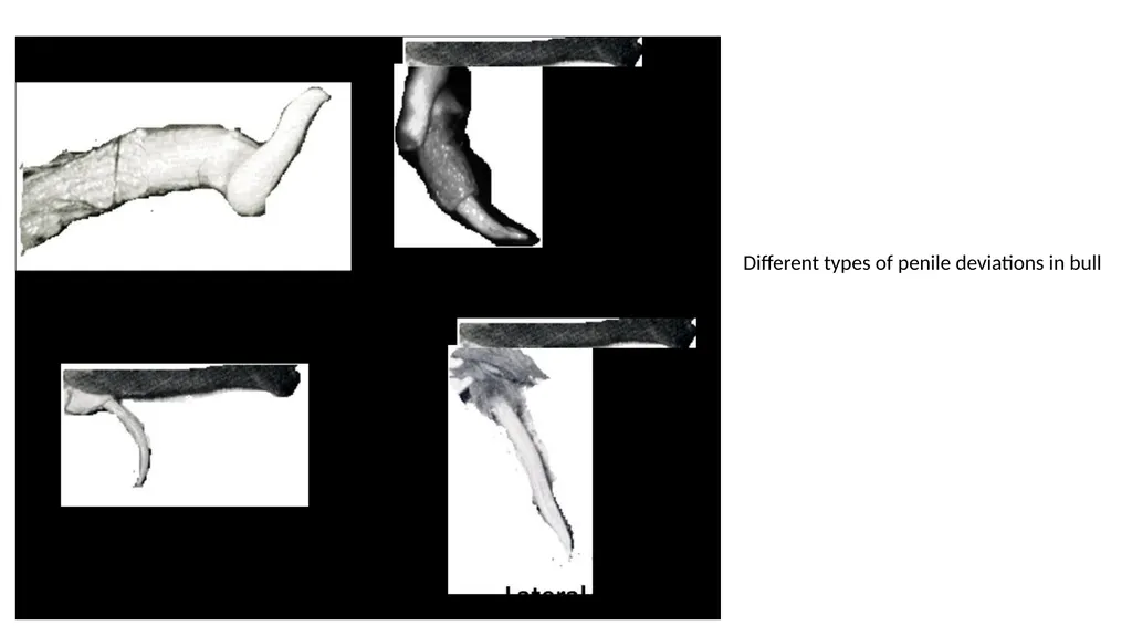
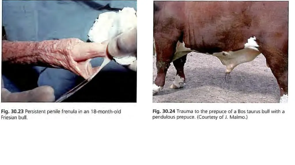
Rupture of the corpus cavernous penis – (ruptured, fractured or broken penis) Prognosis -Poor Persistence of the penile frenulum This is the band of fibrous material that limit the movement of an organ or part. This is results from failure of the separation of the penis and prepuce during puberty. At birth the epithelial surface of the penis and prepuce of bull are fused and ventrally they are united by a band of connective tissue called the frenulum Penile deviation (Phallocampsis) :-This deviation prevent penis from entering the vagina when bulls try to mate
Spiral or corkscrew
Rainbow deviation
S-shaped deviation
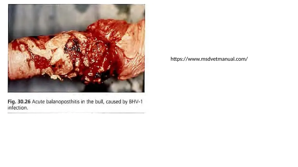
Lateral deviations Balanoposthitis- inflammation of penis and prepuce (common in dog, bull ram); Dourine diseases In equine T. equiperdum
Pizzle rot-ulcerative prosthitis
Phimosis
Paraphimosis (Dog and Stallion) Diphallus- double penis Priapism-Erect penis does not return to its flaccid state Hpospadias and Epispadias-: Defective development of penis(The urethra fails to reach the tip of the penis)<br>
slide16. Treatment: Fascia lata implant technique is used for repair of both spiral and ventral penile deviation. The objective of the technique is to create a firm union of the dorsal apical ligament to the tunica albuginea to prevent its slippage. This technique uses an autogenous or homologous strip of fascia lata from the cranial border of the biceps femoris muscle to provide dorsal reinforcement of the apical ligament and fixation of the ligament to the tunica albuginea. Homologous strips of fascia lata can be harvested from freshly dead cattle and stored in 70% ethyl alcohol until used.<br>
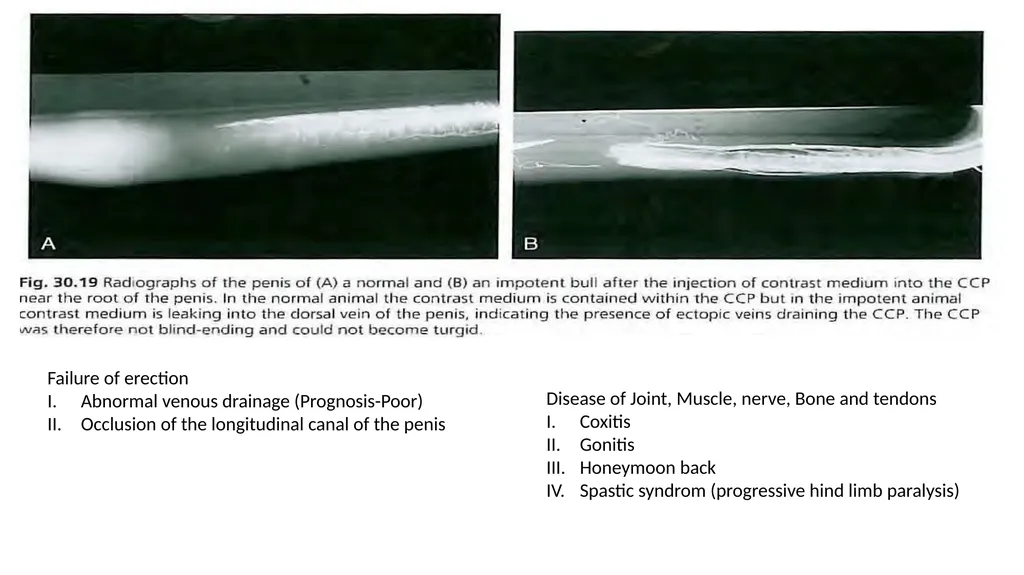
slide18. Failure of erection
Abnormal venous drainage (Prognosis-Poor)
Occlusion of the longitudinal canal of the penis Disease of Joint, Muscle, nerve, Bone and tendons
Coxitis
Gonitis
Honeymoon back
Spastic syndrom (progressive hind limb paralysis)<br>
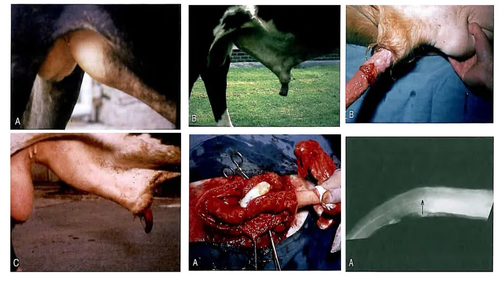
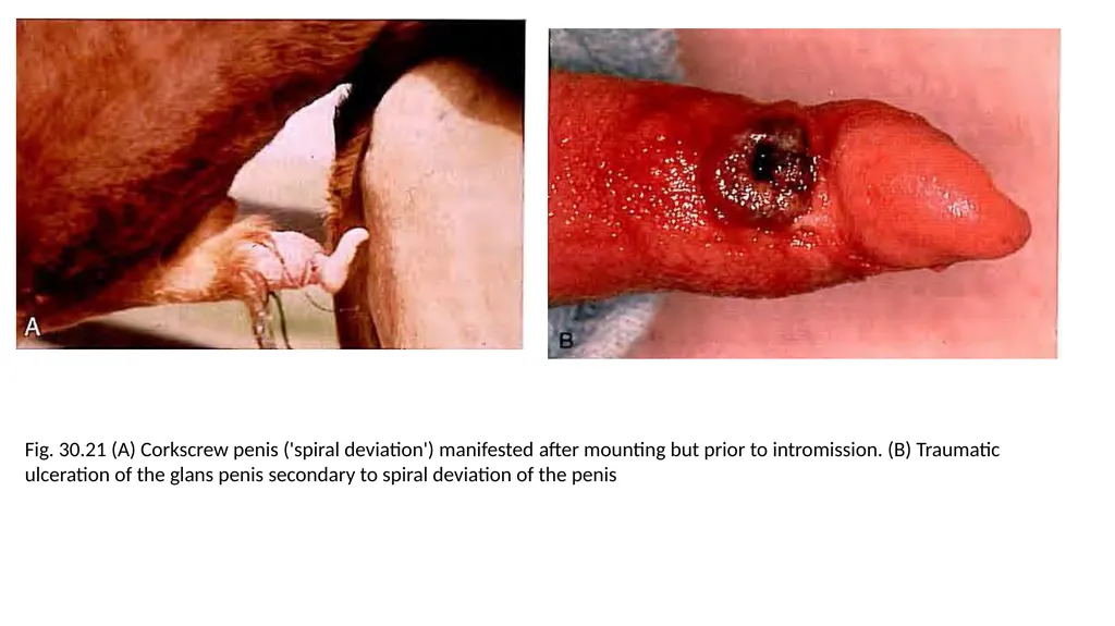
slide19. Fig. 30.21 (A) Corkscrew penis ('spiral deviation') manifested after mounting but prior to intromission. (B) Traumatic ulceration of the glans penis secondary to spiral deviation of the penis<br>
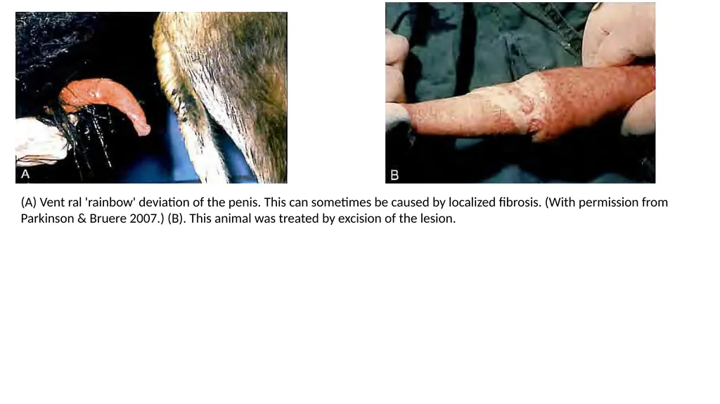
slide20. (A) Vent ral 'rainbow' deviation of the penis. This can sometimes be caused by localized fibrosis. (With permission from Parkinson & Bruere 2007.) (B). This animal was treated by excision of the lesion.<br>
slide21. Different types of penile deviations in bull<br>
slide23. Fig. 30.25 (A) Chronic fibrosis in a prolapse of the distal prepuce in a Hereford bull. (B) Submucosal resection. (C) Final repair of the lesion.<br>
slide24. https://www.msdvetmanual.com/<br>
Asstt. Prof.
Department of Veterinary Clinical Complex UNIT VII VGO 604<br>
slide2. CAUSES OF MALE INFERTILITY IMOTENTIA COEUNDI IMPOTENTIA GENERENDI<br>
slide3. Testicular Hypoplasia: This is a congenital failure in the development of the Spermatogenic epithelium. It is found in two form
Hereditary and non hereditary
Hereditary: Recessive gene
Non-genetic: Extra X chromosome Klinefelter syndrom
Unilateral is more common than bilateral hypoplasia. In bull left side hypoplasia is more than the right
Symptoms
The scrotum of the affected male is smaller than normal male
The spermatic chord is shorter
Increase in proximal protoplasmic droplet
Sexual desire is not affected
Low concentration of semen
Low mortality
High incidence of proximal protoplasmic droplet
Presence of giant cell and medusa cell in semen
Bilateral
Clear watery
Few and no sperm
Diagnosis
Size of testes- small and flabby
Epididymis – small and hard
Libido-normal
Prognosis- poor
Treatment – affected bull should not be used for breeding purpose<br>
slide4. Cryptorchidism :- Failure of testes to descend into the scrotum is called cryptorchidism
High Flanker: When testes are located in the inguinal canal, the male is called high flanker and when cranial to the scrotum is called low flanker
Bilateral
Unilateral
Monorchidic
Anorchidism Incidence : Stallion; Boar; Dog; Unilateral are more common than bilateral Aetiology : Genetic factor : Defect in testis ;Inadequate abdominal pressure : Excessive enlargement of the Gubernaculum Symptoms: Neoplasia and feminization; decrease fertility; behavioural modification eg. Hyper sexuality , excitability & irritability Diagnosis: External palpation ;ultrasonography; Semen picture (low concentration ; abnormal sperm; Azoospermia Prognosis: Poor Treatment : The affected animal should not be used for breeding purpose : Animal should be castrated<br>
slide5. Testicular Degeneration: The partial or complete failure of epithelium of seminiferous tubules to proceed with spermatogenesis. Acquire disorder and is most common cause of infertility in male domestic animals. It may happen very but reaeration is very slow fast Aetiology : Endless : Commons are hyperthermia; Extreme cold; Advance age; Stress; Irradiation; Autoimmune; hormonal imbalance ; Nutritional Disorder
Incidence: Bilateral is very common
Symptoms: Smaller and softer than normal; libido is not affected; Adhesions between testis and scrotum are usually found; fibrosis and calcification also occur in late stage; Testes becomes hard and shrunken and irregular Semen picture: Depend on degree and involvement testicular tissue
Increasing in the incidence of loos head
Immature spermatozoa
Ejaculate volume is nit affected
Motility falls
Abnormality rise Diagnosis: Based on
history
Careful examination of scrotum
Semen picture
Ultrasonography Prognosis- Fair to good Treatment: Correction of cause ; supplementation of nutrition; Supplementation of vit A; Sexual rest; Keep animal in cool<br>
slide6. Orchitis : Inflammation of testis
Inflammation testis and epididymis : Orchiepididymitis
Inflammation of Tunica vaginalis: Periorchitis
Incidence: Unilaterla orchitis is more common than bilateral Orchitis Aetiology: Brucella species and Mycobacterium tuberculosis are most common cause of Orchitis : Physical trauma Symptoms: Acute Orchitis
High fever
Scrotum becomes swollen, hot and painful
Last for 7-14 days after which the scrotal enlargement gradually subside
Fibrosis may develop in last stage and testis become reduce in size, irregular in shape and very hard in consistency Diagnosis:
Scrotal palpation (hot painful and oedematous)
Sex libido: reduce due to pain in scrotal region
Semen picture
Watery
Abnormal spermatozoa
Giant cell
Blood and pus cell
Volume normal Treatment: Sexual rest ,application of ice pack to testes, Systemic administration of antibiotic, Anti inflammatory drug,<br>
slide7. Disease of Accessory Sex Glands Seminal Vasculitis- it is common in bulls, stallion and boar
Aetiology- Bacteria, Virus,
Clinical signs – Most of the affected animal do not show sign of vasculitis, abdominal pain abscess in infected gland, unilateral
Semen picture - Decrease in motility, elevate pH level, Low fructose concentration, brownish colour of semen
Diagnosis- confirmed by per rectal examination ch/by enlarged, tens and painful during the acute phase or lobular fibrous
Prognosis- fair to poor
Treatment- Bull older than 9 years of age seldom recover from vesiculitis. Younger bull recovered within 6 month. Medical treatment of this disease condition is based on long term antibiotic therapy. Amputation of affected gland or removal is only hope for treatment Disease of Prostate Gland Prostatic disease is rare in all animals except dog. In which prostatitis and hyperplasia are common. This may be due to continuous expansion of the gland throughout life that enable development of prostatic hyperplasia in this species.
Prostatitis- Inflammation of the prostrate is common in dog. This is very uncommon in castrated dogs than intact dogs. Prostatitis and Prostatitis hyperplasia often occur together
Aetiology-_Brucella canis , Proteus, Streptococcus
Clinical Sign-Fever, High pulse rate, constipation, arch back, Anorexia, Vomiting, Leucocytosis , weakness, lethargy, Blood in urine,
Treatment- Parental administration of broad range antibiotic for longer period are indicative. If the cause is Brucella then prognosis is poor. In intact male treatment is very difficult. Castration is choice as it prevent the reoccurrences<br>
slide8. Prostatic hyperplasia (BPH)- The most common canine prostatic disorder of intact old dog. It is c/a by an increase n epithelial cell number (hyperplasia) as well as an increase in epithelial cell size (hypertrophy)
Pathogenesis-BPH is androgen dependant and therefore does not occur in castrated dog. This associated with an altered androgen: oestrogen ratio and required the presence of the testes. DHT is the main hormonal mediator for hyperplasia
Clinical Sign-Enlarged and non-painful gland on palpation. Due to becoming of highly vascularization nature of the gland the penis on little pressure bleed or blood in urine. The intensity of bleeding in urethra is so intense that the ejaculate may appear completely red. The size of the prostrate is large enough to cause faecal obstruction rather than urinary obstruction. The enlarged prostrate hang over the pelvic brim
Diagnosis- by symptoms; radiography; ultrasonography; biopsy
Treatment-The most effective treatment is castration; if not castrated then Finasteride or P4 may be used Testicular Neoplasm Testicular neoplasm is rare in the bull, ram, and Boar but common in dog. Testicular tumour are considered one of the most common tumour in old intact male dog. There are mainly three type of testicular tumour in dogs eg. Interstitial CT, Seminoma and Sertoli cell tumour. Crypt orchid dogs are more sensitive ICT (50%) and Seminoma (42%) Sertoli cell tumor (8%) ICT or Ledyding cell tumour led to prostatic hyperplasia and feminization Seminoma – Second most common type of tumour in dog. This arises from the germinal epithelium of the somniferous tubules. This will be found more in crypt orchid dog. Sertoli cell tumour- this rare in occurrence. Canine Sertoli cell tumour is c/b feminization. Excessive E2 production lead to hair loss, brittle hair, thin Skin, female like deposition of body fat, gynaecomastia, penile atrophy, preputial swelling, Anaemia Contd.<br>
slide9. Diagnosis- By symptoms, CBC count, radiography, ultrasonography Treatment- Surgical castration
Prognosis- poor<br>
slide10. SPERM ABNORMALITY A maximum of 20% total sperm abnormalities, with not more than 5 % of any individual class is allowed in bovine semen for use in AI CLASSIFICATIONS OF SPERM ABNORMALITY BASED ON LOCATION OF ABNORMALITY ON THE SPERM- Head defects ; Mid piece defects; Tail defects Based on abnormality arises on different part of the genital tract- Pr. abnormality; Secondary Abnormality; Tertiary abnormality Head abnormality
Microcephalic
Macrocephalic
Elongated
Shot broad head
Pyriform
Double head
Knobed acrosome defects
Diadem defect
Free head Mid piece abnormality
Double mid piece
Abaxially attached
Highly coiled middle piece
Swollen neck
Kinked neck
Swelling of the middle piece
Corkscrew middle piece
Proximal droplets Tail defects
Tightly coiled tail
Double tail
Bent tail
Broken tail BASED ON LOCATION OF ABNORMALITY<br>
slide11. BASED ON ABNORMALITY ARISING DURING PASSAGE IN DIFFERENT PARTS OF GENITAL TRACT Primary abnormality: this defect is arsing during spermatogenesis due to faulty spermatogenesis. This is mostly of testicular in origin eg. head and mid piece defect are mostly pr. Abnormality. The defect such as Knobbed acrosome, pyriform head, micro cephalic sperm . Tail defect such as dag, mitochondrial sheath defect and coiled tail defect. Secondary Abnormality- these occur after the sperm have left the seminiferous tubule and during their passage through the efferent duct, epididymis and vas deferens
Detached normal head
Bent tail
Distal Protoplasmic Droplet Tertiary abnormality- these arises during or after ejaculation. This happen usually due to handling technique eg. Excessive agitation, over heating , too rapid cooling, presence of water and urine
Coiled tail
Bent tail
Acrosome defects GENETIC DEFECTS<br>
slide12. Reproductive abnormalities of male animals Conditions causing failure of normal service (Impotentia Coeundi)
First, conditions causing unwillingness or inability to mount
Prevention of normal copulation from occurring, despite normal libido. conditions causing failure of conception after normal service (Impotentia Generandi) Abnormalities of coitus Disorders associated with failure of coitus can be considered under the following headings:
• immaturity or inexperience
• inability or unwillingness to mount
inability to achieve intromission
• failure of ejaculation Inability or unwillingness to mount<br>
slide13. Honeymoon back-Rupture of lunbodorsal fascia - In this condition, the pain caused by the rupture of the fascia is such that the forelimb cannot be raised in preparation for mounting<br>
slide14. Inability to achieve intromission Failure of erection Ruptured penis and corkscrew penis are the most common abnormalities of the genital system of the bull Occlusion of the longitudinal canals of the penis<br>
slide15. Disease of penis and prepuce
Rupture of the corpus cavernous penis – (ruptured, fractured or broken penis) Prognosis -Poor Persistence of the penile frenulum This is the band of fibrous material that limit the movement of an organ or part. This is results from failure of the separation of the penis and prepuce during puberty. At birth the epithelial surface of the penis and prepuce of bull are fused and ventrally they are united by a band of connective tissue called the frenulum Penile deviation (Phallocampsis) :-This deviation prevent penis from entering the vagina when bulls try to mate
Spiral or corkscrew
Rainbow deviation
S-shaped deviation
Lateral deviations Balanoposthitis- inflammation of penis and prepuce (common in dog, bull ram); Dourine diseases In equine T. equiperdum
Pizzle rot-ulcerative prosthitis
Phimosis
Paraphimosis (Dog and Stallion) Diphallus- double penis Priapism-Erect penis does not return to its flaccid state Hpospadias and Epispadias-: Defective development of penis(The urethra fails to reach the tip of the penis)<br>
slide16. Treatment: Fascia lata implant technique is used for repair of both spiral and ventral penile deviation. The objective of the technique is to create a firm union of the dorsal apical ligament to the tunica albuginea to prevent its slippage. This technique uses an autogenous or homologous strip of fascia lata from the cranial border of the biceps femoris muscle to provide dorsal reinforcement of the apical ligament and fixation of the ligament to the tunica albuginea. Homologous strips of fascia lata can be harvested from freshly dead cattle and stored in 70% ethyl alcohol until used.<br>
slide18. Failure of erection
Abnormal venous drainage (Prognosis-Poor)
Occlusion of the longitudinal canal of the penis Disease of Joint, Muscle, nerve, Bone and tendons
Coxitis
Gonitis
Honeymoon back
Spastic syndrom (progressive hind limb paralysis)<br>
slide19. Fig. 30.21 (A) Corkscrew penis ('spiral deviation') manifested after mounting but prior to intromission. (B) Traumatic ulceration of the glans penis secondary to spiral deviation of the penis<br>
slide20. (A) Vent ral 'rainbow' deviation of the penis. This can sometimes be caused by localized fibrosis. (With permission from Parkinson & Bruere 2007.) (B). This animal was treated by excision of the lesion.<br>
slide21. Different types of penile deviations in bull<br>
slide23. Fig. 30.25 (A) Chronic fibrosis in a prolapse of the distal prepuce in a Hereford bull. (B) Submucosal resection. (C) Final repair of the lesion.<br>
slide24. https://www.msdvetmanual.com/<br>