
Introduction This optimal pathway is primarily
DJ
Published · 15 slides · 0 views
1 / 1

Description
Introduction This optimal pathway is primarily designed to improve outcomes in lung cancer by encouraging best practice, reducing variation, and reducing delays in diagnosis, staging and treatment. Use of guidelines The diagnosis, staging
Related Topics
Download this presentation From Below
"Introduction This optimal pathway is primarily" is the property of its rightful owner. Permission is granted to download and print the materials on this website for personal, non-commercial use only, and to display it on your personal computer provided you do not modify the materials and that you retain all copyright notices contained in the materials. By downloading content from our website, you accept the terms of this agreement.
Share
Embed code
Presentation Transcript
01
Introduction
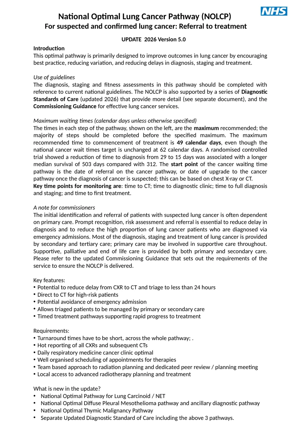
This optimal pathway is primarily designed to improve outcomes in lung cancer by encouraging best practice, reducing variation, and reducing delays in diagnosis, staging and treatment.
Use of guidelines
The diagnosis, staging and fitness assessments in this pathway should be completed with reference to current national guidelines. The NOLCP is also supported by a series of Diagnostic Standards of Care (updated 2026) that provide more detail (see separate document), and the Commissioning Guidance for effective lung cancer services.
Maximum waiting times (calendar days unless otherwise specified)
The times in each step of the pathway, shown on the left, are the maximum recommended; the majority of steps should be completed before the specified maximum. The maximum recommended time to commencement of treatment is 49 calendar days, even though the national cancer wait times target is unchanged at 62 calendar days. A randomised controlled trial showed a reduction of time to diagnosis from 29 to 15 days was associated with a longer median survival of 503 days compared with 312. The start point of the cancer waiting time pathway is the date of referral on the cancer pathway, or date of upgrade to the cancer pathway once the diagnosis of cancer is suspected; this can be based on chest X-ray or CT.
Key time points for monitoring are: time to CT; time to diagnostic clinic; time to full diagnosis and staging; and time to first treatment.
A note for commissioners
The initial identification and referral of patients with suspected lung cancer is often dependent on primary care. Prompt recognition, risk assessment and referral is essential to reduce delay in diagnosis and to reduce the high proportion of lung cancer patients who are diagnosed via emergency admissions. Most of the diagnosis, staging and treatment of lung cancer is provided by secondary and tertiary care; primary care may be involved in supportive care throughout. Supportive, palliative and end of life care is provided by both primary and secondary care. Please refer to the updated Commissioning Guidance that sets out the requirements of the service to ensure the NOLCP is delivered.
Key features:
Potential to reduce delay from CXR to CT and triage to less than 24 hours
Direct to CT for high-risk patients
Potential avoidance of emergency admission
Allows triaged patients to be managed by primary or secondary care
Timed treatment pathways supporting rapid progress to treatment
Requirements:
Turnaround times have to be short, across the whole pathway; .
Hot reporting of all CXRs and subsequent CTs
Daily respiratory medicine cancer clinic optimal
Well organised scheduling of appointments for therapies
Team based approach to radiation planning and dedicated peer review / planning meeting
Local access to advanced radiotherapy planning and treatment
What is new in the update?
National Optimal Pathway for Lung Carcinoid / NET
National Optimal Diffuse Pleural Mesothelioma pathway and ancillary diagnostic pathway
National Optimal Thymic Malignancy Pathway
Separate Updated Diagnostic Standard of Care including the above 3 pathways. UPDATE 2026 Version 5.0 National Optimal Lung Cancer Pathway (NOLCP)
For suspected and confirmed lung cancer: Referral to treatment<br>
This optimal pathway is primarily designed to improve outcomes in lung cancer by encouraging best practice, reducing variation, and reducing delays in diagnosis, staging and treatment.
Use of guidelines
The diagnosis, staging and fitness assessments in this pathway should be completed with reference to current national guidelines. The NOLCP is also supported by a series of Diagnostic Standards of Care (updated 2026) that provide more detail (see separate document), and the Commissioning Guidance for effective lung cancer services.
Maximum waiting times (calendar days unless otherwise specified)
The times in each step of the pathway, shown on the left, are the maximum recommended; the majority of steps should be completed before the specified maximum. The maximum recommended time to commencement of treatment is 49 calendar days, even though the national cancer wait times target is unchanged at 62 calendar days. A randomised controlled trial showed a reduction of time to diagnosis from 29 to 15 days was associated with a longer median survival of 503 days compared with 312. The start point of the cancer waiting time pathway is the date of referral on the cancer pathway, or date of upgrade to the cancer pathway once the diagnosis of cancer is suspected; this can be based on chest X-ray or CT.
Key time points for monitoring are: time to CT; time to diagnostic clinic; time to full diagnosis and staging; and time to first treatment.
A note for commissioners
The initial identification and referral of patients with suspected lung cancer is often dependent on primary care. Prompt recognition, risk assessment and referral is essential to reduce delay in diagnosis and to reduce the high proportion of lung cancer patients who are diagnosed via emergency admissions. Most of the diagnosis, staging and treatment of lung cancer is provided by secondary and tertiary care; primary care may be involved in supportive care throughout. Supportive, palliative and end of life care is provided by both primary and secondary care. Please refer to the updated Commissioning Guidance that sets out the requirements of the service to ensure the NOLCP is delivered.
Key features:
Potential to reduce delay from CXR to CT and triage to less than 24 hours
Direct to CT for high-risk patients
Potential avoidance of emergency admission
Allows triaged patients to be managed by primary or secondary care
Timed treatment pathways supporting rapid progress to treatment
Requirements:
Turnaround times have to be short, across the whole pathway; .
Hot reporting of all CXRs and subsequent CTs
Daily respiratory medicine cancer clinic optimal
Well organised scheduling of appointments for therapies
Team based approach to radiation planning and dedicated peer review / planning meeting
Local access to advanced radiotherapy planning and treatment
What is new in the update?
National Optimal Pathway for Lung Carcinoid / NET
National Optimal Diffuse Pleural Mesothelioma pathway and ancillary diagnostic pathway
National Optimal Thymic Malignancy Pathway
Separate Updated Diagnostic Standard of Care including the above 3 pathways. UPDATE 2026 Version 5.0 National Optimal Lung Cancer Pathway (NOLCP)
For suspected and confirmed lung cancer: Referral to treatment<br>
02
Full MDT discussion of treatment options Day 1-6 Day 28 Day 33 Day 49£ Suitable for potentially curative treatment? # Fast track lung cancer clinic. Assessment by LCNS.
Diagnostic process plan / diagnostic planning meeting prior to clinic. (*4-11) Treatment of co-morbidity and palliation / treatment of symptoms / prehabilitation and smoking cessation. Consider predictive blood biomarker. Curative Intent Management pathway (*4)
Test bundle requested at first OPA incl. at least: PET-CT, spirometry, brain imaging and as required: detailed lung function and cardiac assessment / ECHO.
LCNS clarify/reassure re complex pathway. Day 0-3 No No cancer: Manage/discharge Day 42 Lung cancer unlikely (*1 &2)
Further management according to local protocol with options of further management of CT findings by primary care or secondary care CT within 24h if clinically indicated; inpatients seen within 48h by acute oncology, respiratory and/or supportive/palliative services Yes National Optimal Lung Cancer Pathway
For suspected and confirmed lung cancer: Referral to treatment
UPDATE 2026 Version 5.0 TRIAGE (*1,2) - by radiology or respiratory medicine according to local protocol - Lung cancer suspected? Investigations to yield maximum diagnostic AND staging information with least harm. Results available within 3 working days for subtype and 10 working days / 14 calendar days for molecular markers (see genomic and molecular pathway *5). GP CXR suspicious of lung cancer? (reported before patient leaves department or within 24h.) No Yes Yes No Yes Maximum times
(calendar days) Maximum times Urgent or routine CXR CT same day / within 72h Further investigation(s)? Follow-up Lung Cancer Clinic
Cancer Confirmed and treatment options discussed. Research trial considered.
LCNS Support in practical aspects OPA with treating specialist within 3 working days of completed investigations (full molecular analysis, staging and fitness) Further investigation(s)? No Yes No Yes Clinical diagnosis or patient preference means no biopsy required. Will pathological diagnosis influence treatment and is potential treatment appropriate to patient’s wishes? Day 21
(Day 18 SCLC) No Further investigation(s) indicated? No Yes CT suspicious of lung cancer? No Yes Manage CT not indicated Inpatient referrals for suspected lung cancer Day -3-0 Further discussion needed? Yes No *Refer to separate numbered pathway detail Some or all diagnosis and staging tests may be in a tertiary centre # Low threshold for curative intent pathway; may discuss with wider MDT if unsure Direct biopsy option; (*3) Throughout pathway: • consider entry into a research trial • offer supportive & palliative care, e.g. by LCNS, GP, specialists in palliative care • encourage smoking cessation £ Reflects the aim for reduced time to treatment; the national target remains 62 days + all patients with stage IV cancer should be routinely offered an assessment No Yes High Risk of Lung Cancer? Direct referral route Other referral route E.g. post discharge, incidental finding on imaging (screen detected my go direct to clinic)<br>
Diagnostic process plan / diagnostic planning meeting prior to clinic. (*4-11) Treatment of co-morbidity and palliation / treatment of symptoms / prehabilitation and smoking cessation. Consider predictive blood biomarker. Curative Intent Management pathway (*4)
Test bundle requested at first OPA incl. at least: PET-CT, spirometry, brain imaging and as required: detailed lung function and cardiac assessment / ECHO.
LCNS clarify/reassure re complex pathway. Day 0-3 No No cancer: Manage/discharge Day 42 Lung cancer unlikely (*1 &2)
Further management according to local protocol with options of further management of CT findings by primary care or secondary care CT within 24h if clinically indicated; inpatients seen within 48h by acute oncology, respiratory and/or supportive/palliative services Yes National Optimal Lung Cancer Pathway
For suspected and confirmed lung cancer: Referral to treatment
UPDATE 2026 Version 5.0 TRIAGE (*1,2) - by radiology or respiratory medicine according to local protocol - Lung cancer suspected? Investigations to yield maximum diagnostic AND staging information with least harm. Results available within 3 working days for subtype and 10 working days / 14 calendar days for molecular markers (see genomic and molecular pathway *5). GP CXR suspicious of lung cancer? (reported before patient leaves department or within 24h.) No Yes Yes No Yes Maximum times
(calendar days) Maximum times Urgent or routine CXR CT same day / within 72h Further investigation(s)? Follow-up Lung Cancer Clinic
Cancer Confirmed and treatment options discussed. Research trial considered.
LCNS Support in practical aspects OPA with treating specialist within 3 working days of completed investigations (full molecular analysis, staging and fitness) Further investigation(s)? No Yes No Yes Clinical diagnosis or patient preference means no biopsy required. Will pathological diagnosis influence treatment and is potential treatment appropriate to patient’s wishes? Day 21
(Day 18 SCLC) No Further investigation(s) indicated? No Yes CT suspicious of lung cancer? No Yes Manage CT not indicated Inpatient referrals for suspected lung cancer Day -3-0 Further discussion needed? Yes No *Refer to separate numbered pathway detail Some or all diagnosis and staging tests may be in a tertiary centre # Low threshold for curative intent pathway; may discuss with wider MDT if unsure Direct biopsy option; (*3) Throughout pathway: • consider entry into a research trial • offer supportive & palliative care, e.g. by LCNS, GP, specialists in palliative care • encourage smoking cessation £ Reflects the aim for reduced time to treatment; the national target remains 62 days + all patients with stage IV cancer should be routinely offered an assessment No Yes High Risk of Lung Cancer? Direct referral route Other referral route E.g. post discharge, incidental finding on imaging (screen detected my go direct to clinic)<br>
03
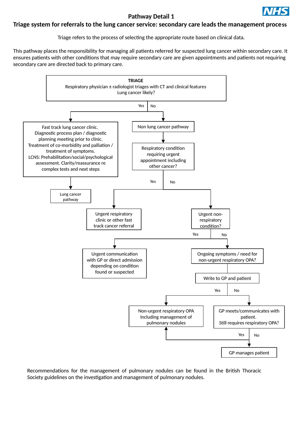
Lung cancer pathway Urgent non-respiratory condition? Respiratory condition requiring urgent appointment including other cancer? GP meets/communicates with patient.
Still requires respiratory OPA? TRIAGE
Respiratory physician ± radiologist triages with CT and clinical features
Lung cancer likely? Yes No GP manages patient No Pathway Detail 1
Triage system for referrals to the lung cancer service: secondary care leads the management process
Triage refers to the process of selecting the appropriate route based on clinical data.
This pathway places the responsibility for managing all patients referred for suspected lung cancer within secondary care. It ensures patients with other conditions that may require secondary care are given appointments and patients not requiring secondary care are directed back to primary care. Fast track lung cancer clinic.
Diagnostic process plan / diagnostic planning meeting prior to clinic.
Treatment of co-morbidity and palliation / treatment of symptoms.
LCNS: Prehabilitation/social/psychological assessment. Clarity/reassurance re complex tests and next steps Non lung cancer pathway Yes No Urgent respiratory clinic or other fast track cancer referral Urgent communication with GP or direct admission depending on condition found or suspected Yes Ongoing symptoms / need for non-urgent respiratory OPA? No Yes No Non-urgent respiratory OPA Including management of pulmonary nodules Write to GP and patient Yes Recommendations for the management of pulmonary nodules can be found in the British Thoracic Society guidelines on the investigation and management of pulmonary nodules.<br>
Still requires respiratory OPA? TRIAGE
Respiratory physician ± radiologist triages with CT and clinical features
Lung cancer likely? Yes No GP manages patient No Pathway Detail 1
Triage system for referrals to the lung cancer service: secondary care leads the management process
Triage refers to the process of selecting the appropriate route based on clinical data.
This pathway places the responsibility for managing all patients referred for suspected lung cancer within secondary care. It ensures patients with other conditions that may require secondary care are given appointments and patients not requiring secondary care are directed back to primary care. Fast track lung cancer clinic.
Diagnostic process plan / diagnostic planning meeting prior to clinic.
Treatment of co-morbidity and palliation / treatment of symptoms.
LCNS: Prehabilitation/social/psychological assessment. Clarity/reassurance re complex tests and next steps Non lung cancer pathway Yes No Urgent respiratory clinic or other fast track cancer referral Urgent communication with GP or direct admission depending on condition found or suspected Yes Ongoing symptoms / need for non-urgent respiratory OPA? No Yes No Non-urgent respiratory OPA Including management of pulmonary nodules Write to GP and patient Yes Recommendations for the management of pulmonary nodules can be found in the British Thoracic Society guidelines on the investigation and management of pulmonary nodules.<br>
04
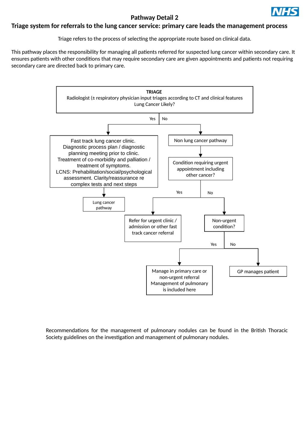
Non-urgent condition? Condition requiring urgent appointment including other cancer? TRIAGE
Radiologist (± respiratory physician input triages according to CT and clinical features
Lung Cancer Likely? Yes No GP manages patient Pathway Detail 2
Triage system for referrals to the lung cancer service: primary care leads the management process
Triage refers to the process of selecting the appropriate route based on clinical data.
This pathway places the responsibility for managing all patients referred for suspected lung cancer within secondary care. It ensures patients with other conditions that may require secondary care are given appointments and patients not requiring secondary care are directed back to primary care. Non lung cancer pathway Yes No Refer for urgent clinic / admission or other fast track cancer referral Manage in primary care or non-urgent referral
Management of pulmonary is included here Yes No Lung cancer pathway Fast track lung cancer clinic.
Diagnostic process plan / diagnostic planning meeting prior to clinic.
Treatment of co-morbidity and palliation / treatment of symptoms.
LCNS: Prehabilitation/social/psychological assessment. Clarity/reassurance re complex tests and next steps Recommendations for the management of pulmonary nodules can be found in the British Thoracic Society guidelines on the investigation and management of pulmonary nodules.<br>
Radiologist (± respiratory physician input triages according to CT and clinical features
Lung Cancer Likely? Yes No GP manages patient Pathway Detail 2
Triage system for referrals to the lung cancer service: primary care leads the management process
Triage refers to the process of selecting the appropriate route based on clinical data.
This pathway places the responsibility for managing all patients referred for suspected lung cancer within secondary care. It ensures patients with other conditions that may require secondary care are given appointments and patients not requiring secondary care are directed back to primary care. Non lung cancer pathway Yes No Refer for urgent clinic / admission or other fast track cancer referral Manage in primary care or non-urgent referral
Management of pulmonary is included here Yes No Lung cancer pathway Fast track lung cancer clinic.
Diagnostic process plan / diagnostic planning meeting prior to clinic.
Treatment of co-morbidity and palliation / treatment of symptoms.
LCNS: Prehabilitation/social/psychological assessment. Clarity/reassurance re complex tests and next steps Recommendations for the management of pulmonary nodules can be found in the British Thoracic Society guidelines on the investigation and management of pulmonary nodules.<br>
05
Suitable for potentially curative treatment? Fast track lung cancer clinic.
Diagnostic process plan / diagnostic planning meeting prior to clinic
Treatment of co-morbidity and palliation / treatment of symptoms
LCNS: Prehabilitation/social/psychological assessment. Clarity/reassurance re complex tests and next steps
Consider predictive blood biomarker National Optimal Pathway No Yes TRIAGE
By radiology or respiratory medicine according to local protocol
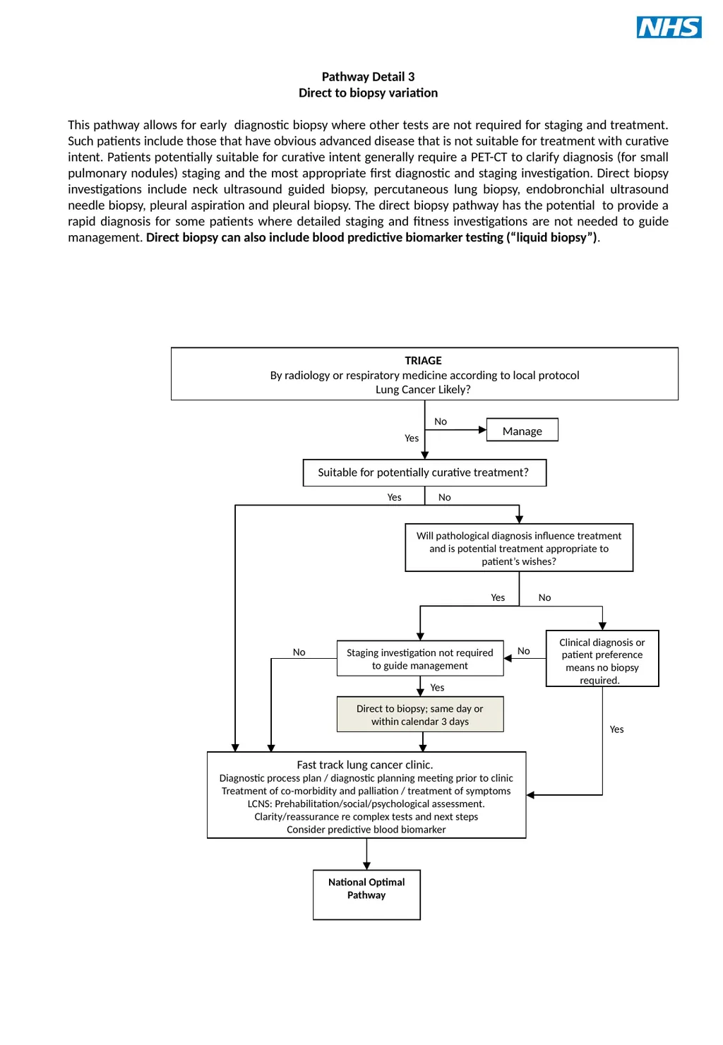
Lung Cancer Likely? Staging investigation not required to guide management Yes Clinical diagnosis or patient preference means no biopsy required. Will pathological diagnosis influence treatment and is potential treatment appropriate to patient’s wishes? No No Yes Manage Pathway Detail 3
Direct to biopsy variation
This pathway allows for early diagnostic biopsy where other tests are not required for staging and treatment. Such patients include those that have obvious advanced disease that is not suitable for treatment with curative intent. Patients potentially suitable for curative intent generally require a PET-CT to clarify diagnosis (for small pulmonary nodules) staging and the most appropriate first diagnostic and staging investigation. Direct biopsy investigations include neck ultrasound guided biopsy, percutaneous lung biopsy, endobronchial ultrasound needle biopsy, pleural aspiration and pleural biopsy. The direct biopsy pathway has the potential to provide a rapid diagnosis for some patients where detailed staging and fitness investigations are not needed to guide management. Direct biopsy can also include blood predictive biomarker testing (“liquid biopsy”). Yes Direct to biopsy; same day or within calendar 3 days No Yes No<br>
Diagnostic process plan / diagnostic planning meeting prior to clinic
Treatment of co-morbidity and palliation / treatment of symptoms
LCNS: Prehabilitation/social/psychological assessment. Clarity/reassurance re complex tests and next steps
Consider predictive blood biomarker National Optimal Pathway No Yes TRIAGE
By radiology or respiratory medicine according to local protocol
Lung Cancer Likely? Staging investigation not required to guide management Yes Clinical diagnosis or patient preference means no biopsy required. Will pathological diagnosis influence treatment and is potential treatment appropriate to patient’s wishes? No No Yes Manage Pathway Detail 3
Direct to biopsy variation
This pathway allows for early diagnostic biopsy where other tests are not required for staging and treatment. Such patients include those that have obvious advanced disease that is not suitable for treatment with curative intent. Patients potentially suitable for curative intent generally require a PET-CT to clarify diagnosis (for small pulmonary nodules) staging and the most appropriate first diagnostic and staging investigation. Direct biopsy investigations include neck ultrasound guided biopsy, percutaneous lung biopsy, endobronchial ultrasound needle biopsy, pleural aspiration and pleural biopsy. The direct biopsy pathway has the potential to provide a rapid diagnosis for some patients where detailed staging and fitness investigations are not needed to guide management. Direct biopsy can also include blood predictive biomarker testing (“liquid biopsy”). Yes Direct to biopsy; same day or within calendar 3 days No Yes No<br>
06
Stage: Potentially T1-3 N0-2 M0 (N2 non-bulky; i.e. <3cm)
Or locally advanced; potential for radical RT?
May include selected patients with oligometastatic disease Simultaneous fast track: Usual diagnosis and staging pathway Patients with borderline fitness$ add:
Preoperative rehabilitation
Shuttle walk test / CPEX / ECHO
Perfusion scan if required
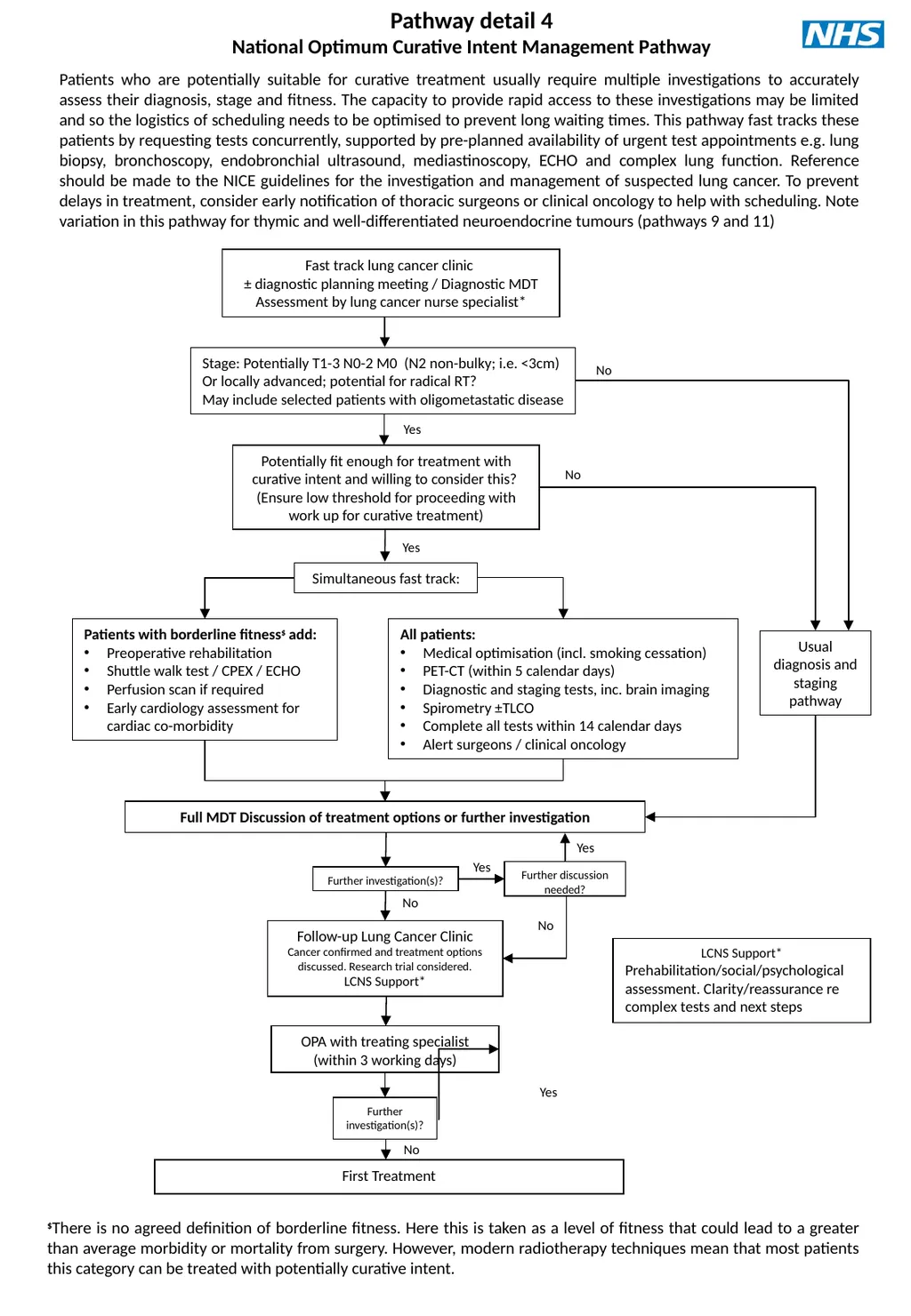
Early cardiology assessment for cardiac co-morbidity Pathway detail 4
National Optimum Curative Intent Management Pathway Potentially fit enough for treatment with curative intent and willing to consider this?
(Ensure low threshold for proceeding with work up for curative treatment) Fast track lung cancer clinic
± diagnostic planning meeting / Diagnostic MDT
Assessment by lung cancer nurse specialist* Full MDT Discussion of treatment options or further investigation OPA with treating specialist (within 3 working days) First Treatment Further investigation(s)? No Yes No Yes All patients:
Medical optimisation (incl. smoking cessation)
PET-CT (within 5 calendar days)
Diagnostic and staging tests, inc. brain imaging
Spirometry ±TLCO
Complete all tests within 14 calendar days
Alert surgeons / clinical oncology Yes No Patients who are potentially suitable for curative treatment usually require multiple investigations to accurately assess their diagnosis, stage and fitness. The capacity to provide rapid access to these investigations may be limited and so the logistics of scheduling needs to be optimised to prevent long waiting times. This pathway fast tracks these patients by requesting tests concurrently, supported by pre-planned availability of urgent test appointments e.g. lung biopsy, bronchoscopy, endobronchial ultrasound, mediastinoscopy, ECHO and complex lung function. Reference should be made to the NICE guidelines for the investigation and management of suspected lung cancer. To prevent delays in treatment, consider early notification of thoracic surgeons or clinical oncology to help with scheduling. Note variation in this pathway for thymic and well-differentiated neuroendocrine tumours (pathways 9 and 11) Further investigation(s)? Follow-up Lung Cancer Clinic
Cancer confirmed and treatment options discussed. Research trial considered.
LCNS Support* No Yes Further discussion needed? Yes No $There is no agreed definition of borderline fitness. Here this is taken as a level of fitness that could lead to a greater than average morbidity or mortality from surgery. However, modern radiotherapy techniques mean that most patients this category can be treated with potentially curative intent. LCNS Support*
Prehabilitation/social/psychological assessment. Clarity/reassurance re complex tests and next steps<br>
Or locally advanced; potential for radical RT?
May include selected patients with oligometastatic disease Simultaneous fast track: Usual diagnosis and staging pathway Patients with borderline fitness$ add:
Preoperative rehabilitation
Shuttle walk test / CPEX / ECHO
Perfusion scan if required
Early cardiology assessment for cardiac co-morbidity Pathway detail 4
National Optimum Curative Intent Management Pathway Potentially fit enough for treatment with curative intent and willing to consider this?
(Ensure low threshold for proceeding with work up for curative treatment) Fast track lung cancer clinic
± diagnostic planning meeting / Diagnostic MDT
Assessment by lung cancer nurse specialist* Full MDT Discussion of treatment options or further investigation OPA with treating specialist (within 3 working days) First Treatment Further investigation(s)? No Yes No Yes All patients:
Medical optimisation (incl. smoking cessation)
PET-CT (within 5 calendar days)
Diagnostic and staging tests, inc. brain imaging
Spirometry ±TLCO
Complete all tests within 14 calendar days
Alert surgeons / clinical oncology Yes No Patients who are potentially suitable for curative treatment usually require multiple investigations to accurately assess their diagnosis, stage and fitness. The capacity to provide rapid access to these investigations may be limited and so the logistics of scheduling needs to be optimised to prevent long waiting times. This pathway fast tracks these patients by requesting tests concurrently, supported by pre-planned availability of urgent test appointments e.g. lung biopsy, bronchoscopy, endobronchial ultrasound, mediastinoscopy, ECHO and complex lung function. Reference should be made to the NICE guidelines for the investigation and management of suspected lung cancer. To prevent delays in treatment, consider early notification of thoracic surgeons or clinical oncology to help with scheduling. Note variation in this pathway for thymic and well-differentiated neuroendocrine tumours (pathways 9 and 11) Further investigation(s)? Follow-up Lung Cancer Clinic
Cancer confirmed and treatment options discussed. Research trial considered.
LCNS Support* No Yes Further discussion needed? Yes No $There is no agreed definition of borderline fitness. Here this is taken as a level of fitness that could lead to a greater than average morbidity or mortality from surgery. However, modern radiotherapy techniques mean that most patients this category can be treated with potentially curative intent. LCNS Support*
Prehabilitation/social/psychological assessment. Clarity/reassurance re complex tests and next steps<br>
07
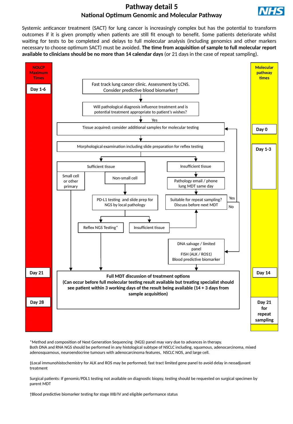
Pathway detail 5
National Optimum Genomic and Molecular Pathway NOLCP
Maximum Times Systemic anticancer treatment (SACT) for lung cancer is increasingly complex but has the potential to transform outcomes if it is given promptly when patients are still fit enough to benefit. Some patients deteriorate whilst waiting for tests to be completed and delays to full molecular analysis (including genomics and other markers necessary to choose optimum SACT) must be avoided. The time from acquisition of sample to full molecular report available to clinicians should be no more than 14 calendar days (or 21 days in the case of repeat sampling). Day 1-6 Fast track lung cancer clinic. Assessment by LCNS.
Consider predictive blood biomarker† Tissue acquired; consider additional samples for molecular testing Yes Will pathological diagnosis influence treatment and is potential treatment appropriate to patient’s wishes? No Day 21 Day 28 Morphological examination including slide preparation for reflex testing Molecular pathway times Day 0 Day 1-3 Insufficient tissue Pathology email / phone lung MDT same day Suitable for repeat sampling? Discuss before next MDT Sufficient tissue Small cell or other primary Non-small cell Full MDT discussion of treatment options
(Can occur before full molecular testing result available but treating specialist should see patient within 3 working days of the result being available (14 + 3 days from sample acquisition) Reflex NGS Testing* PD-L1 testing and slide prep for NGS by local pathology Insufficient tissue Day 14 DNA salvage / limited panel
FISH (ALK / ROS1)
Blood predictive biomarker Day 21 for repeat sampling Yes *Method and composition of Next Generation Sequencing (NGS) panel may vary due to advances in therapy.
Both DNA and RNA NGS should be performed in any histological subtype of NSCLC including, squamous, adenocarcinoma, mixed adenosquamous, neuroendocrine tumours with adenocarcinoma features, NSCLC NOS, and large cell.
‡Local immunohistochemistry for ALK and ROS may be performed; fast tract limited gene panel to avoid delay in neoadjuvant treatment
Surgical patients: If genomic/PDL1 testing not available on diagnostic biopsy, testing should be requested on surgical specimen by parent MDT
†Blood predictive biomarker testing for stage IIIB/IV and eligible performance status<br>
National Optimum Genomic and Molecular Pathway NOLCP
Maximum Times Systemic anticancer treatment (SACT) for lung cancer is increasingly complex but has the potential to transform outcomes if it is given promptly when patients are still fit enough to benefit. Some patients deteriorate whilst waiting for tests to be completed and delays to full molecular analysis (including genomics and other markers necessary to choose optimum SACT) must be avoided. The time from acquisition of sample to full molecular report available to clinicians should be no more than 14 calendar days (or 21 days in the case of repeat sampling). Day 1-6 Fast track lung cancer clinic. Assessment by LCNS.
Consider predictive blood biomarker† Tissue acquired; consider additional samples for molecular testing Yes Will pathological diagnosis influence treatment and is potential treatment appropriate to patient’s wishes? No Day 21 Day 28 Morphological examination including slide preparation for reflex testing Molecular pathway times Day 0 Day 1-3 Insufficient tissue Pathology email / phone lung MDT same day Suitable for repeat sampling? Discuss before next MDT Sufficient tissue Small cell or other primary Non-small cell Full MDT discussion of treatment options
(Can occur before full molecular testing result available but treating specialist should see patient within 3 working days of the result being available (14 + 3 days from sample acquisition) Reflex NGS Testing* PD-L1 testing and slide prep for NGS by local pathology Insufficient tissue Day 14 DNA salvage / limited panel
FISH (ALK / ROS1)
Blood predictive biomarker Day 21 for repeat sampling Yes *Method and composition of Next Generation Sequencing (NGS) panel may vary due to advances in therapy.
Both DNA and RNA NGS should be performed in any histological subtype of NSCLC including, squamous, adenocarcinoma, mixed adenosquamous, neuroendocrine tumours with adenocarcinoma features, NSCLC NOS, and large cell.
‡Local immunohistochemistry for ALK and ROS may be performed; fast tract limited gene panel to avoid delay in neoadjuvant treatment
Surgical patients: If genomic/PDL1 testing not available on diagnostic biopsy, testing should be requested on surgical specimen by parent MDT
†Blood predictive biomarker testing for stage IIIB/IV and eligible performance status<br>
08
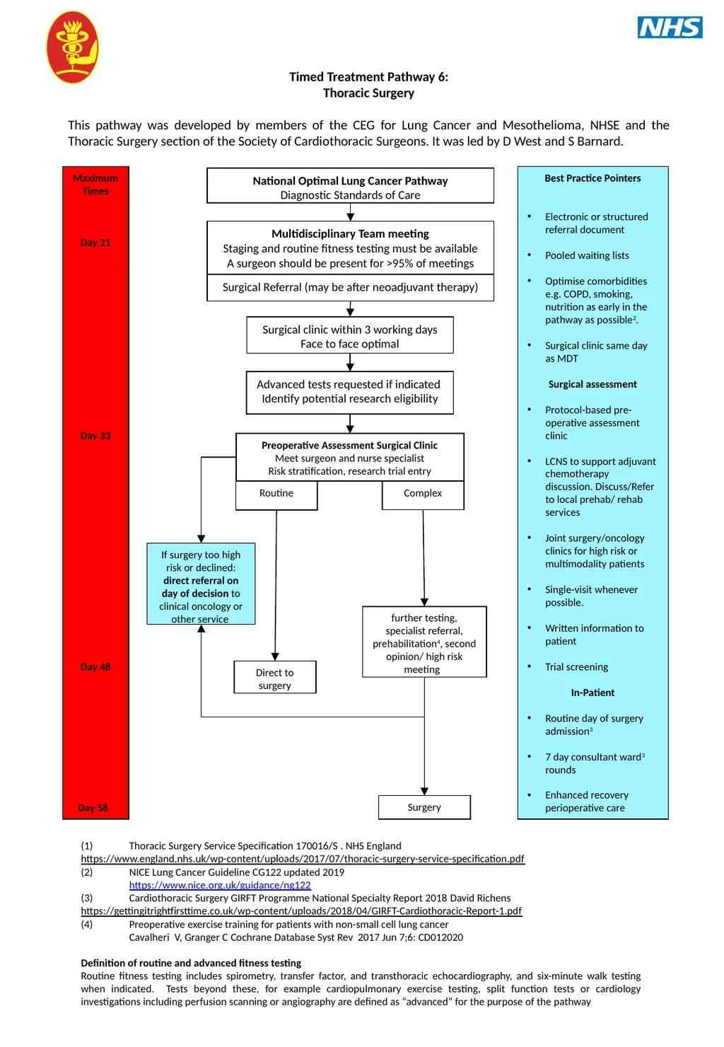
Timed Treatment Pathway 6:
Thoracic Surgery
This pathway was developed by members of the CEG for Lung Cancer and Mesothelioma, NHSE and the Thoracic Surgery section of the Society of Cardiothoracic Surgeons. It was led by D West and S Barnard. Advanced tests requested if indicated
Identify potential research eligibility further testing, specialist referral, prehabilitation4, second opinion/ high risk meeting Maximum Times
Day 21
Day 33
Day 48
Day 58 Multidisciplinary Team meeting
Staging and routine fitness testing must be available
A surgeon should be present for >95% of meetings Routine Preoperative Assessment Surgical Clinic
Meet surgeon and nurse specialist
Risk stratification, research trial entry Surgical Referral (may be after neoadjuvant therapy) Complex Direct to surgery Surgery National Optimal Lung Cancer Pathway
Diagnostic Standards of Care Best Practice Pointers
Electronic or structured referral document
Pooled waiting lists
Optimise comorbidities e.g. COPD, smoking, nutrition as early in the pathway as possible2.
Surgical clinic same day as MDT
Surgical assessment
Protocol-based pre-operative assessment clinic
LCNS to support adjuvant chemotherapy discussion. Discuss/Refer to local prehab/ rehab services
Joint surgery/oncology clinics for high risk or multimodality patients
Single-visit whenever possible.
Written information to patient
Trial screening
In-Patient
Routine day of surgery admission3
7 day consultant ward3 rounds
Enhanced recovery perioperative care (1) Thoracic Surgery Service Specification 170016/S . NHS England
https://www.england.nhs.uk/wp-content/uploads/2017/07/thoracic-surgery-service-specification.pdf
(2) NICE Lung Cancer Guideline CG122 updated 2019
https://www.nice.org.uk/guidance/ng122
(3) Cardiothoracic Surgery GIRFT Programme National Specialty Report 2018 David Richens
https://gettingitrightfirsttime.co.uk/wp-content/uploads/2018/04/GIRFT-Cardiothoracic-Report-1.pdf
(4) Preoperative exercise training for patients with non-small cell lung cancer
Cavalheri V, Granger C Cochrane Database Syst Rev 2017 Jun 7;6: CD012020
Definition of routine and advanced fitness testing
Routine fitness testing includes spirometry, transfer factor, and transthoracic echocardiography, and six-minute walk testing when indicated. Tests beyond these, for example cardiopulmonary exercise testing, split function tests or cardiology investigations including perfusion scanning or angiography are defined as “advanced” for the purpose of the pathway If surgery too high risk or declined: direct referral on day of decision to clinical oncology or other service Surgical clinic within 3 working days Face to face optimal<br>
Thoracic Surgery
This pathway was developed by members of the CEG for Lung Cancer and Mesothelioma, NHSE and the Thoracic Surgery section of the Society of Cardiothoracic Surgeons. It was led by D West and S Barnard. Advanced tests requested if indicated
Identify potential research eligibility further testing, specialist referral, prehabilitation4, second opinion/ high risk meeting Maximum Times
Day 21
Day 33
Day 48
Day 58 Multidisciplinary Team meeting
Staging and routine fitness testing must be available
A surgeon should be present for >95% of meetings Routine Preoperative Assessment Surgical Clinic
Meet surgeon and nurse specialist
Risk stratification, research trial entry Surgical Referral (may be after neoadjuvant therapy) Complex Direct to surgery Surgery National Optimal Lung Cancer Pathway
Diagnostic Standards of Care Best Practice Pointers
Electronic or structured referral document
Pooled waiting lists
Optimise comorbidities e.g. COPD, smoking, nutrition as early in the pathway as possible2.
Surgical clinic same day as MDT
Surgical assessment
Protocol-based pre-operative assessment clinic
LCNS to support adjuvant chemotherapy discussion. Discuss/Refer to local prehab/ rehab services
Joint surgery/oncology clinics for high risk or multimodality patients
Single-visit whenever possible.
Written information to patient
Trial screening
In-Patient
Routine day of surgery admission3
7 day consultant ward3 rounds
Enhanced recovery perioperative care (1) Thoracic Surgery Service Specification 170016/S . NHS England
https://www.england.nhs.uk/wp-content/uploads/2017/07/thoracic-surgery-service-specification.pdf
(2) NICE Lung Cancer Guideline CG122 updated 2019
https://www.nice.org.uk/guidance/ng122
(3) Cardiothoracic Surgery GIRFT Programme National Specialty Report 2018 David Richens
https://gettingitrightfirsttime.co.uk/wp-content/uploads/2018/04/GIRFT-Cardiothoracic-Report-1.pdf
(4) Preoperative exercise training for patients with non-small cell lung cancer
Cavalheri V, Granger C Cochrane Database Syst Rev 2017 Jun 7;6: CD012020
Definition of routine and advanced fitness testing
Routine fitness testing includes spirometry, transfer factor, and transthoracic echocardiography, and six-minute walk testing when indicated. Tests beyond these, for example cardiopulmonary exercise testing, split function tests or cardiology investigations including perfusion scanning or angiography are defined as “advanced” for the purpose of the pathway If surgery too high risk or declined: direct referral on day of decision to clinical oncology or other service Surgical clinic within 3 working days Face to face optimal<br>
09
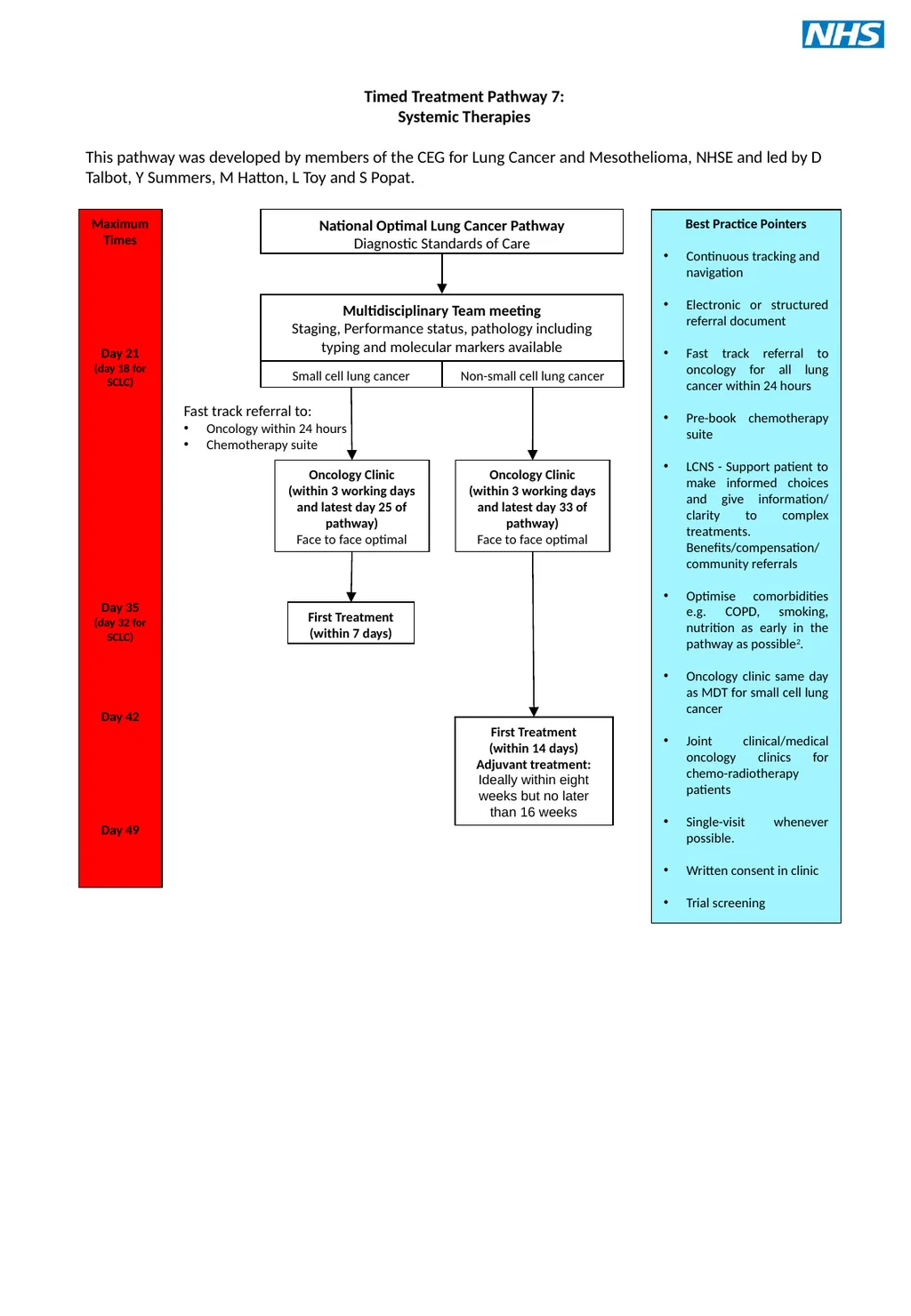
Timed Treatment Pathway 7:
Systemic Therapies
This pathway was developed by members of the CEG for Lung Cancer and Mesothelioma, NHSE and led by D Talbot, Y Summers, M Hatton, L Toy and S Popat. Fast track referral to:
Oncology within 24 hours
Chemotherapy suite Maximum Times
Day 21
(day 18 for SCLC)
Day 35
(day 32 for SCLC)
Day 42
Day 49 Multidisciplinary Team meeting
Staging, Performance status, pathology including typing and molecular markers available Oncology Clinic
(within 3 working days and latest day 25 of pathway)
Face to face optimal National Optimal Lung Cancer Pathway
Diagnostic Standards of Care Best Practice Pointers
Continuous tracking and navigation
Electronic or structured referral document
Fast track referral to oncology for all lung cancer within 24 hours
Pre-book chemotherapy suite
LCNS - Support patient to make informed choices and give information/ clarity to complex treatments. Benefits/compensation/ community referrals
Optimise comorbidities e.g. COPD, smoking, nutrition as early in the pathway as possible2.
Oncology clinic same day as MDT for small cell lung cancer
Joint clinical/medical oncology clinics for chemo-radiotherapy patients
Single-visit whenever possible.
Written consent in clinic
Trial screening Small cell lung cancer Non-small cell lung cancer First Treatment
(within 7 days) Oncology Clinic
(within 3 working days and latest day 33 of pathway)
Face to face optimal First Treatment
(within 14 days)
Adjuvant treatment:
Ideally within eight weeks but no later than 16 weeks<br>
Systemic Therapies
This pathway was developed by members of the CEG for Lung Cancer and Mesothelioma, NHSE and led by D Talbot, Y Summers, M Hatton, L Toy and S Popat. Fast track referral to:
Oncology within 24 hours
Chemotherapy suite Maximum Times
Day 21
(day 18 for SCLC)
Day 35
(day 32 for SCLC)
Day 42
Day 49 Multidisciplinary Team meeting
Staging, Performance status, pathology including typing and molecular markers available Oncology Clinic
(within 3 working days and latest day 25 of pathway)
Face to face optimal National Optimal Lung Cancer Pathway
Diagnostic Standards of Care Best Practice Pointers
Continuous tracking and navigation
Electronic or structured referral document
Fast track referral to oncology for all lung cancer within 24 hours
Pre-book chemotherapy suite
LCNS - Support patient to make informed choices and give information/ clarity to complex treatments. Benefits/compensation/ community referrals
Optimise comorbidities e.g. COPD, smoking, nutrition as early in the pathway as possible2.
Oncology clinic same day as MDT for small cell lung cancer
Joint clinical/medical oncology clinics for chemo-radiotherapy patients
Single-visit whenever possible.
Written consent in clinic
Trial screening Small cell lung cancer Non-small cell lung cancer First Treatment
(within 7 days) Oncology Clinic
(within 3 working days and latest day 33 of pathway)
Face to face optimal First Treatment
(within 14 days)
Adjuvant treatment:
Ideally within eight weeks but no later than 16 weeks<br>
10
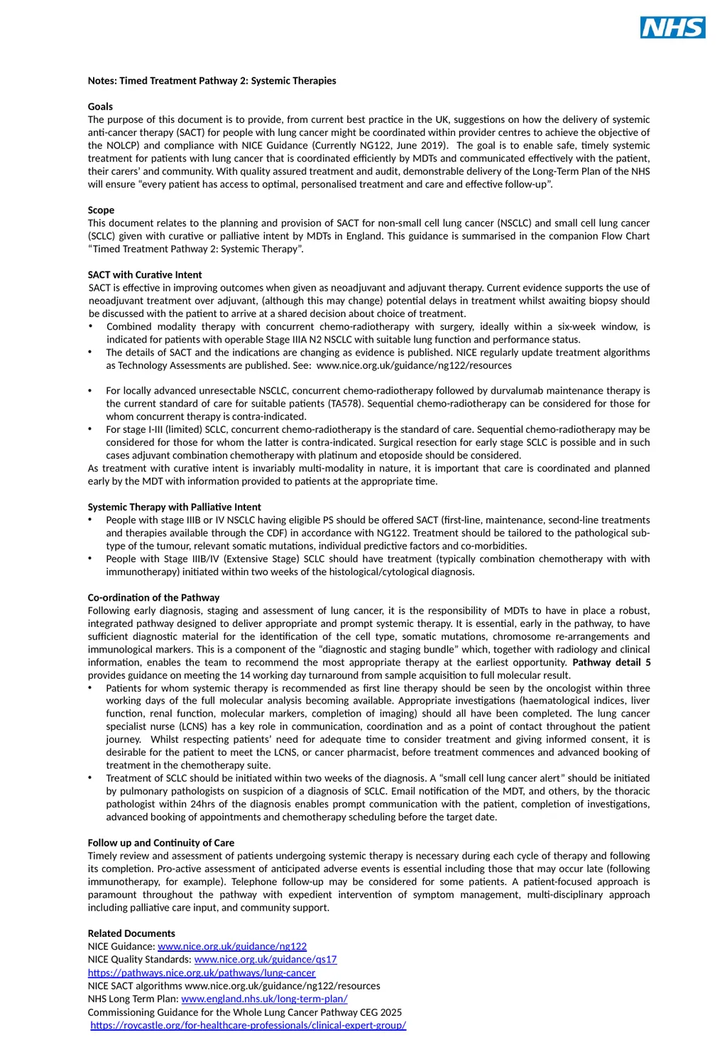
Notes: Timed Treatment Pathway 2: Systemic Therapies
Goals
The purpose of this document is to provide, from current best practice in the UK, suggestions on how the delivery of systemic anti-cancer therapy (SACT) for people with lung cancer might be coordinated within provider centres to achieve the objective of the NOLCP) and compliance with NICE Guidance (Currently NG122, June 2019). The goal is to enable safe, timely systemic treatment for patients with lung cancer that is coordinated efficiently by MDTs and communicated effectively with the patient, their carers’ and community. With quality assured treatment and audit, demonstrable delivery of the Long-Term Plan of the NHS will ensure “every patient has access to optimal, personalised treatment and care and effective follow-up”.
Scope
This document relates to the planning and provision of SACT for non-small cell lung cancer (NSCLC) and small cell lung cancer (SCLC) given with curative or palliative intent by MDTs in England. This guidance is summarised in the companion Flow Chart “Timed Treatment Pathway 2: Systemic Therapy”.
SACT with Curative Intent
SACT is effective in improving outcomes when given as neoadjuvant and adjuvant therapy. Current evidence supports the use of neoadjuvant treatment over adjuvant, (although this may change) potential delays in treatment whilst awaiting biopsy should be discussed with the patient to arrive at a shared decision about choice of treatment.
Combined modality therapy with concurrent chemo-radiotherapy with surgery, ideally within a six-week window, is indicated for patients with operable Stage IIIA N2 NSCLC with suitable lung function and performance status.
The details of SACT and the indications are changing as evidence is published. NICE regularly update treatment algorithms as Technology Assessments are published. See: www.nice.org.uk/guidance/ng122/resources
For locally advanced unresectable NSCLC, concurrent chemo-radiotherapy followed by durvalumab maintenance therapy is the current standard of care for suitable patients (TA578). Sequential chemo-radiotherapy can be considered for those for whom concurrent therapy is contra-indicated.
For stage I-III (limited) SCLC, concurrent chemo-radiotherapy is the standard of care. Sequential chemo-radiotherapy may be considered for those for whom the latter is contra-indicated. Surgical resection for early stage SCLC is possible and in such cases adjuvant combination chemotherapy with platinum and etoposide should be considered.
As treatment with curative intent is invariably multi-modality in nature, it is important that care is coordinated and planned early by the MDT with information provided to patients at the appropriate time.
Systemic Therapy with Palliative Intent
People with stage IIIB or IV NSCLC having eligible PS should be offered SACT (first-line, maintenance, second-line treatments and therapies available through the CDF) in accordance with NG122. Treatment should be tailored to the pathological sub-type of the tumour, relevant somatic mutations, individual predictive factors and co-morbidities.
People with Stage IIIB/IV (Extensive Stage) SCLC should have treatment (typically combination chemotherapy with with immunotherapy) initiated within two weeks of the histological/cytological diagnosis.
Co-ordination of the Pathway
Following early diagnosis, staging and assessment of lung cancer, it is the responsibility of MDTs to have in place a robust, integrated pathway designed to deliver appropriate and prompt systemic therapy. It is essential, early in the pathway, to have sufficient diagnostic material for the identification of the cell type, somatic mutations, chromosome re-arrangements and immunological markers. This is a component of the “diagnostic and staging bundle” which, together with radiology and clinical information, enables the team to recommend the most appropriate therapy at the earliest opportunity. Pathway detail 5 provides guidance on meeting the 14 working day turnaround from sample acquisition to full molecular result.
Patients for whom systemic therapy is recommended as first line therapy should be seen by the oncologist within three working days of the full molecular analysis becoming available. Appropriate investigations (haematological indices, liver function, renal function, molecular markers, completion of imaging) should all have been completed. The lung cancer specialist nurse (LCNS) has a key role in communication, coordination and as a point of contact throughout the patient journey. Whilst respecting patients’ need for adequate time to consider treatment and giving informed consent, it is desirable for the patient to meet the LCNS, or cancer pharmacist, before treatment commences and advanced booking of treatment in the chemotherapy suite.
Treatment of SCLC should be initiated within two weeks of the diagnosis. A “small cell lung cancer alert” should be initiated by pulmonary pathologists on suspicion of a diagnosis of SCLC. Email notification of the MDT, and others, by the thoracic pathologist within 24hrs of the diagnosis enables prompt communication with the patient, completion of investigations, advanced booking of appointments and chemotherapy scheduling before the target date.
Follow up and Continuity of Care
Timely review and assessment of patients undergoing systemic therapy is necessary during each cycle of therapy and following its completion. Pro-active assessment of anticipated adverse events is essential including those that may occur late (following immunotherapy, for example). Telephone follow-up may be considered for some patients. A patient-focused approach is paramount throughout the pathway with expedient intervention of symptom management, multi-disciplinary approach including palliative care input, and community support.
Related Documents
NICE Guidance: www.nice.org.uk/guidance/ng122
NICE Quality Standards: www.nice.org.uk/guidance/qs17
https://pathways.nice.org.uk/pathways/lung-cancer
NICE SACT algorithms www.nice.org.uk/guidance/ng122/resources
NHS Long Term Plan: www.england.nhs.uk/long-term-plan/
Commissioning Guidance for the Whole Lung Cancer Pathway CEG 2025
https://roycastle.org/for-healthcare-professionals/clinical-expert-group/<br>
Goals
The purpose of this document is to provide, from current best practice in the UK, suggestions on how the delivery of systemic anti-cancer therapy (SACT) for people with lung cancer might be coordinated within provider centres to achieve the objective of the NOLCP) and compliance with NICE Guidance (Currently NG122, June 2019). The goal is to enable safe, timely systemic treatment for patients with lung cancer that is coordinated efficiently by MDTs and communicated effectively with the patient, their carers’ and community. With quality assured treatment and audit, demonstrable delivery of the Long-Term Plan of the NHS will ensure “every patient has access to optimal, personalised treatment and care and effective follow-up”.
Scope
This document relates to the planning and provision of SACT for non-small cell lung cancer (NSCLC) and small cell lung cancer (SCLC) given with curative or palliative intent by MDTs in England. This guidance is summarised in the companion Flow Chart “Timed Treatment Pathway 2: Systemic Therapy”.
SACT with Curative Intent
SACT is effective in improving outcomes when given as neoadjuvant and adjuvant therapy. Current evidence supports the use of neoadjuvant treatment over adjuvant, (although this may change) potential delays in treatment whilst awaiting biopsy should be discussed with the patient to arrive at a shared decision about choice of treatment.
Combined modality therapy with concurrent chemo-radiotherapy with surgery, ideally within a six-week window, is indicated for patients with operable Stage IIIA N2 NSCLC with suitable lung function and performance status.
The details of SACT and the indications are changing as evidence is published. NICE regularly update treatment algorithms as Technology Assessments are published. See: www.nice.org.uk/guidance/ng122/resources
For locally advanced unresectable NSCLC, concurrent chemo-radiotherapy followed by durvalumab maintenance therapy is the current standard of care for suitable patients (TA578). Sequential chemo-radiotherapy can be considered for those for whom concurrent therapy is contra-indicated.
For stage I-III (limited) SCLC, concurrent chemo-radiotherapy is the standard of care. Sequential chemo-radiotherapy may be considered for those for whom the latter is contra-indicated. Surgical resection for early stage SCLC is possible and in such cases adjuvant combination chemotherapy with platinum and etoposide should be considered.
As treatment with curative intent is invariably multi-modality in nature, it is important that care is coordinated and planned early by the MDT with information provided to patients at the appropriate time.
Systemic Therapy with Palliative Intent
People with stage IIIB or IV NSCLC having eligible PS should be offered SACT (first-line, maintenance, second-line treatments and therapies available through the CDF) in accordance with NG122. Treatment should be tailored to the pathological sub-type of the tumour, relevant somatic mutations, individual predictive factors and co-morbidities.
People with Stage IIIB/IV (Extensive Stage) SCLC should have treatment (typically combination chemotherapy with with immunotherapy) initiated within two weeks of the histological/cytological diagnosis.
Co-ordination of the Pathway
Following early diagnosis, staging and assessment of lung cancer, it is the responsibility of MDTs to have in place a robust, integrated pathway designed to deliver appropriate and prompt systemic therapy. It is essential, early in the pathway, to have sufficient diagnostic material for the identification of the cell type, somatic mutations, chromosome re-arrangements and immunological markers. This is a component of the “diagnostic and staging bundle” which, together with radiology and clinical information, enables the team to recommend the most appropriate therapy at the earliest opportunity. Pathway detail 5 provides guidance on meeting the 14 working day turnaround from sample acquisition to full molecular result.
Patients for whom systemic therapy is recommended as first line therapy should be seen by the oncologist within three working days of the full molecular analysis becoming available. Appropriate investigations (haematological indices, liver function, renal function, molecular markers, completion of imaging) should all have been completed. The lung cancer specialist nurse (LCNS) has a key role in communication, coordination and as a point of contact throughout the patient journey. Whilst respecting patients’ need for adequate time to consider treatment and giving informed consent, it is desirable for the patient to meet the LCNS, or cancer pharmacist, before treatment commences and advanced booking of treatment in the chemotherapy suite.
Treatment of SCLC should be initiated within two weeks of the diagnosis. A “small cell lung cancer alert” should be initiated by pulmonary pathologists on suspicion of a diagnosis of SCLC. Email notification of the MDT, and others, by the thoracic pathologist within 24hrs of the diagnosis enables prompt communication with the patient, completion of investigations, advanced booking of appointments and chemotherapy scheduling before the target date.
Follow up and Continuity of Care
Timely review and assessment of patients undergoing systemic therapy is necessary during each cycle of therapy and following its completion. Pro-active assessment of anticipated adverse events is essential including those that may occur late (following immunotherapy, for example). Telephone follow-up may be considered for some patients. A patient-focused approach is paramount throughout the pathway with expedient intervention of symptom management, multi-disciplinary approach including palliative care input, and community support.
Related Documents
NICE Guidance: www.nice.org.uk/guidance/ng122
NICE Quality Standards: www.nice.org.uk/guidance/qs17
https://pathways.nice.org.uk/pathways/lung-cancer
NICE SACT algorithms www.nice.org.uk/guidance/ng122/resources
NHS Long Term Plan: www.england.nhs.uk/long-term-plan/
Commissioning Guidance for the Whole Lung Cancer Pathway CEG 2025
https://roycastle.org/for-healthcare-professionals/clinical-expert-group/<br>
11
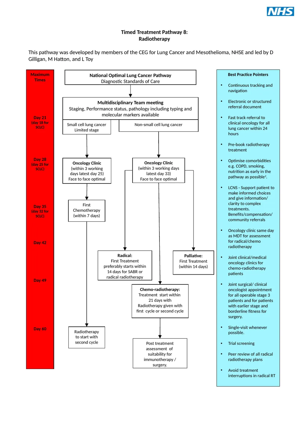
Timed Treatment Pathway 8:
Radiotherapy
This pathway was developed by members of the CEG for Lung Cancer and Mesothelioma, NHSE and led by D Gilligan, M Hatton, and L Toy Maximum Times
Day 21
(day 18 for SCLC)
Day 28
(day 25 for SCLC)
Day 35
(day 32 for SCLC)
Day 42
Day 49
Day 60 Multidisciplinary Team meeting
Staging, Performance status, pathology including typing and molecular markers available Oncology Clinic
(within 3 working days latest day 25)
Face to face optimal National Optimal Lung Cancer Pathway
Diagnostic Standards of Care Best Practice Pointers
Continuous tracking and navigation
Electronic or structured referral document
Fast track referral to clinical oncology for all lung cancer within 24 hours
Pre-book radiotherapy treatment
Optimise comorbidities e.g. COPD, smoking, nutrition as early in the pathway as possible2.
LCNS - Support patient to make informed choices and give information/ clarity to complex treatments. Benefits/compensation/ community referrals
Oncology clinic same day as MDT for assessment for radical/chemo radiotherapy
Joint clinical/medical oncology clinics for chemo-radiotherapy patients
Joint surgical/ clinical oncologist appointment for all operable stage 3 patients and for patients with earlier stage and borderline fitness for surgery.
Single-visit whenever possible.
Trial screening
Peer review of all radical radiotherapy plans
Avoid treatment interruptions in radical RT Small cell lung cancer
Limited stage Non-small cell lung cancer First Chemotherapy
(within 7 days) Oncology Clinic
(within 3 working days latest day 33)
Face to face optimal Palliative:
First Treatment
(within 14 days) Radiotherapy to start with second cycle Radical:
First Treatment preferably starts within 14 days for SABR or radical radiotherapy Chemo-radiotherapy:
Treatment start within 21 days with
Radiotherapy given with first cycle or second cycle Post treatment assessment of suitability for immunotherapy / surgery.<br>
Radiotherapy
This pathway was developed by members of the CEG for Lung Cancer and Mesothelioma, NHSE and led by D Gilligan, M Hatton, and L Toy Maximum Times
Day 21
(day 18 for SCLC)
Day 28
(day 25 for SCLC)
Day 35
(day 32 for SCLC)
Day 42
Day 49
Day 60 Multidisciplinary Team meeting
Staging, Performance status, pathology including typing and molecular markers available Oncology Clinic
(within 3 working days latest day 25)
Face to face optimal National Optimal Lung Cancer Pathway
Diagnostic Standards of Care Best Practice Pointers
Continuous tracking and navigation
Electronic or structured referral document
Fast track referral to clinical oncology for all lung cancer within 24 hours
Pre-book radiotherapy treatment
Optimise comorbidities e.g. COPD, smoking, nutrition as early in the pathway as possible2.
LCNS - Support patient to make informed choices and give information/ clarity to complex treatments. Benefits/compensation/ community referrals
Oncology clinic same day as MDT for assessment for radical/chemo radiotherapy
Joint clinical/medical oncology clinics for chemo-radiotherapy patients
Joint surgical/ clinical oncologist appointment for all operable stage 3 patients and for patients with earlier stage and borderline fitness for surgery.
Single-visit whenever possible.
Trial screening
Peer review of all radical radiotherapy plans
Avoid treatment interruptions in radical RT Small cell lung cancer
Limited stage Non-small cell lung cancer First Chemotherapy
(within 7 days) Oncology Clinic
(within 3 working days latest day 33)
Face to face optimal Palliative:
First Treatment
(within 14 days) Radiotherapy to start with second cycle Radical:
First Treatment preferably starts within 14 days for SABR or radical radiotherapy Chemo-radiotherapy:
Treatment start within 21 days with
Radiotherapy given with first cycle or second cycle Post treatment assessment of suitability for immunotherapy / surgery.<br>
12
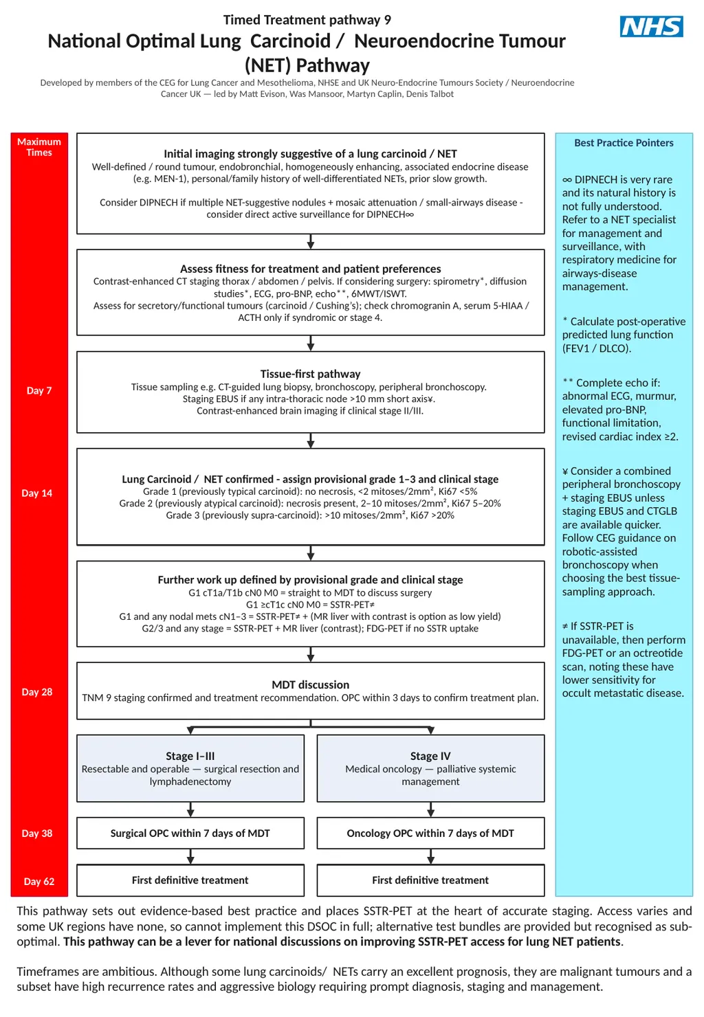
Timed Treatment pathway 9
National Optimal Lung Carcinoid / Neuroendocrine Tumour (NET) Pathway
Developed by members of the CEG for Lung Cancer and Mesothelioma, NHSE and UK Neuro-Endocrine Tumours Society / Neuroendocrine Cancer UK — led by Matt Evison, Was Mansoor, Martyn Caplin, Denis Talbot Maximum
Times Best Practice Pointers ∞ DIPNECH is very rare and its natural history is not fully understood. Refer to a NET specialist for management and surveillance, with respiratory medicine for airways-disease management.
* Calculate post-operative predicted lung function (FEV1 / DLCO).
** Complete echo if: abnormal ECG, murmur, elevated pro-BNP, functional limitation, revised cardiac index ≥2.
¥ Consider a combined peripheral bronchoscopy + staging EBUS unless staging EBUS and CTGLB are available quicker. Follow CEG guidance on robotic-assisted bronchoscopy when choosing the best tissue-sampling approach.
≠ If SSTR-PET is unavailable, then perform FDG-PET or an octreotide scan, noting these have lower sensitivity for occult metastatic disease. Initial imaging strongly suggestive of a lung carcinoid / NET
Well-defined / round tumour, endobronchial, homogeneously enhancing, associated endocrine disease (e.g. MEN-1), personal/family history of well-differentiated NETs, prior slow growth.
Consider DIPNECH if multiple NET-suggestive nodules + mosaic attenuation / small-airways disease - consider direct active surveillance for DIPNECH∞ Assess fitness for treatment and patient preferences
Contrast-enhanced CT staging thorax / abdomen / pelvis. If considering surgery: spirometry*, diffusion studies*, ECG, pro-BNP, echo**, 6MWT/ISWT.
Assess for secretory/functional tumours (carcinoid / Cushing’s); check chromogranin A, serum 5-HIAA / ACTH only if syndromic or stage 4. Tissue-first pathway
Tissue sampling e.g. CT-guided lung biopsy, bronchoscopy, peripheral bronchoscopy.
Staging EBUS if any intra-thoracic node >10 mm short axis¥.
Contrast-enhanced brain imaging if clinical stage II/III. Day 7 Lung Carcinoid / NET confirmed - assign provisional grade 1–3 and clinical stage
Grade 1 (previously typical carcinoid): no necrosis, <2 mitoses/2mm², Ki67 <5%
Grade 2 (previously atypical carcinoid): necrosis present, 2–10 mitoses/2mm², Ki67 5–20%
Grade 3 (previously supra-carcinoid): >10 mitoses/2mm², Ki67 >20% Day 14 Further work up defined by provisional grade and clinical stage
G1 cT1a/T1b cN0 M0 = straight to MDT to discuss surgery
G1 ≥cT1c cN0 M0 = SSTR-PET≠
G1 and any nodal mets cN1–3 = SSTR-PET≠ + (MR liver with contrast is option as low yield)
G2/3 and any stage = SSTR-PET + MR liver (contrast); FDG-PET if no SSTR uptake MDT discussion
TNM 9 staging confirmed and treatment recommendation. OPC within 3 days to confirm treatment plan. Day 28 Stage I–III
Resectable and operable — surgical resection and lymphadenectomy Stage IV
Medical oncology — palliative systemic management Surgical OPC within 7 days of MDT Oncology OPC within 7 days of MDT Day 38 First definitive treatment First definitive treatment Day 62 This pathway sets out evidence-based best practice and places SSTR-PET at the heart of accurate staging. Access varies and some UK regions have none, so cannot implement this DSOC in full; alternative test bundles are provided but recognised as sub-optimal. This pathway can be a lever for national discussions on improving SSTR-PET access for lung NET patients.
Timeframes are ambitious. Although some lung carcinoids/ NETs carry an excellent prognosis, they are malignant tumours and a subset have high recurrence rates and aggressive biology requiring prompt diagnosis, staging and management.<br>
National Optimal Lung Carcinoid / Neuroendocrine Tumour (NET) Pathway
Developed by members of the CEG for Lung Cancer and Mesothelioma, NHSE and UK Neuro-Endocrine Tumours Society / Neuroendocrine Cancer UK — led by Matt Evison, Was Mansoor, Martyn Caplin, Denis Talbot Maximum
Times Best Practice Pointers ∞ DIPNECH is very rare and its natural history is not fully understood. Refer to a NET specialist for management and surveillance, with respiratory medicine for airways-disease management.
* Calculate post-operative predicted lung function (FEV1 / DLCO).
** Complete echo if: abnormal ECG, murmur, elevated pro-BNP, functional limitation, revised cardiac index ≥2.
¥ Consider a combined peripheral bronchoscopy + staging EBUS unless staging EBUS and CTGLB are available quicker. Follow CEG guidance on robotic-assisted bronchoscopy when choosing the best tissue-sampling approach.
≠ If SSTR-PET is unavailable, then perform FDG-PET or an octreotide scan, noting these have lower sensitivity for occult metastatic disease. Initial imaging strongly suggestive of a lung carcinoid / NET
Well-defined / round tumour, endobronchial, homogeneously enhancing, associated endocrine disease (e.g. MEN-1), personal/family history of well-differentiated NETs, prior slow growth.
Consider DIPNECH if multiple NET-suggestive nodules + mosaic attenuation / small-airways disease - consider direct active surveillance for DIPNECH∞ Assess fitness for treatment and patient preferences
Contrast-enhanced CT staging thorax / abdomen / pelvis. If considering surgery: spirometry*, diffusion studies*, ECG, pro-BNP, echo**, 6MWT/ISWT.
Assess for secretory/functional tumours (carcinoid / Cushing’s); check chromogranin A, serum 5-HIAA / ACTH only if syndromic or stage 4. Tissue-first pathway
Tissue sampling e.g. CT-guided lung biopsy, bronchoscopy, peripheral bronchoscopy.
Staging EBUS if any intra-thoracic node >10 mm short axis¥.
Contrast-enhanced brain imaging if clinical stage II/III. Day 7 Lung Carcinoid / NET confirmed - assign provisional grade 1–3 and clinical stage
Grade 1 (previously typical carcinoid): no necrosis, <2 mitoses/2mm², Ki67 <5%
Grade 2 (previously atypical carcinoid): necrosis present, 2–10 mitoses/2mm², Ki67 5–20%
Grade 3 (previously supra-carcinoid): >10 mitoses/2mm², Ki67 >20% Day 14 Further work up defined by provisional grade and clinical stage
G1 cT1a/T1b cN0 M0 = straight to MDT to discuss surgery
G1 ≥cT1c cN0 M0 = SSTR-PET≠
G1 and any nodal mets cN1–3 = SSTR-PET≠ + (MR liver with contrast is option as low yield)
G2/3 and any stage = SSTR-PET + MR liver (contrast); FDG-PET if no SSTR uptake MDT discussion
TNM 9 staging confirmed and treatment recommendation. OPC within 3 days to confirm treatment plan. Day 28 Stage I–III
Resectable and operable — surgical resection and lymphadenectomy Stage IV
Medical oncology — palliative systemic management Surgical OPC within 7 days of MDT Oncology OPC within 7 days of MDT Day 38 First definitive treatment First definitive treatment Day 62 This pathway sets out evidence-based best practice and places SSTR-PET at the heart of accurate staging. Access varies and some UK regions have none, so cannot implement this DSOC in full; alternative test bundles are provided but recognised as sub-optimal. This pathway can be a lever for national discussions on improving SSTR-PET access for lung NET patients.
Timeframes are ambitious. Although some lung carcinoids/ NETs carry an excellent prognosis, they are malignant tumours and a subset have high recurrence rates and aggressive biology requiring prompt diagnosis, staging and management.<br>
13
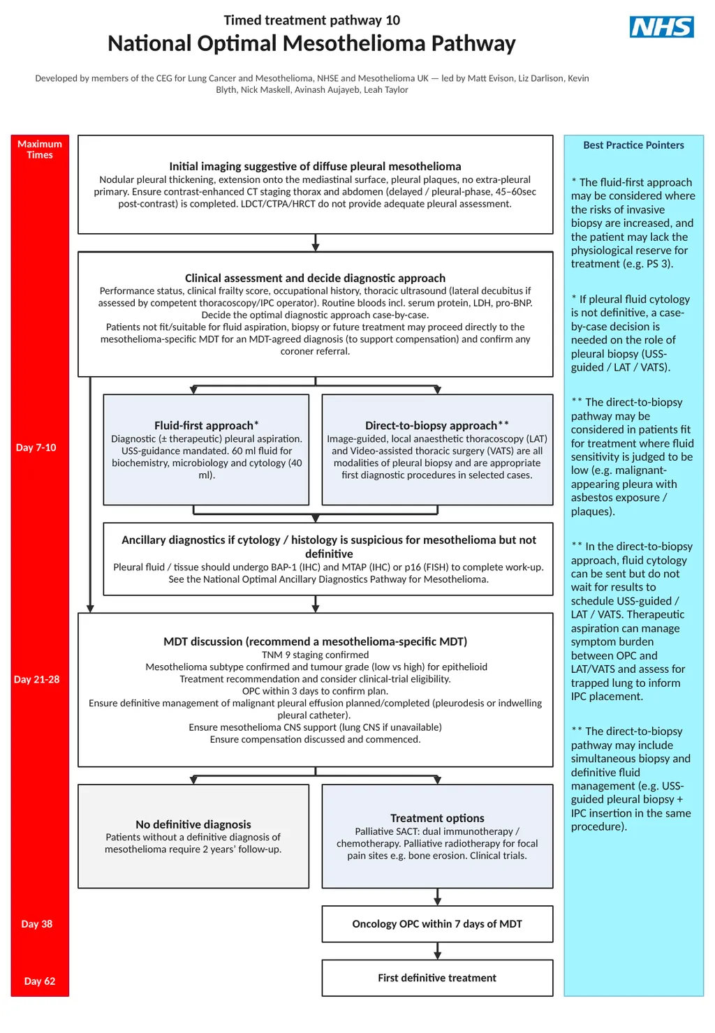
Timed treatment pathway 10
National Optimal Mesothelioma Pathway
Developed by members of the CEG for Lung Cancer and Mesothelioma, NHSE and Mesothelioma UK — led by Matt Evison, Liz Darlison, Kevin Blyth, Nick Maskell, Avinash Aujayeb, Leah Taylor Maximum
Times Best Practice Pointers * The fluid-first approach may be considered where the risks of invasive biopsy are increased, and the patient may lack the physiological reserve for treatment (e.g. PS 3).
* If pleural fluid cytology is not definitive, a case-by-case decision is needed on the role of pleural biopsy (USS-guided / LAT / VATS).
** The direct-to-biopsy pathway may be considered in patients fit for treatment where fluid sensitivity is judged to be low (e.g. malignant-appearing pleura with asbestos exposure / plaques).
** In the direct-to-biopsy approach, fluid cytology can be sent but do not wait for results to schedule USS-guided / LAT / VATS. Therapeutic aspiration can manage symptom burden between OPC and LAT/VATS and assess for trapped lung to inform IPC placement.
** The direct-to-biopsy pathway may include simultaneous biopsy and definitive fluid management (e.g. USS-guided pleural biopsy + IPC insertion in the same procedure). Initial imaging suggestive of diffuse pleural mesothelioma
Nodular pleural thickening, extension onto the mediastinal surface, pleural plaques, no extra-pleural primary. Ensure contrast-enhanced CT staging thorax and abdomen (delayed / pleural-phase, 45–60sec post-contrast) is completed. LDCT/CTPA/HRCT do not provide adequate pleural assessment. Clinical assessment and decide diagnostic approach
Performance status, clinical frailty score, occupational history, thoracic ultrasound (lateral decubitus if assessed by competent thoracoscopy/IPC operator). Routine bloods incl. serum protein, LDH, pro-BNP. Decide the optimal diagnostic approach case-by-case.
Patients not fit/suitable for fluid aspiration, biopsy or future treatment may proceed directly to the mesothelioma-specific MDT for an MDT-agreed diagnosis (to support compensation) and confirm any coroner referral. Day 7-10 Fluid-first approach*
Diagnostic (± therapeutic) pleural aspiration. USS-guidance mandated. 60 ml fluid for biochemistry, microbiology and cytology (40 ml). Direct-to-biopsy approach**
Image-guided, local anaesthetic thoracoscopy (LAT) and Video-assisted thoracic surgery (VATS) are all modalities of pleural biopsy and are appropriate first diagnostic procedures in selected cases. Ancillary diagnostics if cytology / histology is suspicious for mesothelioma but not definitive
Pleural fluid / tissue should undergo BAP-1 (IHC) and MTAP (IHC) or p16 (FISH) to complete work-up. See the National Optimal Ancillary Diagnostics Pathway for Mesothelioma. MDT discussion (recommend a mesothelioma-specific MDT)
TNM 9 staging confirmed
Mesothelioma subtype confirmed and tumour grade (low vs high) for epithelioid
Treatment recommendation and consider clinical-trial eligibility.
OPC within 3 days to confirm plan.
Ensure definitive management of malignant pleural effusion planned/completed (pleurodesis or indwelling pleural catheter).
Ensure mesothelioma CNS support (lung CNS if unavailable)
Ensure compensation discussed and commenced. Day 21-28 No definitive diagnosis
Patients without a definitive diagnosis of mesothelioma require 2 years’ follow-up. Treatment options
Palliative SACT: dual immunotherapy / chemotherapy. Palliative radiotherapy for focal pain sites e.g. bone erosion. Clinical trials. Oncology OPC within 7 days of MDT First definitive treatment Day 38 Day 62<br>
National Optimal Mesothelioma Pathway
Developed by members of the CEG for Lung Cancer and Mesothelioma, NHSE and Mesothelioma UK — led by Matt Evison, Liz Darlison, Kevin Blyth, Nick Maskell, Avinash Aujayeb, Leah Taylor Maximum
Times Best Practice Pointers * The fluid-first approach may be considered where the risks of invasive biopsy are increased, and the patient may lack the physiological reserve for treatment (e.g. PS 3).
* If pleural fluid cytology is not definitive, a case-by-case decision is needed on the role of pleural biopsy (USS-guided / LAT / VATS).
** The direct-to-biopsy pathway may be considered in patients fit for treatment where fluid sensitivity is judged to be low (e.g. malignant-appearing pleura with asbestos exposure / plaques).
** In the direct-to-biopsy approach, fluid cytology can be sent but do not wait for results to schedule USS-guided / LAT / VATS. Therapeutic aspiration can manage symptom burden between OPC and LAT/VATS and assess for trapped lung to inform IPC placement.
** The direct-to-biopsy pathway may include simultaneous biopsy and definitive fluid management (e.g. USS-guided pleural biopsy + IPC insertion in the same procedure). Initial imaging suggestive of diffuse pleural mesothelioma
Nodular pleural thickening, extension onto the mediastinal surface, pleural plaques, no extra-pleural primary. Ensure contrast-enhanced CT staging thorax and abdomen (delayed / pleural-phase, 45–60sec post-contrast) is completed. LDCT/CTPA/HRCT do not provide adequate pleural assessment. Clinical assessment and decide diagnostic approach
Performance status, clinical frailty score, occupational history, thoracic ultrasound (lateral decubitus if assessed by competent thoracoscopy/IPC operator). Routine bloods incl. serum protein, LDH, pro-BNP. Decide the optimal diagnostic approach case-by-case.
Patients not fit/suitable for fluid aspiration, biopsy or future treatment may proceed directly to the mesothelioma-specific MDT for an MDT-agreed diagnosis (to support compensation) and confirm any coroner referral. Day 7-10 Fluid-first approach*
Diagnostic (± therapeutic) pleural aspiration. USS-guidance mandated. 60 ml fluid for biochemistry, microbiology and cytology (40 ml). Direct-to-biopsy approach**
Image-guided, local anaesthetic thoracoscopy (LAT) and Video-assisted thoracic surgery (VATS) are all modalities of pleural biopsy and are appropriate first diagnostic procedures in selected cases. Ancillary diagnostics if cytology / histology is suspicious for mesothelioma but not definitive
Pleural fluid / tissue should undergo BAP-1 (IHC) and MTAP (IHC) or p16 (FISH) to complete work-up. See the National Optimal Ancillary Diagnostics Pathway for Mesothelioma. MDT discussion (recommend a mesothelioma-specific MDT)
TNM 9 staging confirmed
Mesothelioma subtype confirmed and tumour grade (low vs high) for epithelioid
Treatment recommendation and consider clinical-trial eligibility.
OPC within 3 days to confirm plan.
Ensure definitive management of malignant pleural effusion planned/completed (pleurodesis or indwelling pleural catheter).
Ensure mesothelioma CNS support (lung CNS if unavailable)
Ensure compensation discussed and commenced. Day 21-28 No definitive diagnosis
Patients without a definitive diagnosis of mesothelioma require 2 years’ follow-up. Treatment options
Palliative SACT: dual immunotherapy / chemotherapy. Palliative radiotherapy for focal pain sites e.g. bone erosion. Clinical trials. Oncology OPC within 7 days of MDT First definitive treatment Day 38 Day 62<br>
14
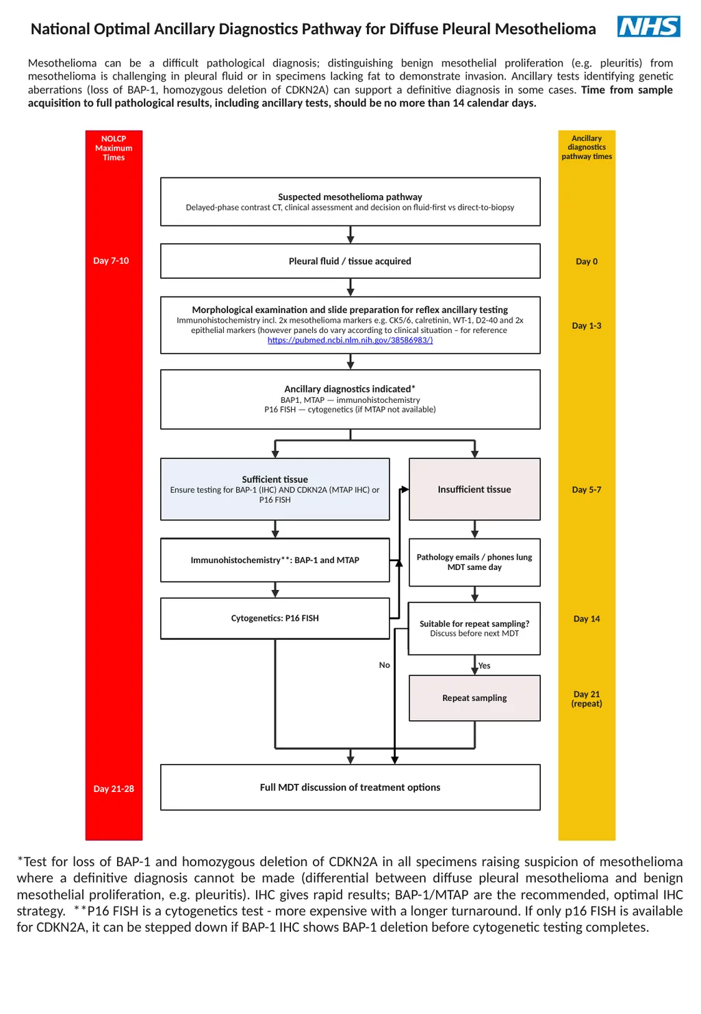
National Optimal Ancillary Diagnostics Pathway for Diffuse Pleural Mesothelioma Mesothelioma can be a difficult pathological diagnosis; distinguishing benign mesothelial proliferation (e.g. pleuritis) from mesothelioma is challenging in pleural fluid or in specimens lacking fat to demonstrate invasion. Ancillary tests identifying genetic aberrations (loss of BAP-1, homozygous deletion of CDKN2A) can support a definitive diagnosis in some cases. Time from sample acquisition to full pathological results, including ancillary tests, should be no more than 14 calendar days. NOLCP
Maximum Times Ancillary
diagnostics
pathway times Suspected mesothelioma pathway
Delayed-phase contrast CT, clinical assessment and decision on fluid-first vs direct-to-biopsy Day 7-10 Pleural fluid / tissue acquired Day 0 Morphological examination and slide preparation for reflex ancillary testing
Immunohistochemistry incl. 2x mesothelioma markers e.g. CK5/6, calretinin, WT-1, D2-40 and 2x epithelial markers (however panels do vary according to clinical situation – for reference https://pubmed.ncbi.nlm.nih.gov/38586983/) Day 1-3 Ancillary diagnostics indicated*
BAP1, MTAP — immunohistochemistry
P16 FISH — cytogenetics (if MTAP not available) Sufficient tissue
Ensure testing for BAP-1 (IHC) AND CDKN2A (MTAP IHC) or P16 FISH Insufficient tissue Day 5-7 Immunohistochemistry**: BAP-1 and MTAP Cytogenetics: P16 FISH Day 14 Pathology emails / phones lung MDT same day Suitable for repeat sampling?
Discuss before next MDT Yes Repeat sampling Day 21
(repeat) Full MDT discussion of treatment options Day 21-28 *Test for loss of BAP-1 and homozygous deletion of CDKN2A in all specimens raising suspicion of mesothelioma where a definitive diagnosis cannot be made (differential between diffuse pleural mesothelioma and benign mesothelial proliferation, e.g. pleuritis). IHC gives rapid results; BAP-1/MTAP are the recommended, optimal IHC strategy. **P16 FISH is a cytogenetics test - more expensive with a longer turnaround. If only p16 FISH is available for CDKN2A, it can be stepped down if BAP-1 IHC shows BAP-1 deletion before cytogenetic testing completes. No<br>
Maximum Times Ancillary
diagnostics
pathway times Suspected mesothelioma pathway
Delayed-phase contrast CT, clinical assessment and decision on fluid-first vs direct-to-biopsy Day 7-10 Pleural fluid / tissue acquired Day 0 Morphological examination and slide preparation for reflex ancillary testing
Immunohistochemistry incl. 2x mesothelioma markers e.g. CK5/6, calretinin, WT-1, D2-40 and 2x epithelial markers (however panels do vary according to clinical situation – for reference https://pubmed.ncbi.nlm.nih.gov/38586983/) Day 1-3 Ancillary diagnostics indicated*
BAP1, MTAP — immunohistochemistry
P16 FISH — cytogenetics (if MTAP not available) Sufficient tissue
Ensure testing for BAP-1 (IHC) AND CDKN2A (MTAP IHC) or P16 FISH Insufficient tissue Day 5-7 Immunohistochemistry**: BAP-1 and MTAP Cytogenetics: P16 FISH Day 14 Pathology emails / phones lung MDT same day Suitable for repeat sampling?
Discuss before next MDT Yes Repeat sampling Day 21
(repeat) Full MDT discussion of treatment options Day 21-28 *Test for loss of BAP-1 and homozygous deletion of CDKN2A in all specimens raising suspicion of mesothelioma where a definitive diagnosis cannot be made (differential between diffuse pleural mesothelioma and benign mesothelial proliferation, e.g. pleuritis). IHC gives rapid results; BAP-1/MTAP are the recommended, optimal IHC strategy. **P16 FISH is a cytogenetics test - more expensive with a longer turnaround. If only p16 FISH is available for CDKN2A, it can be stepped down if BAP-1 IHC shows BAP-1 deletion before cytogenetic testing completes. No<br>
15
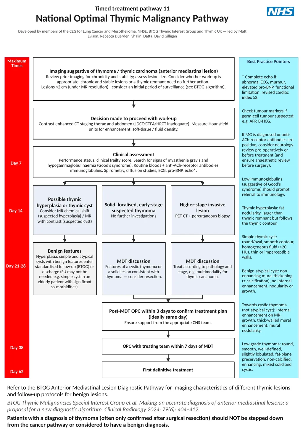
Timed treatment pathway 11
National Optimal Thymic Malignancy Pathway
Developed by members of the CEG for Lung Cancer and Mesothelioma, NHSE, BTOG Thymic Interest Group and Thymic UK — led by Matt Evison, Rebecca Duerden, Shalini Datta, David Gilligan Maximum
Times Best Practice Pointers * Complete echo if: abnormal ECG, murmur, elevated pro-BNP, functional limitation, revised cardiac index ≥2.
Check tumour markers if germ-cell tumour suspected: e.g. AFP, B-HCG.
If MG is diagnosed or anti-ACh-receptor antibodies are positive, consider neurology review pre-operatively or before treatment (and ensure anaesthetic review before surgery).
Low immunoglobulins (suggestive of Good’s syndrome) should prompt referral to immunology.
Thymic hyperplasia: fat nodularity, larger than thymic remnant but follows the thymic contour.
Simple thymic cyst: round/oval, smooth contour, homogeneous fluid (<20 HU), thin or imperceptible walls.
Benign atypical cyst: non-enhancing mural thickening (± calcification), no internal enhancement, nodularity or growth.
Towards cystic thymoma (not atypical cyst): internal enhancement on MR, growth, thick-walled mural enhancement, mural nodularity.
Low-grade thymoma: round, smooth, well-defined, slightly lobulated, fat-plane preservation, non-calcified, enhancing, mixed solid and cystic. Imaging suggestive of thymoma / thymic carcinoma (anterior mediastinal lesion)
Review prior imaging for chronicity and stability; assess lesion size. Consider whether work-up is appropriate: chronic and stable lesions or a thymic remnant need no further action.
Lesions <2 cm (under MR resolution) - consider an initial period of surveillance (see BTOG algorithm). Decision made to proceed with work-up
Contrast-enhanced CT staging thorax and abdomen (LDCT/CTPA/HRCT inadequate). Measure Hounsfield units for enhancement, soft-tissue / fluid density. Day 7 Clinical assessment
Performance status, clinical frailty score. Search for signs of myasthenia gravis and hypogammaglobulinaemia (Good’s syndrome). Routine bloods + anti-ACh-receptor antibodies, immunoglobulins. Spirometry, diffusion studies, ECG, pro-BNP, echo*. Possible thymic hyperplasia or thymic cyst
Consider MR chemical shift (suspected hyperplasia) / MR with contrast (suspected cyst) Solid, localised, early-stage suspected thymoma
No further investigations Higher-stage invasive lesion
PET-CT + percutaneous biopsy Day 14 Benign features
Hyperplasia, simple and atypical cysts with benign features enter standardised follow-up (BTOG) or discharge (FU may not be needed e.g. simple cyst in an elderly patient with significant co-morbidities). MDT discussion
Features of a cystic thymoma or a solid lesion consistent with thymoma — consider resection. MDT discussion
Treat according to pathology and stage, e.g. multimodality for thymic carcinoma. Day 21-28 Post-MDT OPC within 3 days to confirm treatment plan (ideally same day)
Ensure support from the appropriate CNS team. OPC with treating team within 7 days of MDT Day 38 First definitive treatment Day 62 Refer to the BTOG Anterior Mediastinal Lesion Diagnostic Pathway for imaging characteristics of different thymic lesions and follow-up protocols for benign lesions.
BTOG Thymic Malignancies Special Interest Group et al. Making an accurate diagnosis of anterior mediastinal lesions: a proposal for a new diagnostic algorithm. Clinical Radiology 2024; 79(6): 404–412.
Patients with a diagnosis of thymoma (often only confirmed after surgical resection) should NOT be stepped down from the cancer pathway or considered to have a benign diagnosis.<br>
National Optimal Thymic Malignancy Pathway
Developed by members of the CEG for Lung Cancer and Mesothelioma, NHSE, BTOG Thymic Interest Group and Thymic UK — led by Matt Evison, Rebecca Duerden, Shalini Datta, David Gilligan Maximum
Times Best Practice Pointers * Complete echo if: abnormal ECG, murmur, elevated pro-BNP, functional limitation, revised cardiac index ≥2.
Check tumour markers if germ-cell tumour suspected: e.g. AFP, B-HCG.
If MG is diagnosed or anti-ACh-receptor antibodies are positive, consider neurology review pre-operatively or before treatment (and ensure anaesthetic review before surgery).
Low immunoglobulins (suggestive of Good’s syndrome) should prompt referral to immunology.
Thymic hyperplasia: fat nodularity, larger than thymic remnant but follows the thymic contour.
Simple thymic cyst: round/oval, smooth contour, homogeneous fluid (<20 HU), thin or imperceptible walls.
Benign atypical cyst: non-enhancing mural thickening (± calcification), no internal enhancement, nodularity or growth.
Towards cystic thymoma (not atypical cyst): internal enhancement on MR, growth, thick-walled mural enhancement, mural nodularity.
Low-grade thymoma: round, smooth, well-defined, slightly lobulated, fat-plane preservation, non-calcified, enhancing, mixed solid and cystic. Imaging suggestive of thymoma / thymic carcinoma (anterior mediastinal lesion)
Review prior imaging for chronicity and stability; assess lesion size. Consider whether work-up is appropriate: chronic and stable lesions or a thymic remnant need no further action.
Lesions <2 cm (under MR resolution) - consider an initial period of surveillance (see BTOG algorithm). Decision made to proceed with work-up
Contrast-enhanced CT staging thorax and abdomen (LDCT/CTPA/HRCT inadequate). Measure Hounsfield units for enhancement, soft-tissue / fluid density. Day 7 Clinical assessment
Performance status, clinical frailty score. Search for signs of myasthenia gravis and hypogammaglobulinaemia (Good’s syndrome). Routine bloods + anti-ACh-receptor antibodies, immunoglobulins. Spirometry, diffusion studies, ECG, pro-BNP, echo*. Possible thymic hyperplasia or thymic cyst
Consider MR chemical shift (suspected hyperplasia) / MR with contrast (suspected cyst) Solid, localised, early-stage suspected thymoma
No further investigations Higher-stage invasive lesion
PET-CT + percutaneous biopsy Day 14 Benign features
Hyperplasia, simple and atypical cysts with benign features enter standardised follow-up (BTOG) or discharge (FU may not be needed e.g. simple cyst in an elderly patient with significant co-morbidities). MDT discussion
Features of a cystic thymoma or a solid lesion consistent with thymoma — consider resection. MDT discussion
Treat according to pathology and stage, e.g. multimodality for thymic carcinoma. Day 21-28 Post-MDT OPC within 3 days to confirm treatment plan (ideally same day)
Ensure support from the appropriate CNS team. OPC with treating team within 7 days of MDT Day 38 First definitive treatment Day 62 Refer to the BTOG Anterior Mediastinal Lesion Diagnostic Pathway for imaging characteristics of different thymic lesions and follow-up protocols for benign lesions.
BTOG Thymic Malignancies Special Interest Group et al. Making an accurate diagnosis of anterior mediastinal lesions: a proposal for a new diagnostic algorithm. Clinical Radiology 2024; 79(6): 404–412.
Patients with a diagnosis of thymoma (often only confirmed after surgical resection) should NOT be stepped down from the cancer pathway or considered to have a benign diagnosis.<br>