
Introduction To Pathology Dr. Raya D. Marji, M.D.



Description: Introduction To Pathology Dr. Raya D. Marji, M.D. Email: r.marjibau.edu.jo Office: O-505 HEMOSTASIS AND THROMBOSIS Normal hemostasis: Series of regulated processes that culminate in the formation of a blood clot that limits bleeding from
Related Topics
Download Presentation
"Introduction To Pathology Dr. Raya D. Marji, M.D." is the property of its rightful owner. Permission is granted to download and print the materials on this website for personal, non-commercial use only, and to display it on your personal computer provided you do not modify the materials and that you retain all copyright notices contained in the materials. By downloading content from our website, you accept the terms of this agreement.
Presentation Transcript
slide1. Introduction To Pathology Dr. Raya D. Marji, M.D.
Email: r.marji@bau.edu.jo
Office: O-505<br>
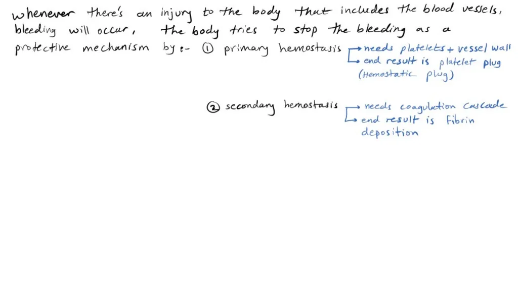
slide7. HEMOSTASIS AND THROMBOSIS Normal hemostasis: Series of regulated processes that culminate in the formation of a blood clot that limits bleeding from an injured vessel.
The pathologic counterpart of hemostasis is thrombosis, the formation of blood clot (thrombus) within non-traumatized, intact vessels.<br>
slide8. Normal Hemostasis Involving platelets, clotting factors, and endothelium.
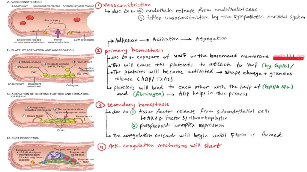
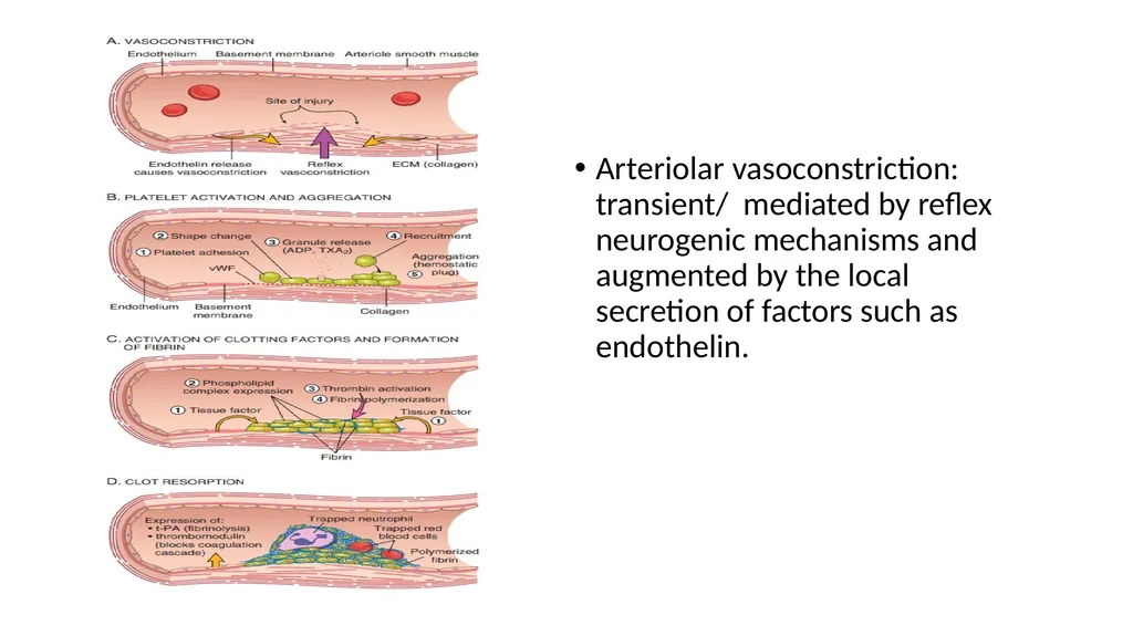
Arteriolar vasoconstriction occurs immediately and markedly reduces blood flow to the injured area. It is mediated by reflex neurogenic mechanisms and augmented by the local secretion of factors such as endothelin, a potent endothelium-derived vasoconstrictor.
Transient.<br>
slide9. Arteriolar vasoconstriction: transient/ mediated by reflex neurogenic mechanisms and augmented by the local secretion of factors such as endothelin.<br>
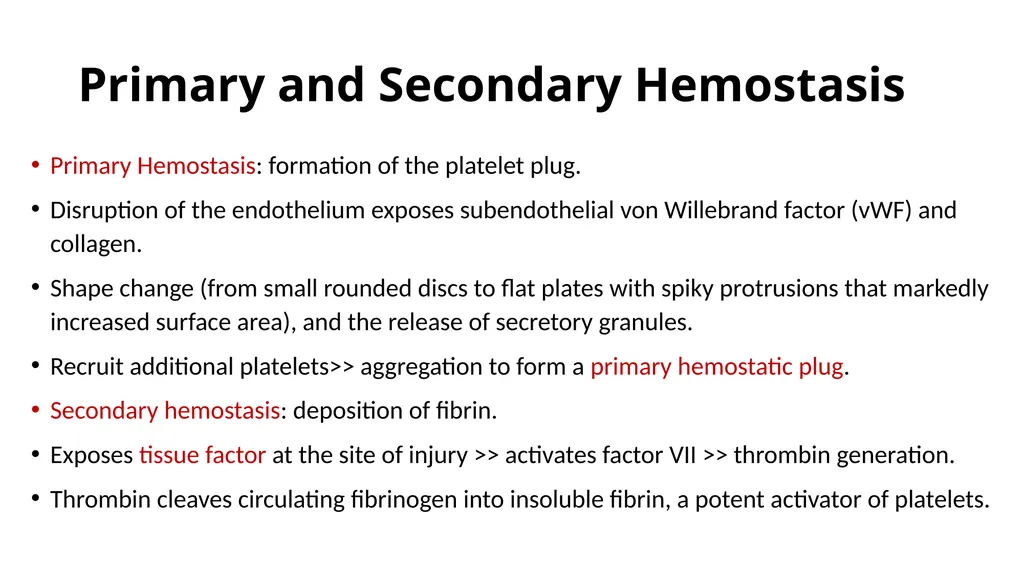
slide10. Primary and Secondary Hemostasis Primary Hemostasis: formation of the platelet plug.
Disruption of the endothelium exposes subendothelial von Willebrand factor (vWF) and collagen.
Shape change (from small rounded discs to flat plates with spiky protrusions that markedly increased surface area), and the release of secretory granules.
Recruit additional platelets>> aggregation to form a primary hemostatic plug.
Secondary hemostasis: deposition of fibrin.
Exposes tissue factor at the site of injury >> activates factor VII >> thrombin generation.
Thrombin cleaves circulating fibrinogen into insoluble fibrin, a potent activator of platelets.<br>
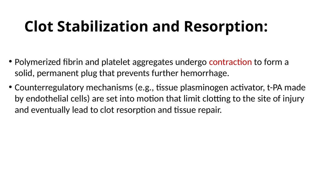
slide11. Clot Stabilization and Resorption: Polymerized fibrin and platelet aggregates undergo contraction to form a solid, permanent plug that prevents further hemorrhage.
Counterregulatory mechanisms (e.g., tissue plasminogen activator, t-PA made by endothelial cells) are set into motion that limit clotting to the site of injury and eventually lead to clot resorption and tissue repair.<br>
slide12. Platelets Disc-shaped anucleate cell fragments that are shed from megakaryocytes in the bone marrow into the bloodstream.
Their function depends on several glycoprotein receptors, a contractile cytoskeleton, and two types of cytoplasmic granules.
α-Granules.
Dense (or δ) granules.<br>
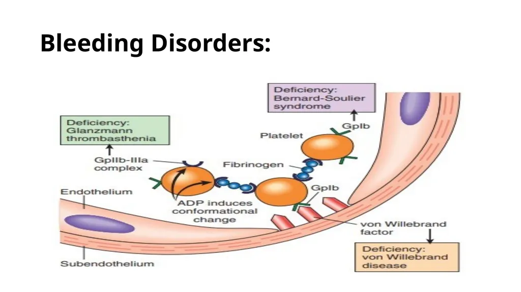
slide13. Platelet adhesion: interactions with vWF, which acts as a bridge between the platelet surface receptor glycoprotein Ib (GpIb) and exposed collagen.
Genetic deficiencies of vWF (von Willebrand disease) or GpIb (BernardSoulier syndrome) result in bleeding disorders, attesting to the importance of these factors.
Platelets rapidly change shape following adhesion, being converted from smooth discs to spiky “sea urchins” with greatly increased surface area. This change is accompanied by alterations in glycoprotein IIb/IIIa that increase its affinity for fibrinogen, and by the translocation of negatively charged phospholipids to the platelet surface.
Phospholipids bind calcium and serve as nucleation sites for the assembly of coagulation factor complexes.<br>
slide14. Secretion (release reaction) of granule contents occurs along with changes in shape; these two events are often referred to together as platelet activation.
Thrombin activates platelets through a special type of G-protein– coupled receptor referred to as a protease-activated receptor (PAR).
Activated platelets also produce the prostaglandin thromboxane A2 (TXA2), a potent inducer of platelet aggregation.<br>
slide15. Platelet aggregation: glycoprotein IIb/IIIa allows binding of fibrinogen.
Inherited deficiency of GpIIb-IIIa results in a bleeding disorder called Glanzmann thrombasthenia.<br>
slide16. Bleeding Disorders:<br>
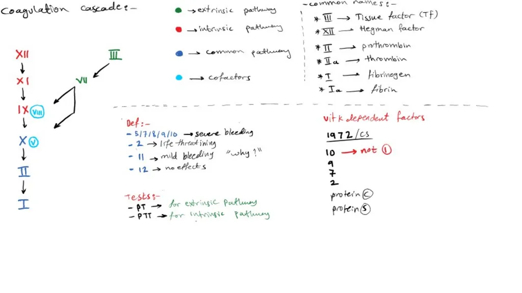
slide17. Coagulation Cascade Deposition of an insoluble fibrin clot.
Each reaction step involves an enzyme (an activated coagulation factor), a substrate (an inactive proenzyme form of a coagulation factor), and a cofactor (a reaction accelerator).
Components are assembled on a negatively charged phospholipid surface.<br>
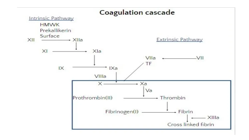
slide18. The coagulation cascade is divided into the extrinsic and intrinsic pathways.
The prothrombin time (PT) assay assesses the function of the proteins in the extrinsic pathway (factors VII, X, V, II (prothrombin), and fibrinogen).
The partial thromboplastin time (PTT) assay screens the function of the proteins in the intrinsic pathway (factors XII, XI, IX, VIII, X, V, II, and fibrinogen).<br>
slide20. Thrombin: Conversion of fibrinogen into crosslinked fibrin.
Platelet activation.
Proinflammatory effects.
Anti-coagulant effects.<br>
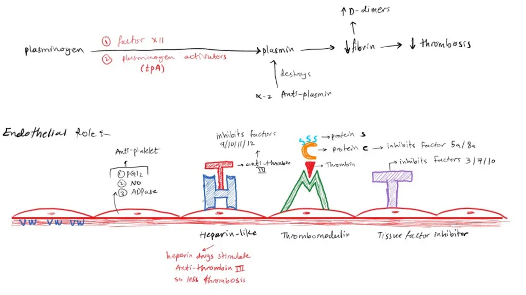
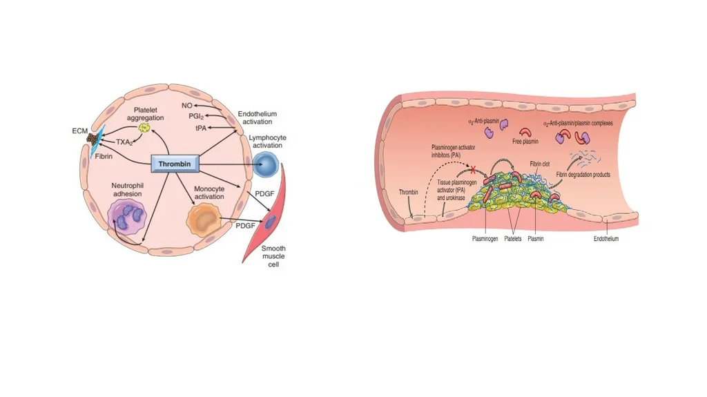
slide21. Fibrinolysis: The enzymatic activity of plasmin, which breaks down fibrin and interferes with its polymerization.
An elevated level of breakdown products of fibrinogen (fibrin-derived D-dimers).
Plasmin is generated by enzymatic catabolism of the inactive circulating precursor plasminogen, either by a factor XII–dependent pathway or by plasminogen activators (t-PA).<br>
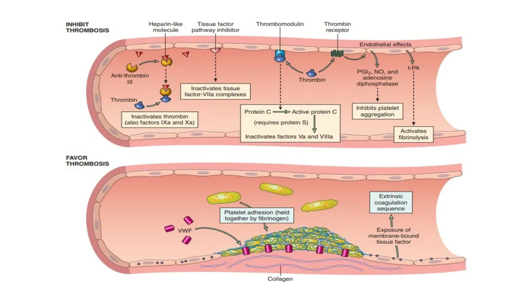
slide24. Endothelium Normal endothelial cells express a multitude of factors that inhibit the procoagulant activities of platelets and coagulation factors and that augment fibrinolysis.
Platelet inhibitory effects.
Anticoagulant effects.
Fibrinolytic effects.<br>
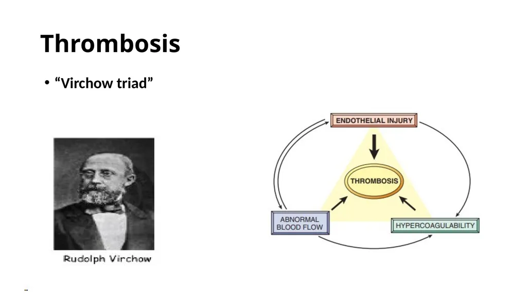
slide25. Thrombosis “Virchow triad”<br>
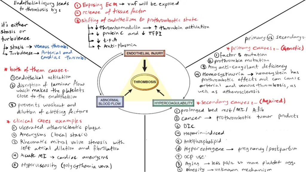
slide27. Endothelial Injury Severe endothelial injury may trigger thrombosis by exposing VWF and tissue factor.
Inflammation and other noxious stimuli also promote thrombosis by shifting the pattern of gene expression in endothelium to one that is “prothrombotic.”
Endothelial activation or dysfunction: physical injury, infectious agents, abnormal blood flow, inflammatory mediators, metabolic abnormalities, such as hypercholesterolemia or homocystinemia, and toxins absorbed from cigarette smoke.<br>
slide28. Abnormal Blood Flow Turbulence (chaotic blood flow) contributes to arterial and cardiac thrombosis by causing endothelial injury or dysfunction, and stasis.
Ulcerated atherosclerotic plaques.
Abnormal aortic and arterial dilations called aneurysms.
Acute myocardial infarction.
Mitral valve stenosis.
Atrial Fibrillation.
Hyperviscosity syndromes.<br>
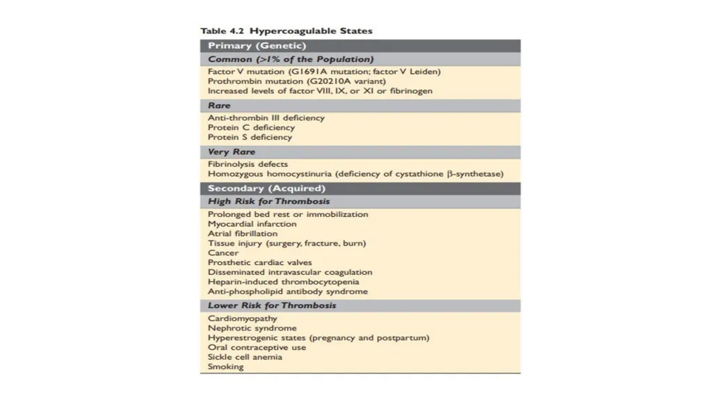
slide29. Hypercoagulability Abnormally high tendency of the blood to clot.
Divided into:
Primary (genetic): mostly caused by mutations in the factor V (Leiden mutation) and prothrombin genes, (2% to 15% of whites carry a specific factor V mutation)
secondary (acquired) disorders.<br>
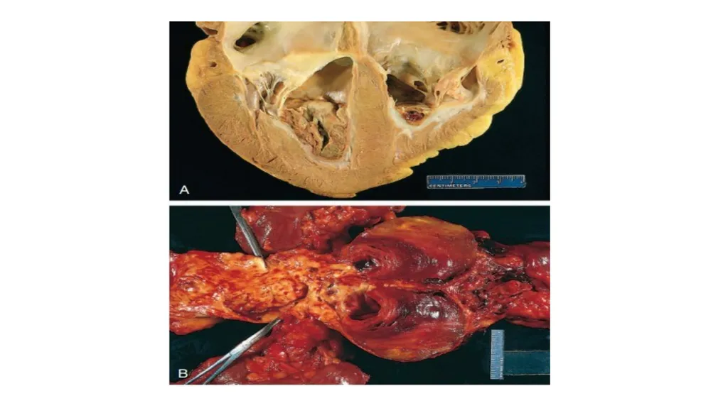
slide31. MORPHOLOGY Thrombi can have grossly (and microscopically) apparent laminations called lines of Zahn; these represent pale platelet and fibrin layers alternating with darker red cell–rich layers.<br>
slide32. Arterial thrombi are frequently occlusive.
Rich in platelets. Venous thrombi (phlebothrombosis) are almost invariably occlusive;
Contain more enmeshed red cells.
Lower extremities (90% of venous thromboses) > the upper extremities, periprostatic plexus, or ovarian and periuterine veins, and under special circumstances they may be found in the dural sinuses, portal vein, or hepatic vein.<br>
slide33. Thrombi on heart valves are called vegetations.
Bacterial or fungal bloodborne infections= infective endocarditis.
Sterile vegetations =nonbacterial thrombotic endocarditis, verrucous endocarditis (LibmanSacks endocarditis).<br>
slide35. Fate of the Thrombus Propagation.
Embolization.
Dissolution.
Organization and recanalization.<br>
slide36. Venous Thrombosis (Phlebothrombosis): In the superficial or the deep veins of the leg.
Superficial venous: (saphenous system) varicosities; these rarely embolize but they can be painful and can cause local congestion and swelling from impaired venous outflow, predisposing the overlying skin to the development of infections and varicose ulcers.
Deep venous thromboses (DVTs) in the larger leg veins at or above the knee joint (e.g., popliteal, femoral, and iliac veins) are more serious because they are prone to embolize. Although such DVTs may cause local pain and edema, collateral channels often circumvent the venous obstruction. Consequently, DVTs are entirely asymptomatic in approximately 50% of patients and are recognized only after they have embolized to the lungs.
Thus, common predisposing factors include congestive heart failure, bed rest, and immobilization, Trauma, surgery, and burns, pregnancy, Tumors.<br>
slide38. Arterial and Cardiac Thrombosis: Atherosclerosis is a major cause of arterial thromboses.
Embolization: the brain, kidneys, and spleen.<br>
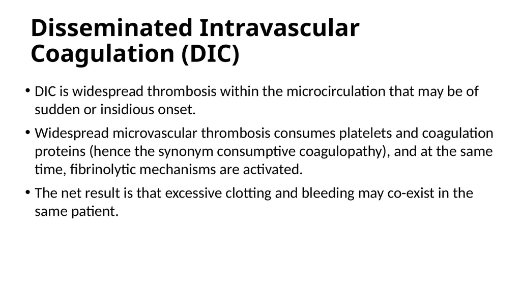
slide39. Disseminated Intravascular Coagulation (DIC) DIC is widespread thrombosis within the microcirculation that may be of sudden or insidious onset.
Widespread microvascular thrombosis consumes platelets and coagulation proteins (hence the synonym consumptive coagulopathy), and at the same time, fibrinolytic mechanisms are activated.
The net result is that excessive clotting and bleeding may co-exist in the same patient.<br>
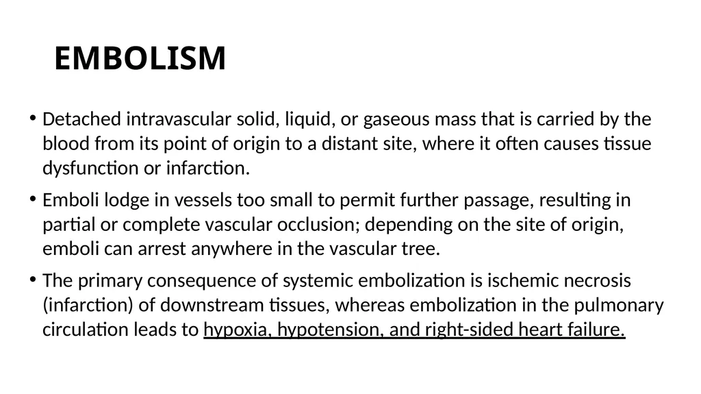
slide40. EMBOLISM Detached intravascular solid, liquid, or gaseous mass that is carried by the blood from its point of origin to a distant site, where it often causes tissue dysfunction or infarction.
Emboli lodge in vessels too small to permit further passage, resulting in partial or complete vascular occlusion; depending on the site of origin, emboli can arrest anywhere in the vascular tree.
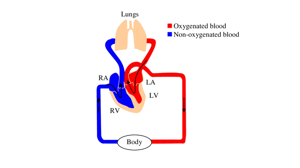
The primary consequence of systemic embolization is ischemic necrosis (infarction) of downstream tissues, whereas embolization in the pulmonary circulation leads to hypoxia, hypotension, and right-sided heart failure.<br>
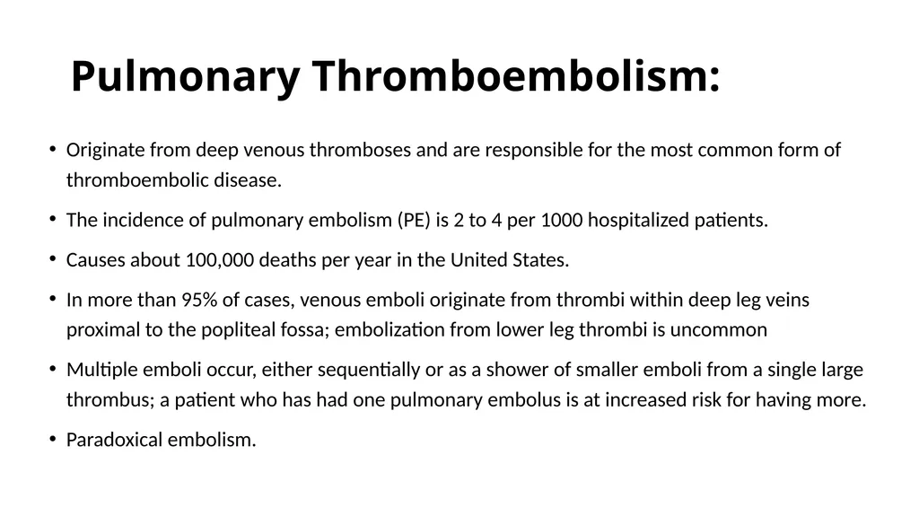
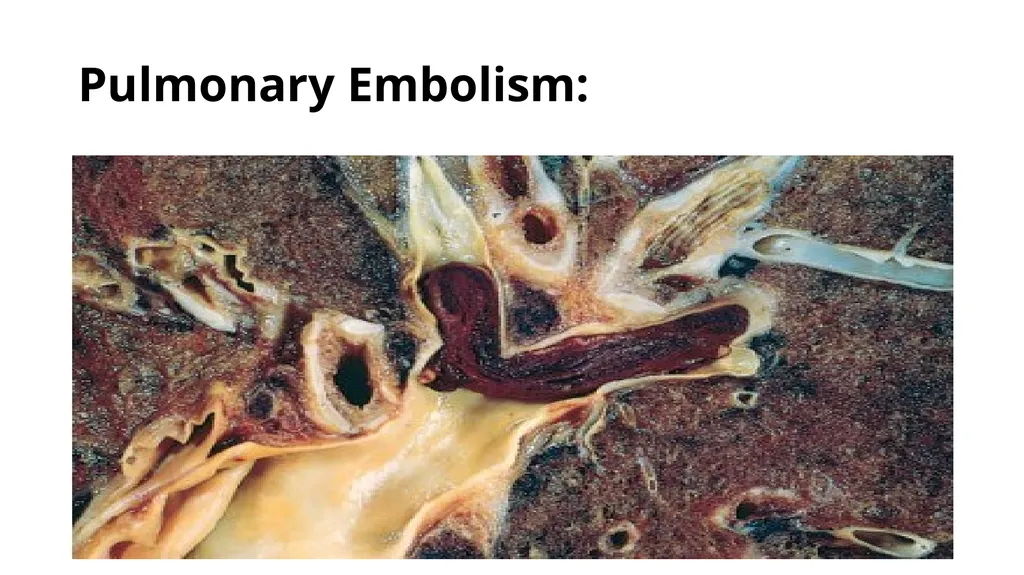
slide41. Pulmonary Thromboembolism: Originate from deep venous thromboses and are responsible for the most common form of thromboembolic disease.
The incidence of pulmonary embolism (PE) is 2 to 4 per 1000 hospitalized patients.
Causes about 100,000 deaths per year in the United States.
In more than 95% of cases, venous emboli originate from thrombi within deep leg veins proximal to the popliteal fossa; embolization from lower leg thrombi is uncommon
Multiple emboli occur, either sequentially or as a shower of smaller emboli from a single large thrombus; a patient who has had one pulmonary embolus is at increased risk for having more.
Paradoxical embolism.<br>
slide43. Pulmonary Embolism:<br>
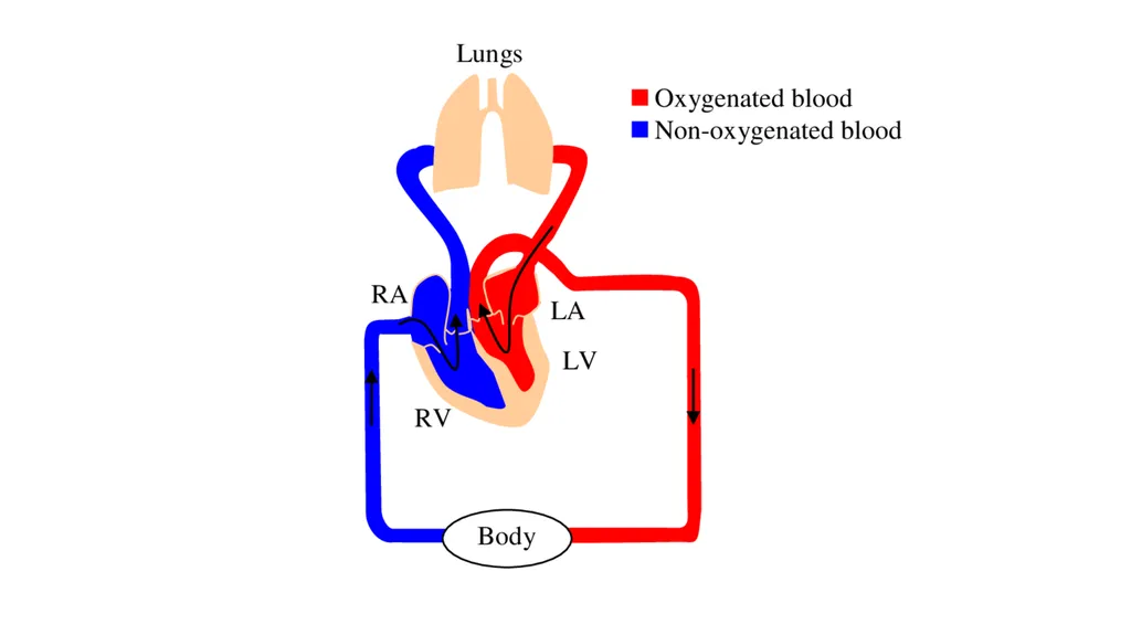
slide44. Systemic Thromboembolism: 80% arise from intracardiac mural thrombi.
Two-thirds of these are associated with left ventricular infarcts and another 25% with dilated left atria (e.g., secondary to mitral valve disease).
The remainder originate from aortic aneurysms, thrombi overlying ulcerated atherosclerotic plaques, fragmented valvular vegetations, or the venous system (paradoxical emboli).
10% to 15% of systemic emboli are of unknown origin.
The lower extremities (75%) and central nervous system (10%); intestines, kidneys, and spleen are less common targets.<br>
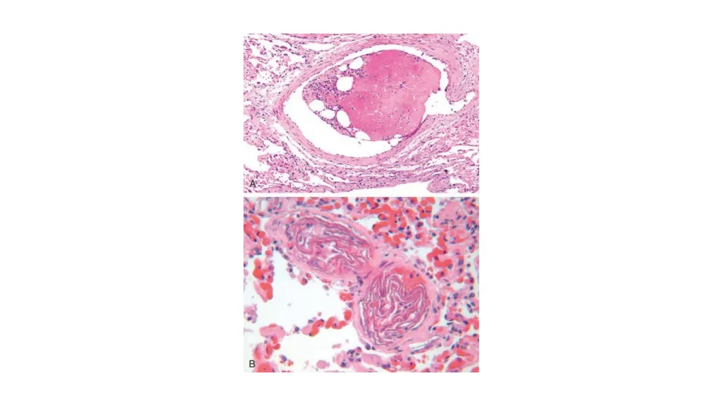
slide45. Others … Fat Embolism.
Amniotic Fluid Embolism.
Air Embolism.<br>
Email: r.marji@bau.edu.jo
Office: O-505<br>
slide7. HEMOSTASIS AND THROMBOSIS Normal hemostasis: Series of regulated processes that culminate in the formation of a blood clot that limits bleeding from an injured vessel.
The pathologic counterpart of hemostasis is thrombosis, the formation of blood clot (thrombus) within non-traumatized, intact vessels.<br>
slide8. Normal Hemostasis Involving platelets, clotting factors, and endothelium.
Arteriolar vasoconstriction occurs immediately and markedly reduces blood flow to the injured area. It is mediated by reflex neurogenic mechanisms and augmented by the local secretion of factors such as endothelin, a potent endothelium-derived vasoconstrictor.
Transient.<br>
slide9. Arteriolar vasoconstriction: transient/ mediated by reflex neurogenic mechanisms and augmented by the local secretion of factors such as endothelin.<br>
slide10. Primary and Secondary Hemostasis Primary Hemostasis: formation of the platelet plug.
Disruption of the endothelium exposes subendothelial von Willebrand factor (vWF) and collagen.
Shape change (from small rounded discs to flat plates with spiky protrusions that markedly increased surface area), and the release of secretory granules.
Recruit additional platelets>> aggregation to form a primary hemostatic plug.
Secondary hemostasis: deposition of fibrin.
Exposes tissue factor at the site of injury >> activates factor VII >> thrombin generation.
Thrombin cleaves circulating fibrinogen into insoluble fibrin, a potent activator of platelets.<br>
slide11. Clot Stabilization and Resorption: Polymerized fibrin and platelet aggregates undergo contraction to form a solid, permanent plug that prevents further hemorrhage.
Counterregulatory mechanisms (e.g., tissue plasminogen activator, t-PA made by endothelial cells) are set into motion that limit clotting to the site of injury and eventually lead to clot resorption and tissue repair.<br>
slide12. Platelets Disc-shaped anucleate cell fragments that are shed from megakaryocytes in the bone marrow into the bloodstream.
Their function depends on several glycoprotein receptors, a contractile cytoskeleton, and two types of cytoplasmic granules.
α-Granules.
Dense (or δ) granules.<br>
slide13. Platelet adhesion: interactions with vWF, which acts as a bridge between the platelet surface receptor glycoprotein Ib (GpIb) and exposed collagen.
Genetic deficiencies of vWF (von Willebrand disease) or GpIb (BernardSoulier syndrome) result in bleeding disorders, attesting to the importance of these factors.
Platelets rapidly change shape following adhesion, being converted from smooth discs to spiky “sea urchins” with greatly increased surface area. This change is accompanied by alterations in glycoprotein IIb/IIIa that increase its affinity for fibrinogen, and by the translocation of negatively charged phospholipids to the platelet surface.
Phospholipids bind calcium and serve as nucleation sites for the assembly of coagulation factor complexes.<br>
slide14. Secretion (release reaction) of granule contents occurs along with changes in shape; these two events are often referred to together as platelet activation.
Thrombin activates platelets through a special type of G-protein– coupled receptor referred to as a protease-activated receptor (PAR).
Activated platelets also produce the prostaglandin thromboxane A2 (TXA2), a potent inducer of platelet aggregation.<br>
slide15. Platelet aggregation: glycoprotein IIb/IIIa allows binding of fibrinogen.
Inherited deficiency of GpIIb-IIIa results in a bleeding disorder called Glanzmann thrombasthenia.<br>
slide16. Bleeding Disorders:<br>
slide17. Coagulation Cascade Deposition of an insoluble fibrin clot.
Each reaction step involves an enzyme (an activated coagulation factor), a substrate (an inactive proenzyme form of a coagulation factor), and a cofactor (a reaction accelerator).
Components are assembled on a negatively charged phospholipid surface.<br>
slide18. The coagulation cascade is divided into the extrinsic and intrinsic pathways.
The prothrombin time (PT) assay assesses the function of the proteins in the extrinsic pathway (factors VII, X, V, II (prothrombin), and fibrinogen).
The partial thromboplastin time (PTT) assay screens the function of the proteins in the intrinsic pathway (factors XII, XI, IX, VIII, X, V, II, and fibrinogen).<br>
slide20. Thrombin: Conversion of fibrinogen into crosslinked fibrin.
Platelet activation.
Proinflammatory effects.
Anti-coagulant effects.<br>
slide21. Fibrinolysis: The enzymatic activity of plasmin, which breaks down fibrin and interferes with its polymerization.
An elevated level of breakdown products of fibrinogen (fibrin-derived D-dimers).
Plasmin is generated by enzymatic catabolism of the inactive circulating precursor plasminogen, either by a factor XII–dependent pathway or by plasminogen activators (t-PA).<br>
slide24. Endothelium Normal endothelial cells express a multitude of factors that inhibit the procoagulant activities of platelets and coagulation factors and that augment fibrinolysis.
Platelet inhibitory effects.
Anticoagulant effects.
Fibrinolytic effects.<br>
slide25. Thrombosis “Virchow triad”<br>
slide27. Endothelial Injury Severe endothelial injury may trigger thrombosis by exposing VWF and tissue factor.
Inflammation and other noxious stimuli also promote thrombosis by shifting the pattern of gene expression in endothelium to one that is “prothrombotic.”
Endothelial activation or dysfunction: physical injury, infectious agents, abnormal blood flow, inflammatory mediators, metabolic abnormalities, such as hypercholesterolemia or homocystinemia, and toxins absorbed from cigarette smoke.<br>
slide28. Abnormal Blood Flow Turbulence (chaotic blood flow) contributes to arterial and cardiac thrombosis by causing endothelial injury or dysfunction, and stasis.
Ulcerated atherosclerotic plaques.
Abnormal aortic and arterial dilations called aneurysms.
Acute myocardial infarction.
Mitral valve stenosis.
Atrial Fibrillation.
Hyperviscosity syndromes.<br>
slide29. Hypercoagulability Abnormally high tendency of the blood to clot.
Divided into:
Primary (genetic): mostly caused by mutations in the factor V (Leiden mutation) and prothrombin genes, (2% to 15% of whites carry a specific factor V mutation)
secondary (acquired) disorders.<br>
slide31. MORPHOLOGY Thrombi can have grossly (and microscopically) apparent laminations called lines of Zahn; these represent pale platelet and fibrin layers alternating with darker red cell–rich layers.<br>
slide32. Arterial thrombi are frequently occlusive.
Rich in platelets. Venous thrombi (phlebothrombosis) are almost invariably occlusive;
Contain more enmeshed red cells.
Lower extremities (90% of venous thromboses) > the upper extremities, periprostatic plexus, or ovarian and periuterine veins, and under special circumstances they may be found in the dural sinuses, portal vein, or hepatic vein.<br>
slide33. Thrombi on heart valves are called vegetations.
Bacterial or fungal bloodborne infections= infective endocarditis.
Sterile vegetations =nonbacterial thrombotic endocarditis, verrucous endocarditis (LibmanSacks endocarditis).<br>
slide35. Fate of the Thrombus Propagation.
Embolization.
Dissolution.
Organization and recanalization.<br>
slide36. Venous Thrombosis (Phlebothrombosis): In the superficial or the deep veins of the leg.
Superficial venous: (saphenous system) varicosities; these rarely embolize but they can be painful and can cause local congestion and swelling from impaired venous outflow, predisposing the overlying skin to the development of infections and varicose ulcers.
Deep venous thromboses (DVTs) in the larger leg veins at or above the knee joint (e.g., popliteal, femoral, and iliac veins) are more serious because they are prone to embolize. Although such DVTs may cause local pain and edema, collateral channels often circumvent the venous obstruction. Consequently, DVTs are entirely asymptomatic in approximately 50% of patients and are recognized only after they have embolized to the lungs.
Thus, common predisposing factors include congestive heart failure, bed rest, and immobilization, Trauma, surgery, and burns, pregnancy, Tumors.<br>
slide38. Arterial and Cardiac Thrombosis: Atherosclerosis is a major cause of arterial thromboses.
Embolization: the brain, kidneys, and spleen.<br>
slide39. Disseminated Intravascular Coagulation (DIC) DIC is widespread thrombosis within the microcirculation that may be of sudden or insidious onset.
Widespread microvascular thrombosis consumes platelets and coagulation proteins (hence the synonym consumptive coagulopathy), and at the same time, fibrinolytic mechanisms are activated.
The net result is that excessive clotting and bleeding may co-exist in the same patient.<br>
slide40. EMBOLISM Detached intravascular solid, liquid, or gaseous mass that is carried by the blood from its point of origin to a distant site, where it often causes tissue dysfunction or infarction.
Emboli lodge in vessels too small to permit further passage, resulting in partial or complete vascular occlusion; depending on the site of origin, emboli can arrest anywhere in the vascular tree.
The primary consequence of systemic embolization is ischemic necrosis (infarction) of downstream tissues, whereas embolization in the pulmonary circulation leads to hypoxia, hypotension, and right-sided heart failure.<br>
slide41. Pulmonary Thromboembolism: Originate from deep venous thromboses and are responsible for the most common form of thromboembolic disease.
The incidence of pulmonary embolism (PE) is 2 to 4 per 1000 hospitalized patients.
Causes about 100,000 deaths per year in the United States.
In more than 95% of cases, venous emboli originate from thrombi within deep leg veins proximal to the popliteal fossa; embolization from lower leg thrombi is uncommon
Multiple emboli occur, either sequentially or as a shower of smaller emboli from a single large thrombus; a patient who has had one pulmonary embolus is at increased risk for having more.
Paradoxical embolism.<br>
slide43. Pulmonary Embolism:<br>
slide44. Systemic Thromboembolism: 80% arise from intracardiac mural thrombi.
Two-thirds of these are associated with left ventricular infarcts and another 25% with dilated left atria (e.g., secondary to mitral valve disease).
The remainder originate from aortic aneurysms, thrombi overlying ulcerated atherosclerotic plaques, fragmented valvular vegetations, or the venous system (paradoxical emboli).
10% to 15% of systemic emboli are of unknown origin.
The lower extremities (75%) and central nervous system (10%); intestines, kidneys, and spleen are less common targets.<br>
slide45. Others … Fat Embolism.
Amniotic Fluid Embolism.
Air Embolism.<br>