Lactobacillus species (Enlarged view) Slide 1.
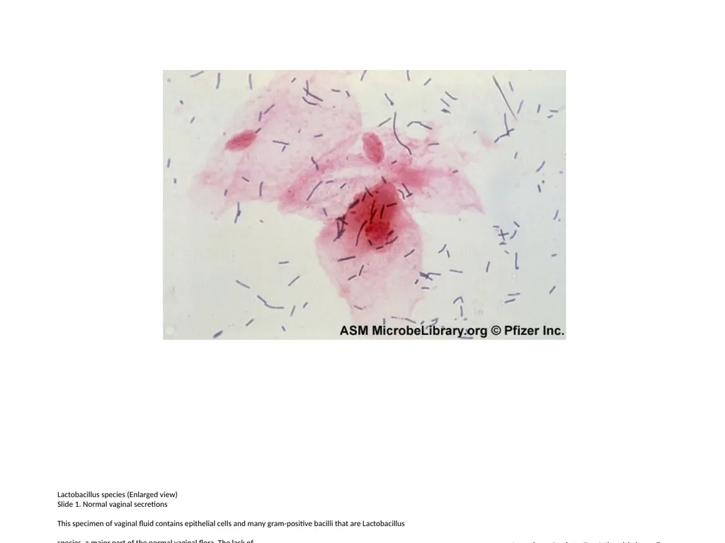
Description: Lactobacillus species (Enlarged view) Slide 1. Normal vaginal secretions This specimen of vaginal fluid contains epithelial cells and many gram-positive bacilli that are Lactobacillus species, a major part of the normal vaginal flora. The
Related Topics
Download Presentation
"Lactobacillus species (Enlarged view) Slide 1." is the property of its rightful owner. Permission is granted to download and print the materials on this website for personal, non-commercial use only, and to display it on your personal computer provided you do not modify the materials and that you retain all copyright notices contained in the materials. By downloading content from our website, you accept the terms of this agreement.
Presentation Transcript
slide1. Lactobacillus species (Enlarged view)
Slide 1. Normal vaginal secretions
This specimen of vaginal fluid contains epithelial cells and many gram-positive bacilli that are Lactobacillus

species, a major part of the normal vaginal flora. The lack of
neutrophils and the presence of normal epithelial cells suggest that
inflammation is absent. American Society For Microbiology ©<br>
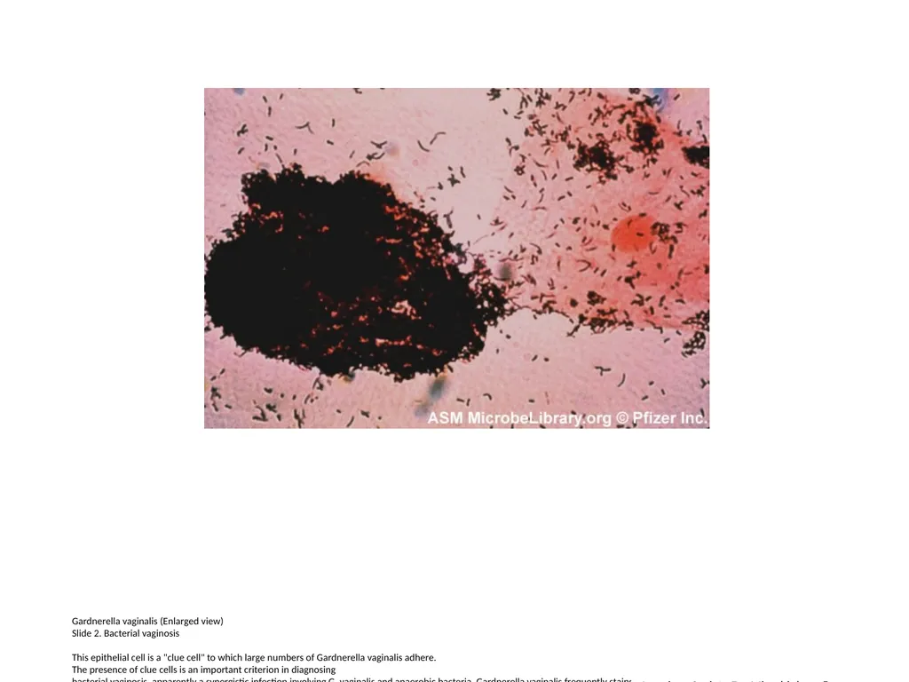
slide2. Gardnerella vaginalis (Enlarged view)
Slide 2. Bacterial vaginosis
This epithelial cell is a "clue cell" to which large numbers of Gardnerella vaginalis adhere.
The presence of clue cells is an important criterion in diagnosing
bacterial vaginosis, apparently a synergistic infection involving G. vaginalis and anaerobic bacteria. Gardnerella vaginalis frequently stains gram-variable, as does Mobiluncus curtisii, an anaerobic bacterium often associated with bacterial vaginosis and visible here as gram-positive curved rods. American Society For Microbiology ©<br>
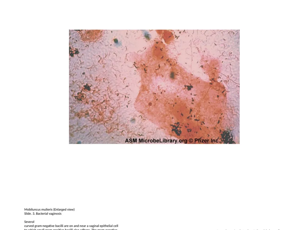
slide3. Mobiluncus mulieris (Enlarged view)
Slide. 3. Bacterial vaginosis
Several
curved gram-negative bacilli are on and near a vaginal epithelial cell
to which small gram-positive bacilli also adhere. The gram-negative
rods are Mobiluncus mulieris, bacteria often present in the secretions of patients with bacterial vaginosis. American Society For Microbiology ©<br>
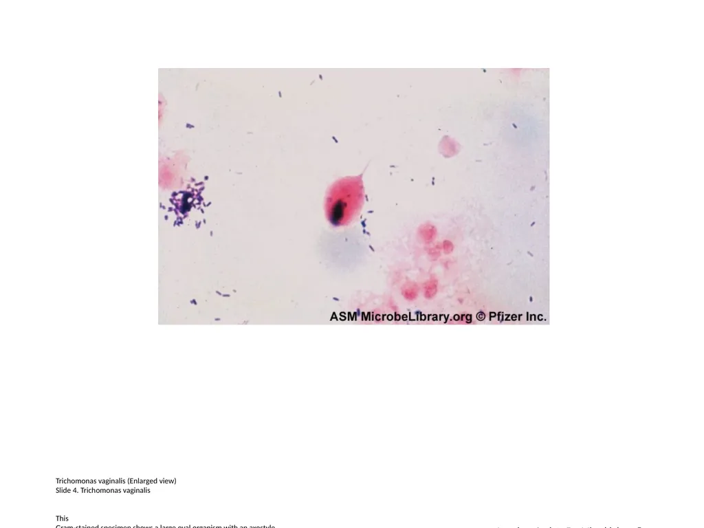
slide4. Trichomonas vaginalis (Enlarged view)
Slide 4. Trichomonas vaginalis
This
Gram-stained specimen shows a large oval organism with an axostyle,
which is a supporting rod running through the body of a trichomonad and
protruding posteriorly. Trichomonas vaginalis, a protozoan that causes vaginitis, is usually more easily detected on a wet mount than on a Gram stain. American Society For Microbiology ©<br>
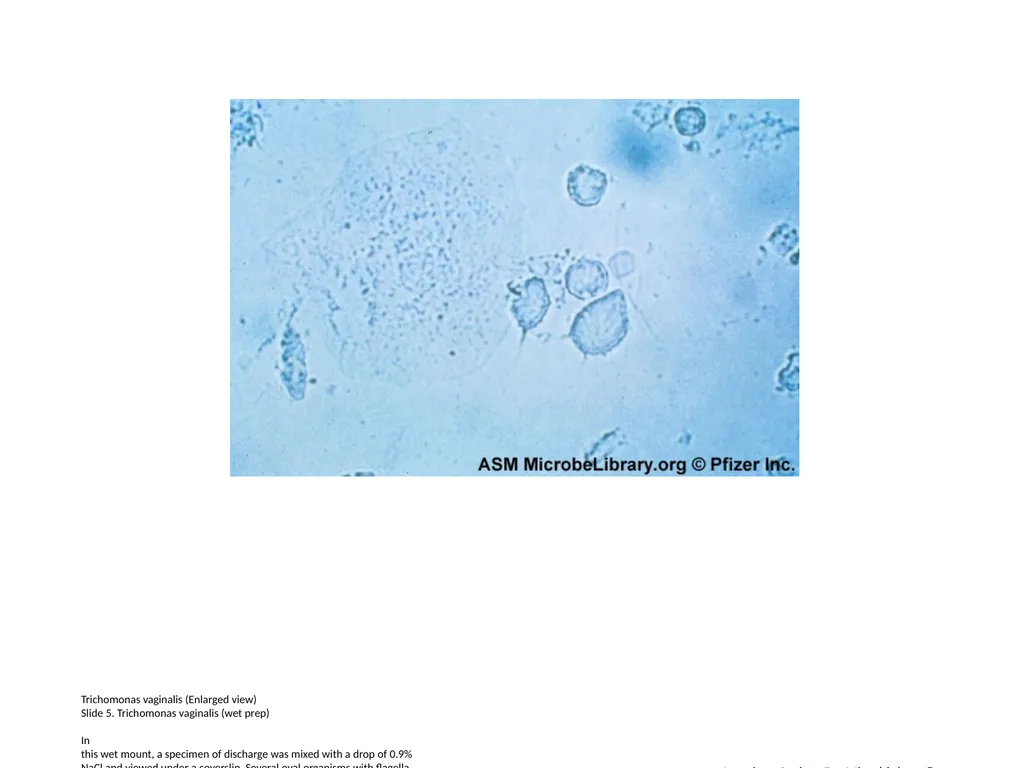
slide5. Trichomonas vaginalis (Enlarged view)
Slide 5. Trichomonas vaginalis (wet prep)
In
this wet mount, a specimen of discharge was mixed with a drop of 0.9%
NaCl and viewed under a coverslip. Several oval organisms with flagella
are visible. Trichomonads are often larger than neutrophils (white
blood cells) and, on a wet mount, often move with a jerky motion across
the microscopic field. American Society For Microbiology ©<br>
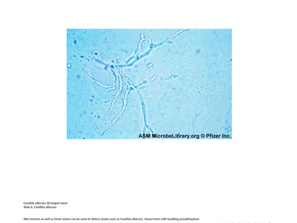
slide6. Candida albicans (Enlarged view)
Slide 6. Candida albicans
Wet mounts as well as Gram stains can be used to detect yeasts such as Candida albicans, shown here with budding pseudohyphae. American Society For Microbiology ©<br>
Slide 1. Normal vaginal secretions
This specimen of vaginal fluid contains epithelial cells and many gram-positive bacilli that are Lactobacillus
species, a major part of the normal vaginal flora. The lack of
neutrophils and the presence of normal epithelial cells suggest that
inflammation is absent. American Society For Microbiology ©<br>
slide2. Gardnerella vaginalis (Enlarged view)
Slide 2. Bacterial vaginosis
This epithelial cell is a "clue cell" to which large numbers of Gardnerella vaginalis adhere.
The presence of clue cells is an important criterion in diagnosing
bacterial vaginosis, apparently a synergistic infection involving G. vaginalis and anaerobic bacteria. Gardnerella vaginalis frequently stains gram-variable, as does Mobiluncus curtisii, an anaerobic bacterium often associated with bacterial vaginosis and visible here as gram-positive curved rods. American Society For Microbiology ©<br>
slide3. Mobiluncus mulieris (Enlarged view)
Slide. 3. Bacterial vaginosis
Several
curved gram-negative bacilli are on and near a vaginal epithelial cell
to which small gram-positive bacilli also adhere. The gram-negative
rods are Mobiluncus mulieris, bacteria often present in the secretions of patients with bacterial vaginosis. American Society For Microbiology ©<br>
slide4. Trichomonas vaginalis (Enlarged view)
Slide 4. Trichomonas vaginalis
This
Gram-stained specimen shows a large oval organism with an axostyle,
which is a supporting rod running through the body of a trichomonad and
protruding posteriorly. Trichomonas vaginalis, a protozoan that causes vaginitis, is usually more easily detected on a wet mount than on a Gram stain. American Society For Microbiology ©<br>
slide5. Trichomonas vaginalis (Enlarged view)
Slide 5. Trichomonas vaginalis (wet prep)
In
this wet mount, a specimen of discharge was mixed with a drop of 0.9%
NaCl and viewed under a coverslip. Several oval organisms with flagella
are visible. Trichomonads are often larger than neutrophils (white
blood cells) and, on a wet mount, often move with a jerky motion across
the microscopic field. American Society For Microbiology ©<br>
slide6. Candida albicans (Enlarged view)
Slide 6. Candida albicans
Wet mounts as well as Gram stains can be used to detect yeasts such as Candida albicans, shown here with budding pseudohyphae. American Society For Microbiology ©<br>