Lecture 5: navicular disease, side bones and quittor Dr Gyan Dev Singh, Assistant professor, VCC, BVC, Patna-14 QUITTOR: Necrosis of the lateral cartilage and is noticed by the presence of sinus openings in the coronet region. Fore or hind
"Lecture 5: navicular disease, side bones and" is the property of its rightful owner. Permission is granted to
download and print the materials on this website for personal, non-commercial use only, and to display it
on your personal computer provided you do not modify the materials and that you retain all copyright
notices contained in the materials. By downloading content from our website, you accept the terms of this
agreement.
Presentation Transcript
01
Lecture 5: navicular disease, side bones and quittor
Dr Gyan Dev Singh, Assistant professor, VCC, BVC, Patna-14<br>
02
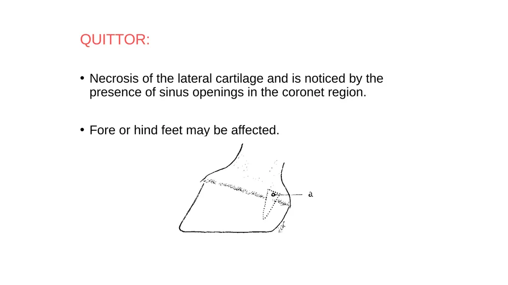
QUITTOR:
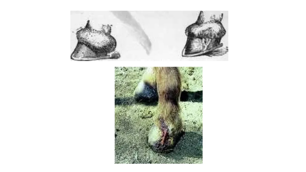
Necrosis of the lateral cartilage and is noticed by the presence of sinus openings in the coronet region.
Fore or hind feet may be affected.<br>
03
Etiology
1) Infection getting into the cartilage through an
opening in the coronet region, e.g., tread injuries.
2) Necrosis of skin in the pastern region due to constant accumulation of mud.
3) Suppuration extending from neighboring lesions like suppurating corn, sand crack, penetrating wounds of the foot, etc.<br>
04
Symptoms
Coronet appears swollen and one or more sinus openings may be noticed in the region.
Sinus openings arise from the necrosed portion of the cartilage.
Complications
1) Septic arthritis of the corono-pedal joint due to
extension of the lesion.
2) Necrosis of os pedis.
3) Necrosis of the sensitive lamina or plantar surface
4) Gangrene - affected region of skin.<br>
05
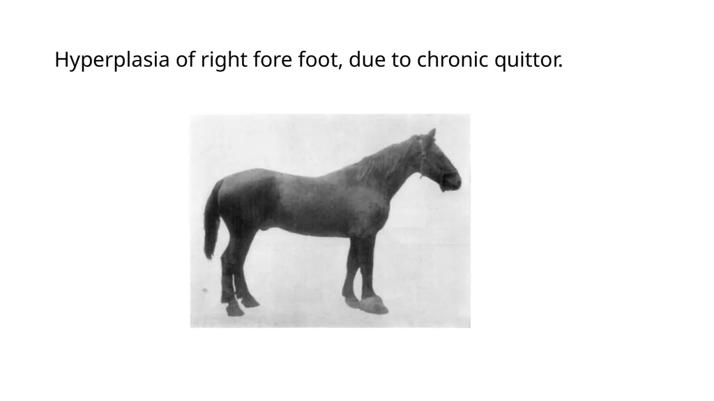
Hyperplasia of right fore foot, due to chronic quittor.<br>
06
Prognosis
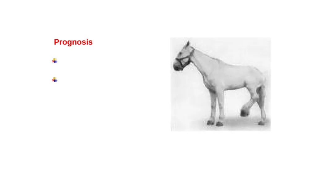
Guarded.
Quittor in the hind limbs
responds to treatment
better than of fore
limbs because the lateral
cartilages are thinner in
the hind limbs.<br>
07
Treatment
On general principles as for sinuses.
Surgical treatment consists of removal of lateral cartilage.
1) Irrigation of the sinus tracts with antiseptic
lotions to remove pus and necrotic tissue.
But this may cause spread of the lesion to surrounding tissues because it is difficult to remove fluid completely.<br>
08
2) Solutions injected into the sinus tracts to control
bacterial infection and facilitate separation of necrotic tissue:
Corrosive sublimate in alcohol (1 in 10%),
Zinc chloride solution (5 to 10%),
Zinc sulphate solution (5 to 10%).
3) A solid caustic like silver nitrate is powdered and introduced through the sinus openings. This is called Coring.<br>
09
4) Applying a red-hot iron through the sinus openings
5) A combination of treatment with red-hot iron and use of caustics.
6) A seton passed through opening in the coronet and through wall of the hoof in level with the bottom of the suppurating tract, provides good drainage and helps quicker healing.
7) The operation for removal of cartilage (excision of the lateral cartilage) is done under general anesthesia.<br>
10
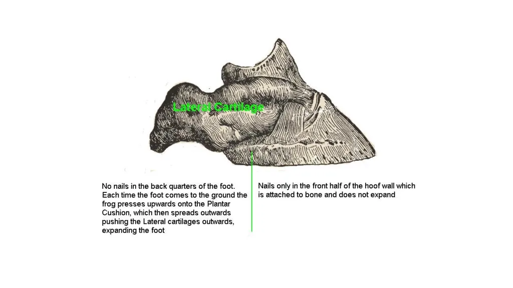
SIDE BONES Ossified lateral Cartilages of the foot are called side bones.
Incidence
Ossification of the lateral cartilage is more commonly noticed in the fore-feet.<br>
11
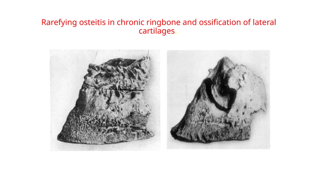
Rarefying osteitis in chronic ringbone and ossification of lateral cartilages.<br>
12
ETIOLOGY Hereditory predisposition,
Natural tendency of cartilages in contact with bone to ossify.
Bad conformation.
Defective shoeing.
Concussion.
Direct injury to the cartilage resulting in inflammation and subsequent invasion of cartilage with osteoblasts<br>
13
SYMPTOMS
Lameness may or may not be present.
In horses having good open feet and doing only slow work,
Side bones do not usually cause lameness.<br>
14
DIAGNOSIS
In the initial stages difficult because at this stage the palpable portion of the cartilage might not have ossified.
When ossification is complete, diagnosis is easy.<br>
15
TREATMENT
If there is no pain, or lameness treatment is not advised.
If side bones are causing lameness the quarters of the hoof may be grooved or thinned. This facilitates expansion of the foot and relieves pain.
Partial removal of the cartilage is recommended only if it has become very prominent
Flat wide webbed shoe is recommended
Digital neurectomy may be tried to relieve lameness (medial and lateral palmar nerve block).<br>
16
NAVICULAR DISEASE Navicular disease is a chronic ostitis of the navicular bone, associated with navicular bursitis and inflammation of the plantar aponeurosis.
INCIDENCE
More common in light horses than in heavy horses.
Common in horses about seven years old.
Young horses, donkeys and mules are only rarely affected.
More common in the fore-feet.<br>
17
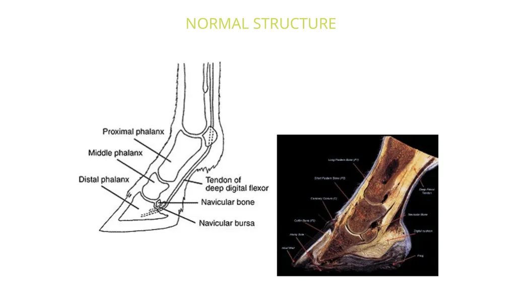
NORMAL STRUCTURE<br>
18
ETIOLOGY
Predisposing causes:
Heredity. Abnormal, conformation of foot,
e.g., upright and narrow foot, contracted foot, foot having small frog etc.
Excessively sloping pastern and long toe
Defective shoeing
Thick-heeled shoe, etc;
Exciting cause: Severe and repeated concussion,<br>
19
PATHOLOGY
There is inflammation of the navicular bone and the navicular bursa.
Exostosis may develop. Friction of exostosis on the flexor tendon may cause sprain of the tendon.
There may be adhesions formed between the tendon and bone.
Sometimes there is rarefaction of navicular bone and it may fracture easily.<br>
20
SYMPTOMS
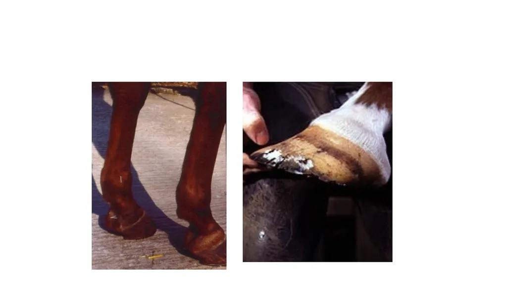
Pointing of the foot at rest.
In the initial stage lameness is intermittent and the animal goes without lameness after period of rest.
As the disease progresses lameness becomes more pronounced.
The stride is shortened, the toe is dragged and there is stumbling.
The gait may be "pottery" or "stilted". A "groggy gait" or "shuffling gait" is rather characteristic.
When the horse is turned, the animal "screws" on the fore-limbs; rather than having them lifted normally.<br>
21
SEQUELAE
In chronic cases changes in the foot are marked by contraction of heels, concavity of the sole, etc.
The foot becomes "boxy" (i.e., contracted and high at the heels with very concave soles).
There may be atrophy of the frog. Bulging of the hollow of the heels due to enlargement of the navicular bursa may be seen.
Rarely, however, a foot affected with navicular disease may not show any abnormality in shape.<br>
22
DIAGNOSIS
Diagnosis is made from the symptoms.
The animal may evince pain when pressure is applied on the central third of the frog.
Digital nerve block and radiography are helpful for confirming the diagnosis.