
MacConkey Agar Plates Author: Mary E. Allen
NS
Published · 36 slides · 0 views
1 / 1















Description
MacConkey Agar Plates Author: Mary E. Allen Citation: Mary E. Allen. 2005. Macconkey agar plates. Publication Date: September 2005 MacConkey Agar Plate (Enlarged view) FIG. 1. Uninoculated MacConkey agar plate. (David Miller and Patrick
Related Topics
Share
Embed code
Download this presentation From Below
"MacConkey Agar Plates Author: Mary E. Allen" is the property of its rightful owner. Permission is granted to download and print the materials on this website for personal, non-commercial use only, and to display it on your personal computer provided you do not modify the materials and that you retain all copyright notices contained in the materials. By downloading content from our website, you accept the terms of this agreement.
Presentation Transcript
01
MacConkey Agar Plates Author: Mary E. Allen
Citation: Mary E. Allen. 2005. Macconkey agar plates.
Publication Date: September 2005<br>
Citation: Mary E. Allen. 2005. Macconkey agar plates.
Publication Date: September 2005<br>
02
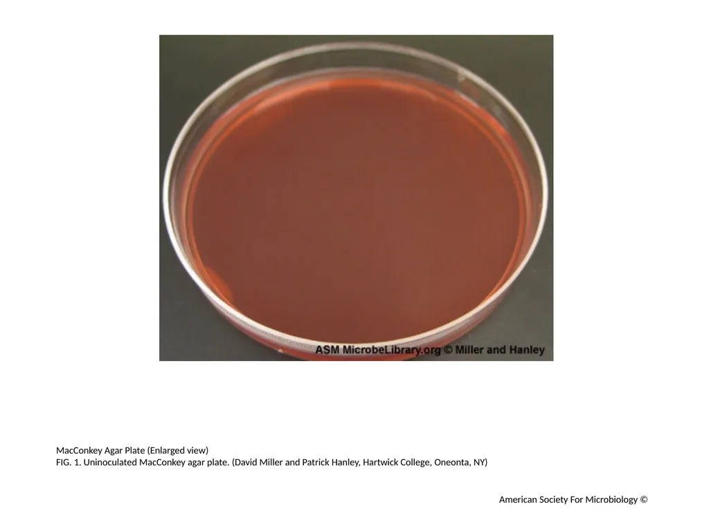
MacConkey Agar Plate (Enlarged view)
FIG. 1. Uninoculated MacConkey agar plate. (David Miller and Patrick Hanley, Hartwick College, Oneonta, NY) American Society For Microbiology ©<br>
FIG. 1. Uninoculated MacConkey agar plate. (David Miller and Patrick Hanley, Hartwick College, Oneonta, NY) American Society For Microbiology ©<br>
03
Escherichia coli and Serratia marcesens(Enlarged view)
FIG. 2. MacConkey agar plate inoculated with the gram-negative lactose fermenter Escherichia coli and the gram-negative non-lactose fermenter Serratia marcescens. (David Miller and Patrick Hanley, Hartwick College, Oneonta, NY) American Society For Microbiology ©<br>
FIG. 2. MacConkey agar plate inoculated with the gram-negative lactose fermenter Escherichia coli and the gram-negative non-lactose fermenter Serratia marcescens. (David Miller and Patrick Hanley, Hartwick College, Oneonta, NY) American Society For Microbiology ©<br>
04
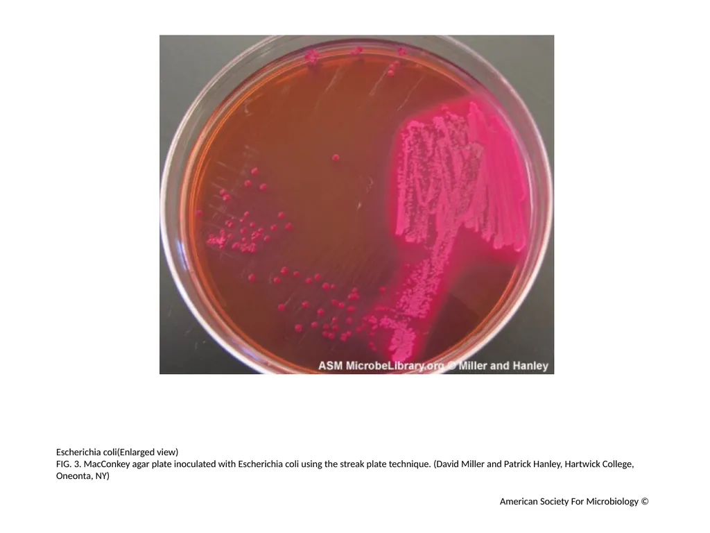
Escherichia coli(Enlarged view)
FIG. 3. MacConkey agar plate inoculated with Escherichia coli using the streak plate technique. (David Miller and Patrick Hanley, Hartwick College, Oneonta, NY) American Society For Microbiology ©<br>
FIG. 3. MacConkey agar plate inoculated with Escherichia coli using the streak plate technique. (David Miller and Patrick Hanley, Hartwick College, Oneonta, NY) American Society For Microbiology ©<br>
05
Escherichia coli (Enlarged view)
FIG. 4. MacConkey agar plate inoculated with Escherichia coli using the streak plate technique. (Anne Y. Tsang, University of Maryland, College Park, MD) American Society For Microbiology ©<br>
FIG. 4. MacConkey agar plate inoculated with Escherichia coli using the streak plate technique. (Anne Y. Tsang, University of Maryland, College Park, MD) American Society For Microbiology ©<br>
06
Escherichia coli (Enlarged view)
FIG. 5. MacConkey agar plate inoculated with Escherichia coli. (David Miller and Patrick Hanley, Hartwick College, Oneonta, NY) American Society For Microbiology ©<br>
FIG. 5. MacConkey agar plate inoculated with Escherichia coli. (David Miller and Patrick Hanley, Hartwick College, Oneonta, NY) American Society For Microbiology ©<br>
07
Escherichia coli and Serratia marcesens (Enlarged view)
FIG. 6. MacConkey agar plate inoculated with the gram-negative lactose non-fermenter Serratia marcescens and Escherichia coli using the streak plate technique. (David Miller and Patrick Hanley, Hartwick College, Oneonta, NY) American Society For Microbiology ©<br>
FIG. 6. MacConkey agar plate inoculated with the gram-negative lactose non-fermenter Serratia marcescens and Escherichia coli using the streak plate technique. (David Miller and Patrick Hanley, Hartwick College, Oneonta, NY) American Society For Microbiology ©<br>
08
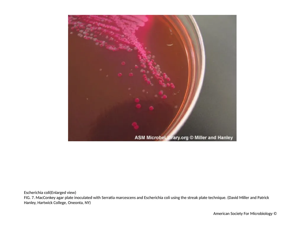
Escherichia coli(Enlarged view)
FIG. 7. MacConkey agar plate inoculated with Serratia marcescens and Escherichia coli using the streak plate technique. (David Miller and Patrick Hanley, Hartwick College, Oneonta, NY) American Society For Microbiology ©<br>
FIG. 7. MacConkey agar plate inoculated with Serratia marcescens and Escherichia coli using the streak plate technique. (David Miller and Patrick Hanley, Hartwick College, Oneonta, NY) American Society For Microbiology ©<br>
09
Escherichia coli & Salmonella typhimurium (Enlarged view)
FIG. 8. MacConkey agar plate inoculated with Escherichia coli and Salmonella typhimurium using the streak plate technique. (Anne Y. Tsang, University of Maryland, College Park, MD) American Society For Microbiology ©<br>
FIG. 8. MacConkey agar plate inoculated with Escherichia coli and Salmonella typhimurium using the streak plate technique. (Anne Y. Tsang, University of Maryland, College Park, MD) American Society For Microbiology ©<br>
10
Klebsiella pneumoniae (Enlarged view)
FIG. 9. MacConkey agar plate inoculated with the gram-negative lactose fermenter Klebsiella pneumoniae using the streak plate technique. (Mary Allen, Hartwick College, Oneonta, NY) American Society For Microbiology ©<br>
FIG. 9. MacConkey agar plate inoculated with the gram-negative lactose fermenter Klebsiella pneumoniae using the streak plate technique. (Mary Allen, Hartwick College, Oneonta, NY) American Society For Microbiology ©<br>
11
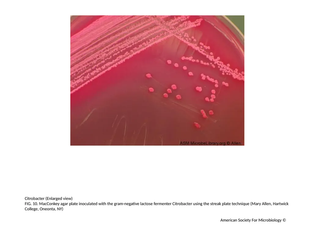
Citrobacter (Enlarged view)
FIG. 10. MacConkey agar plate inoculated with the gram-negative lactose fermenter Citrobacter using the streak plate technique (Mary Allen, Hartwick College, Oneonta, NY) American Society For Microbiology ©<br>
FIG. 10. MacConkey agar plate inoculated with the gram-negative lactose fermenter Citrobacter using the streak plate technique (Mary Allen, Hartwick College, Oneonta, NY) American Society For Microbiology ©<br>
12
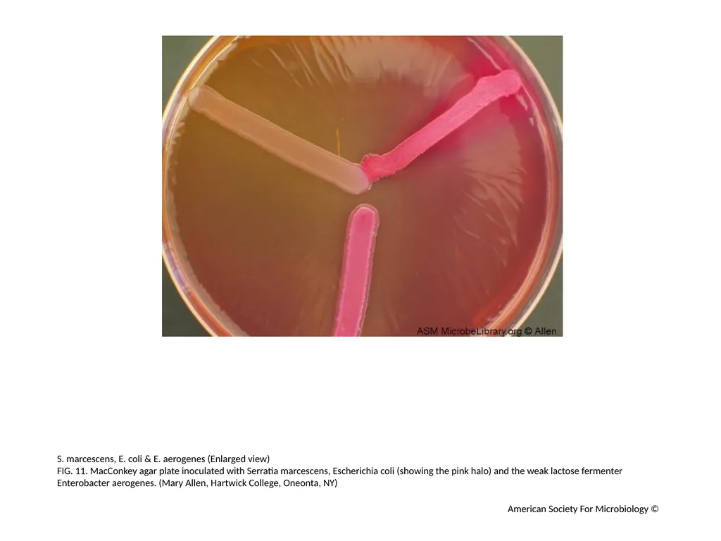
S. marcescens, E. coli & E. aerogenes (Enlarged view)
FIG. 11. MacConkey agar plate inoculated with Serratia marcescens, Escherichia coli (showing the pink halo) and the weak lactose fermenter Enterobacter aerogenes. (Mary Allen, Hartwick College, Oneonta, NY) American Society For Microbiology ©<br>
FIG. 11. MacConkey agar plate inoculated with Serratia marcescens, Escherichia coli (showing the pink halo) and the weak lactose fermenter Enterobacter aerogenes. (Mary Allen, Hartwick College, Oneonta, NY) American Society For Microbiology ©<br>
13
Shigella sonnei, Serratia marcescens, E. coli( Enlarged view)
FIG. 12. MacConkey agar plate inoculated with the delayed lactose fermenter Shigella sonnei (light pink growth), Serratia marcescens, and Escherichia coli (showing the pink halo). (Mary Allen, Hartwick College, Oneonta, NY) American Society For Microbiology ©<br>
FIG. 12. MacConkey agar plate inoculated with the delayed lactose fermenter Shigella sonnei (light pink growth), Serratia marcescens, and Escherichia coli (showing the pink halo). (Mary Allen, Hartwick College, Oneonta, NY) American Society For Microbiology ©<br>
14
Shigella sonnei, Serratia marcescens, E. coli (Enlarged view)
FIG. 13. MacConkey agar plate inoculated with the delayed lactose fermenter Shigella sonnei (light pink growth), Serratia marcescens, and Escherichia coli (showing the pink halo). (Mary Allen, Hartwick College, Oneonta, NY) American Society For Microbiology ©<br>
FIG. 13. MacConkey agar plate inoculated with the delayed lactose fermenter Shigella sonnei (light pink growth), Serratia marcescens, and Escherichia coli (showing the pink halo). (Mary Allen, Hartwick College, Oneonta, NY) American Society For Microbiology ©<br>
15
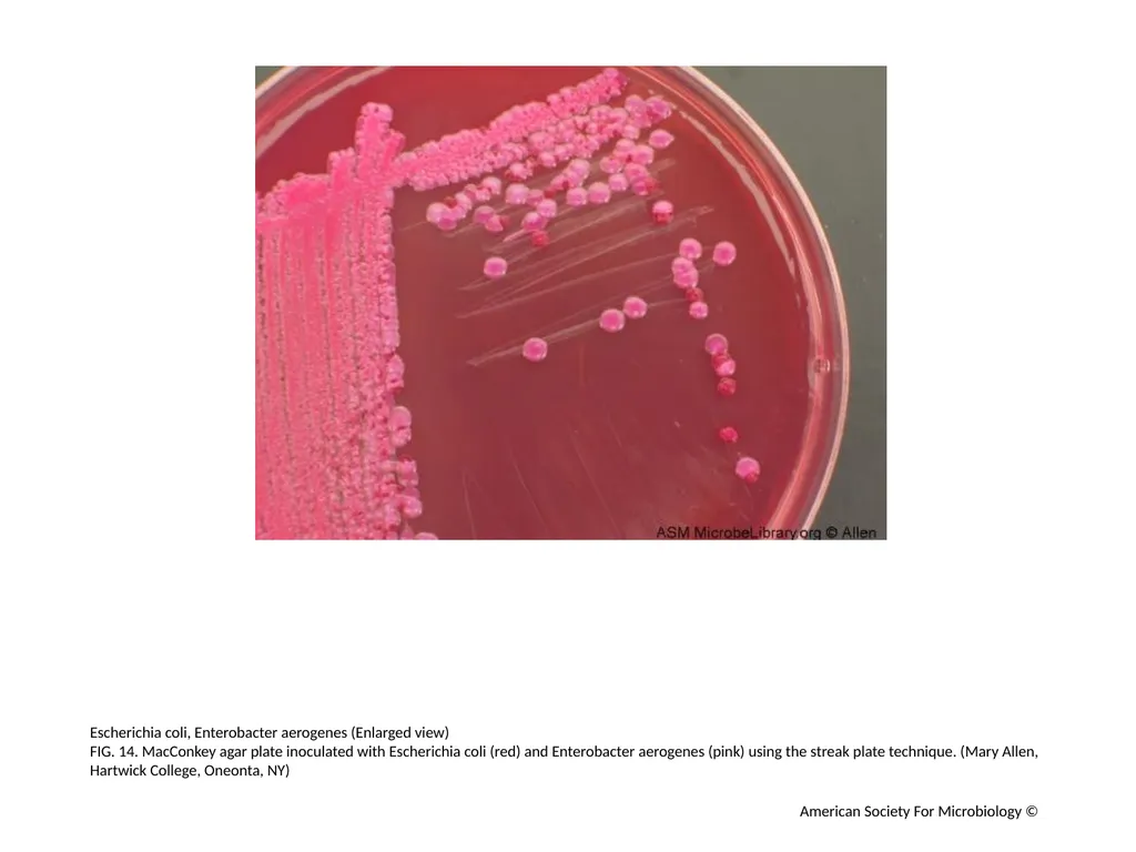
Escherichia coli, Enterobacter aerogenes (Enlarged view)
FIG. 14. MacConkey agar plate inoculated with Escherichia coli (red) and Enterobacter aerogenes (pink) using the streak plate technique. (Mary Allen, Hartwick College, Oneonta, NY) American Society For Microbiology ©<br>
FIG. 14. MacConkey agar plate inoculated with Escherichia coli (red) and Enterobacter aerogenes (pink) using the streak plate technique. (Mary Allen, Hartwick College, Oneonta, NY) American Society For Microbiology ©<br>
16
Escherichia coli, Klebsiella pneumoniae (Enlarged view)
FIG. 15. MacConkey agar plate inoculated with Escherichia coli (red) and Klebsiella pneumoniae (pink) using the streak plate technique. (Mary Allen, Hartwick College, Oneonta, NY) American Society For Microbiology ©<br>
FIG. 15. MacConkey agar plate inoculated with Escherichia coli (red) and Klebsiella pneumoniae (pink) using the streak plate technique. (Mary Allen, Hartwick College, Oneonta, NY) American Society For Microbiology ©<br>
17
Escherichia coli, Klebsiella pneumoniae (Enlarged view)
FIG. 16. MacConkey agar plate inoculated with Escherichia coli (red) and Klebsiella pneumoniae (pink) using the streak plate technique. (Mary Allen, Hartwick College, Oneonta, NY) American Society For Microbiology ©<br>
FIG. 16. MacConkey agar plate inoculated with Escherichia coli (red) and Klebsiella pneumoniae (pink) using the streak plate technique. (Mary Allen, Hartwick College, Oneonta, NY) American Society For Microbiology ©<br>
18
Burkholderia cepacian (Enlarged view)
FIG. 17. Burkholderia cepacia grows as tiny pinpoints on MacConkey agar in 24 hours at 37°C. (Rebecca Buxton, University of Utah, Salt Lake City, UT) American Society For Microbiology ©<br>
FIG. 17. Burkholderia cepacia grows as tiny pinpoints on MacConkey agar in 24 hours at 37°C. (Rebecca Buxton, University of Utah, Salt Lake City, UT) American Society For Microbiology ©<br>
19
Burkholderia cepacian (Enlarged view)
FIG. 18. Same plate as FIG. 17 at 48 hours, Burkholderia cepacia displays small non-lactose fermenting colonies. Some strains appear somewhat purple due to strong lactose oxidation. (Rebecca Buxton, University of Utah, Salt Lake City, UT) American Society For Microbiology ©<br>
FIG. 18. Same plate as FIG. 17 at 48 hours, Burkholderia cepacia displays small non-lactose fermenting colonies. Some strains appear somewhat purple due to strong lactose oxidation. (Rebecca Buxton, University of Utah, Salt Lake City, UT) American Society For Microbiology ©<br>
20
Stenotrophomonas maltophilia (Enlarged view)
FIG. 19. Stenotrophomonas maltophilia grows as tiny pinpoints on MacConkey agar in 24 hours at 37°C. (Rebecca Buxton, University of Utah, Salt Lake City, UT) American Society For Microbiology ©<br>
FIG. 19. Stenotrophomonas maltophilia grows as tiny pinpoints on MacConkey agar in 24 hours at 37°C. (Rebecca Buxton, University of Utah, Salt Lake City, UT) American Society For Microbiology ©<br>
21
Stenotrophomonas maltophilia (Enlarged view)
FIG. 20. Same plate as FIG. 19 at 48 hours, Stenotrophomonas has distinct non-lactose fermenting colonies. The indicator has turned an alkaline tan color. (Rebecca Buxton, University of Utah, Salt Lake City, UT) American Society For Microbiology ©<br>
FIG. 20. Same plate as FIG. 19 at 48 hours, Stenotrophomonas has distinct non-lactose fermenting colonies. The indicator has turned an alkaline tan color. (Rebecca Buxton, University of Utah, Salt Lake City, UT) American Society For Microbiology ©<br>
22
Pseudomonas aeruginosa (Enlarged view)
FIG. 21. Typical spreading non-lactose fermenting colonies of Pseudomonas aeruginosa. The heavy growth in the primary innoculum has begun to display a blue-green pigment. (Rebecca Buxton, University of Utah, Salt Lake City, UT) American Society For Microbiology ©<br>
FIG. 21. Typical spreading non-lactose fermenting colonies of Pseudomonas aeruginosa. The heavy growth in the primary innoculum has begun to display a blue-green pigment. (Rebecca Buxton, University of Utah, Salt Lake City, UT) American Society For Microbiology ©<br>
23
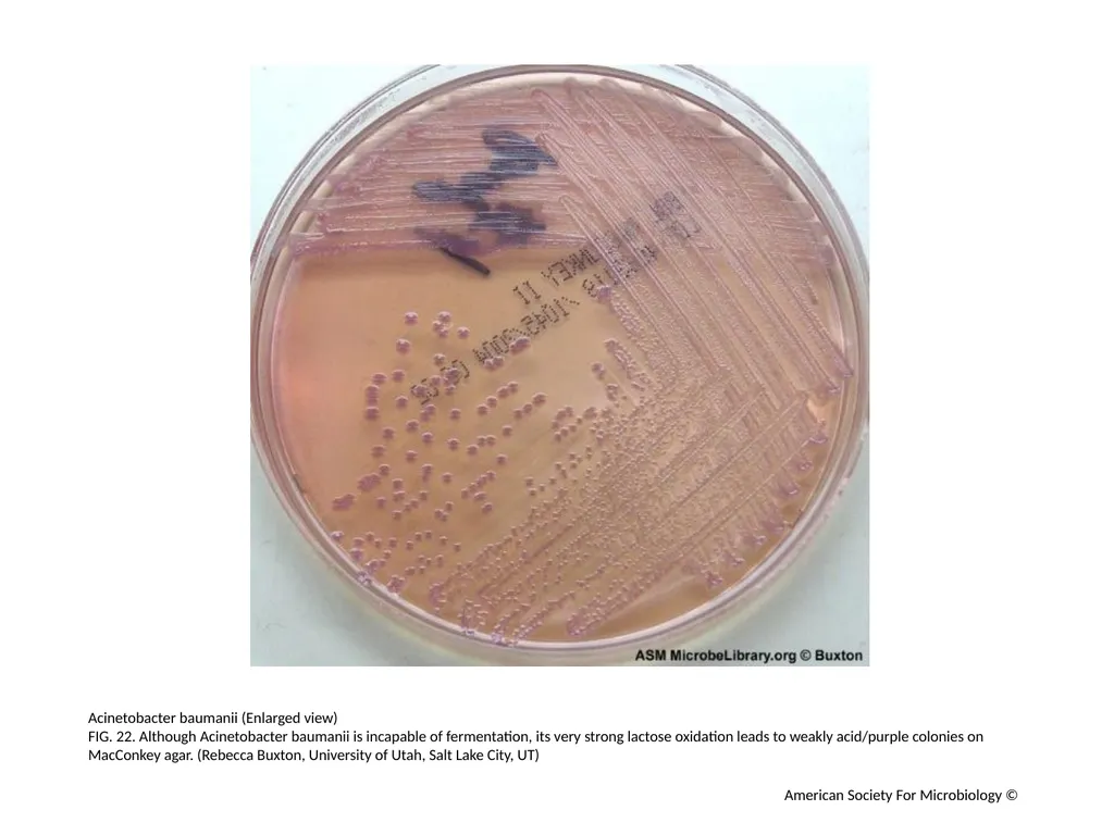
Acinetobacter baumanii (Enlarged view)
FIG. 22. Although Acinetobacter baumanii is incapable of fermentation, its very strong lactose oxidation leads to weakly acid/purple colonies on MacConkey agar. (Rebecca Buxton, University of Utah, Salt Lake City, UT) American Society For Microbiology ©<br>
FIG. 22. Although Acinetobacter baumanii is incapable of fermentation, its very strong lactose oxidation leads to weakly acid/purple colonies on MacConkey agar. (Rebecca Buxton, University of Utah, Salt Lake City, UT) American Society For Microbiology ©<br>
24
Pseudomonas aeruginosa (Enlarged view)
FIG. 23. An encapsulated strain of Pseudomonas aeruginosa recovered from a cystic fibrosis patient at 24 hours. (Rebecca Buxton, University of Utah, Salt Lake City, UT) American Society For Microbiology ©<br>
FIG. 23. An encapsulated strain of Pseudomonas aeruginosa recovered from a cystic fibrosis patient at 24 hours. (Rebecca Buxton, University of Utah, Salt Lake City, UT) American Society For Microbiology ©<br>
25
Pseudomonas aeruginosa (Enlarged view)
FIG. 24. Same plate as FIG. 23 at 48 hours, this strain of Pseudomonas aeruginosa make abundant, mucoid capsular material. (Rebecca Buxton, University of Utah, Salt Lake City, UT) American Society For Microbiology ©<br>
FIG. 24. Same plate as FIG. 23 at 48 hours, this strain of Pseudomonas aeruginosa make abundant, mucoid capsular material. (Rebecca Buxton, University of Utah, Salt Lake City, UT) American Society For Microbiology ©<br>
26
Fecal flora with Salmonella (Enlarged view)
FIG. 25. Normal fecal flora mixed with Salmonella. This unusual Salmonella strain ferments lactose. (The colonies were distinguished by producing abundant H2S on Hektoen Agar). (Rebecca Buxton, University of Utah, Salt Lake City, UT) American Society For Microbiology ©<br>
FIG. 25. Normal fecal flora mixed with Salmonella. This unusual Salmonella strain ferments lactose. (The colonies were distinguished by producing abundant H2S on Hektoen Agar). (Rebecca Buxton, University of Utah, Salt Lake City, UT) American Society For Microbiology ©<br>
27
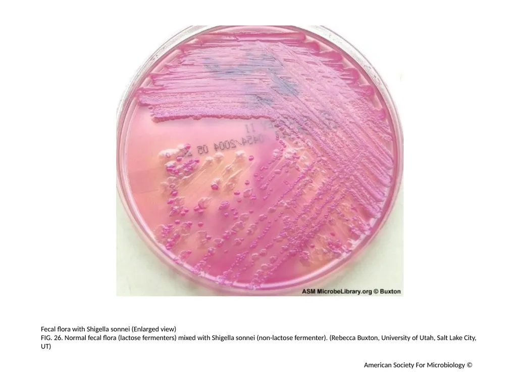
Fecal flora with Shigella sonnei (Enlarged view)
FIG. 26. Normal fecal flora (lactose fermenters) mixed with Shigella sonnei (non-lactose fermenter). (Rebecca Buxton, University of Utah, Salt Lake City, UT) American Society For Microbiology ©<br>
FIG. 26. Normal fecal flora (lactose fermenters) mixed with Shigella sonnei (non-lactose fermenter). (Rebecca Buxton, University of Utah, Salt Lake City, UT) American Society For Microbiology ©<br>
28
Escherichia coli (Enlarged view)
FIG. 27. This appears to be a pure culture of E.coli. (The patient was suffering from significant diarrhea due to Campylobacter jejuni, which doesn't grow on MacConkey agar). (Rebecca Buxton, University of Utah, Salt Lake City, UT) American Society For Microbiology ©<br>
FIG. 27. This appears to be a pure culture of E.coli. (The patient was suffering from significant diarrhea due to Campylobacter jejuni, which doesn't grow on MacConkey agar). (Rebecca Buxton, University of Utah, Salt Lake City, UT) American Society For Microbiology ©<br>
29
Vibrio species (Enlarged view)
FIG. 28. On very close observation, the tiny pinpoints of a Vibrio species are visible among the large, normal fecal lactose fermenters. On Thiosulfate Citrate Bile Sucrose (TCBS) agar, this sample grew yellow, sucrose-fermenting colonies of V. alginolyticus. (Rebecca Buxton, University of Utah, Salt Lake City, UT) American Society For Microbiology ©<br>
FIG. 28. On very close observation, the tiny pinpoints of a Vibrio species are visible among the large, normal fecal lactose fermenters. On Thiosulfate Citrate Bile Sucrose (TCBS) agar, this sample grew yellow, sucrose-fermenting colonies of V. alginolyticus. (Rebecca Buxton, University of Utah, Salt Lake City, UT) American Society For Microbiology ©<br>
30
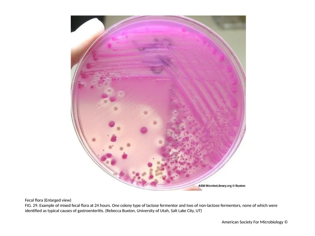
Fecal flora (Enlarged view)
FIG. 29. Example of mixed fecal flora at 24 hours. One colony type of lactose fermentor and two of non-lactose fermentors, none of which were identified as typical causes of gastroenteritis. (Rebecca Buxton, University of Utah, Salt Lake City, UT) American Society For Microbiology ©<br>
FIG. 29. Example of mixed fecal flora at 24 hours. One colony type of lactose fermentor and two of non-lactose fermentors, none of which were identified as typical causes of gastroenteritis. (Rebecca Buxton, University of Utah, Salt Lake City, UT) American Society For Microbiology ©<br>
31
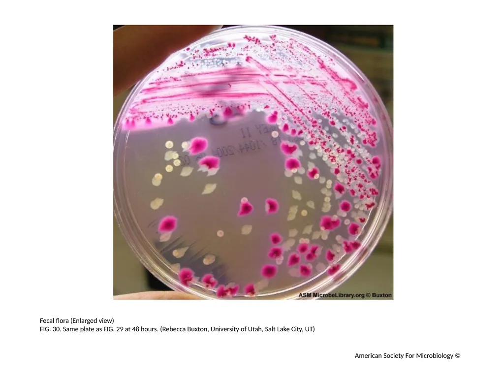
Fecal flora (Enlarged view)
FIG. 30. Same plate as FIG. 29 at 48 hours. (Rebecca Buxton, University of Utah, Salt Lake City, UT) American Society For Microbiology ©<br>
FIG. 30. Same plate as FIG. 29 at 48 hours. (Rebecca Buxton, University of Utah, Salt Lake City, UT) American Society For Microbiology ©<br>
32
Serratia marcescens (Enlarged view)
FIG. 31. Red-pigmented Serratia marcescens. Students often mistake the red pigment for lactose fermentation. (Rebecca Buxton, University of Utah, Salt Lake City, UT) American Society For Microbiology ©<br>
FIG. 31. Red-pigmented Serratia marcescens. Students often mistake the red pigment for lactose fermentation. (Rebecca Buxton, University of Utah, Salt Lake City, UT) American Society For Microbiology ©<br>
33
Klebsiella pneumoniae (Enlarged view)
FIG. 32. Klebsiella pneumoniae: Mucoid, lactose-fermenting colonies are typical of Klebsiella and Enterobacter species. (Rebecca Buxton, University of Utah, Salt Lake City, UT) American Society For Microbiology ©<br>
FIG. 32. Klebsiella pneumoniae: Mucoid, lactose-fermenting colonies are typical of Klebsiella and Enterobacter species. (Rebecca Buxton, University of Utah, Salt Lake City, UT) American Society For Microbiology ©<br>
34
Proteus vulgaris (Enlarged view)
FIG. 33. Proteus vulgaris: Non-lactose fermenters with slight swarming. (Rebecca Buxton, University of Utah, Salt Lake City, UT) American Society For Microbiology ©<br>
FIG. 33. Proteus vulgaris: Non-lactose fermenters with slight swarming. (Rebecca Buxton, University of Utah, Salt Lake City, UT) American Society For Microbiology ©<br>
35
Growth results of three gram-negative bacilli grown on MacConkey agar
FIG. 34.
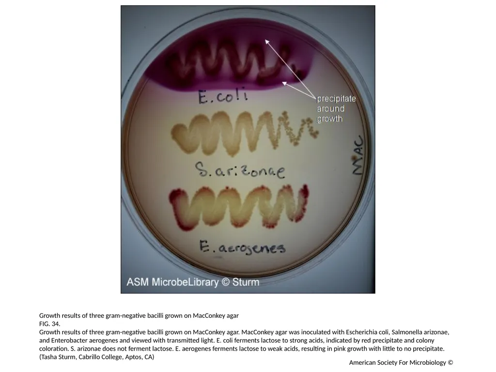
Growth results of three gram-negative bacilli grown on MacConkey agar. MacConkey agar was inoculated with Escherichia coli, Salmonella arizonae, and Enterobacter aerogenes and viewed with transmitted light. E. coli ferments lactose to strong acids, indicated by red precipitate and colony coloration. S. arizonae does not ferment lactose. E. aerogenes ferments lactose to weak acids, resulting in pink growth with little to no precipitate. (Tasha Sturm, Cabrillo College, Aptos, CA) American Society For Microbiology ©<br>
FIG. 34.
Growth results of three gram-negative bacilli grown on MacConkey agar. MacConkey agar was inoculated with Escherichia coli, Salmonella arizonae, and Enterobacter aerogenes and viewed with transmitted light. E. coli ferments lactose to strong acids, indicated by red precipitate and colony coloration. S. arizonae does not ferment lactose. E. aerogenes ferments lactose to weak acids, resulting in pink growth with little to no precipitate. (Tasha Sturm, Cabrillo College, Aptos, CA) American Society For Microbiology ©<br>
36
Organisms grown on MacConkey Agar
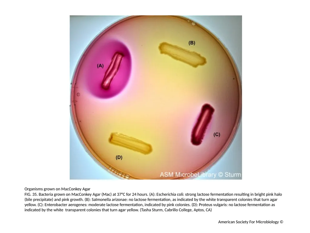
FIG. 35. Bacteria grown on MacConkey Agar (Mac) at 37°C for 24 hours. (A): Escherichia coli: strong lactose fermentation resulting in bright pink halo (bile precipitate) and pink growth. (B): Salmonella arizonae: no lactose fermentation, as indicated by the white transparent colonies that turn agar yellow. (C): Enterobacter aerogenes: moderate lactose fermentation, indicated by pink colonies. (D): Proteus vulgaris: no lactose fermentation as indicated by the white transparent colonies that turn agar yellow. (Tasha Sturm, Cabrillo College, Aptos, CA) American Society For Microbiology ©<br>
FIG. 35. Bacteria grown on MacConkey Agar (Mac) at 37°C for 24 hours. (A): Escherichia coli: strong lactose fermentation resulting in bright pink halo (bile precipitate) and pink growth. (B): Salmonella arizonae: no lactose fermentation, as indicated by the white transparent colonies that turn agar yellow. (C): Enterobacter aerogenes: moderate lactose fermentation, indicated by pink colonies. (D): Proteus vulgaris: no lactose fermentation as indicated by the white transparent colonies that turn agar yellow. (Tasha Sturm, Cabrillo College, Aptos, CA) American Society For Microbiology ©<br>