
Main pathogenic bacteria of Spirochetaceae
LS
Published · 14 slides · 0 views

1 / 1


Description
Main pathogenic bacteria of Spirochetaceae Classification of Human Terponema Non-pathogenic T.denticola T.macrodenticum T.orale Conditionally pathogenic T.vincentii Pathogenic T.pallidum pallidum (syphilis) T.pallidum endemicum (bejel)
Related Topics
Share
Embed code
Download this presentation From Below
"Main pathogenic bacteria of Spirochetaceae" is the property of its rightful owner. Permission is granted to download and print the materials on this website for personal, non-commercial use only, and to display it on your personal computer provided you do not modify the materials and that you retain all copyright notices contained in the materials. By downloading content from our website, you accept the terms of this agreement.
Presentation Transcript
01
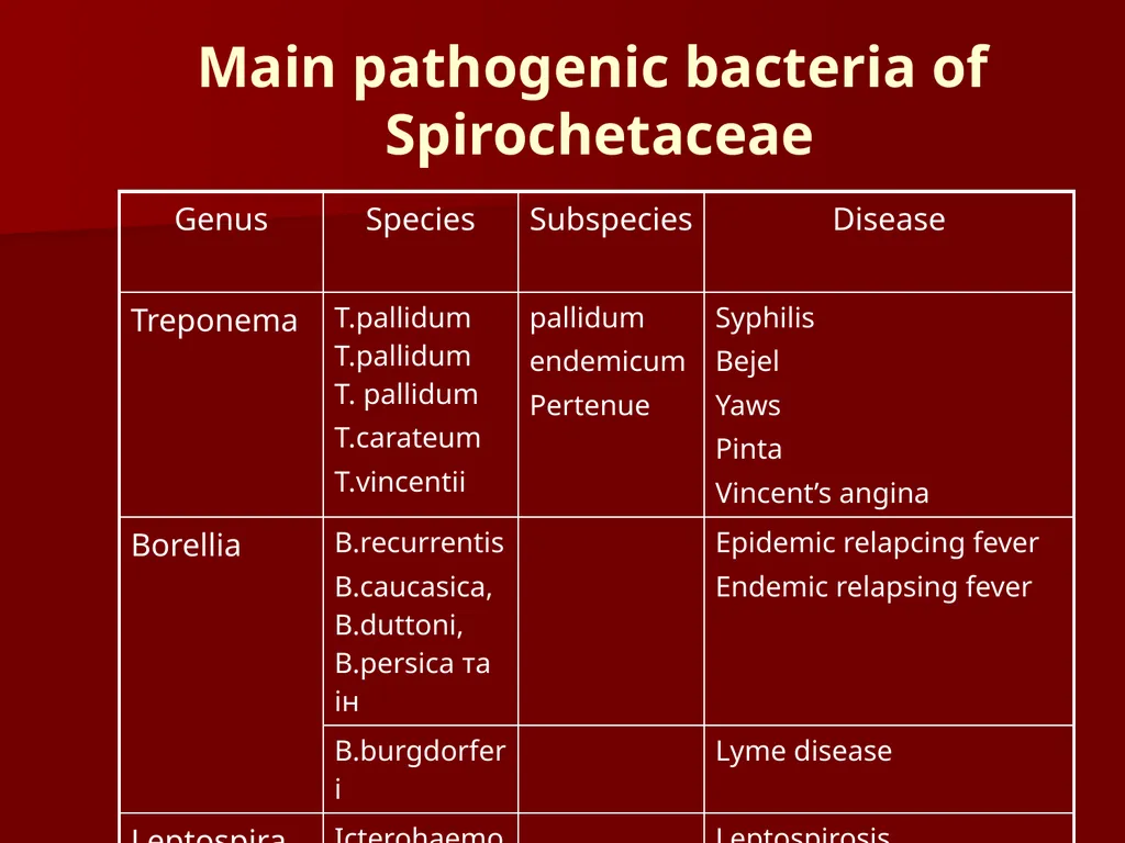
Main pathogenic bacteria of Spirochetaceae<br>
02
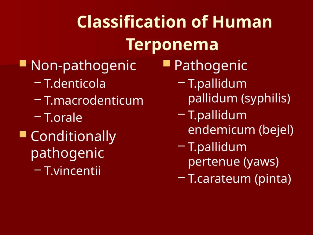
Classification of Human Terponema Non-pathogenic
T.denticola
T.macrodenticum
T.orale
Conditionally pathogenic
T.vincentii Pathogenic
T.pallidum pallidum (syphilis)
T.pallidum endemicum (bejel)
T.pallidum pertenue (yaws)
T.carateum (pinta)<br>
T.denticola
T.macrodenticum
T.orale
Conditionally pathogenic
T.vincentii Pathogenic
T.pallidum pallidum (syphilis)
T.pallidum endemicum (bejel)
T.pallidum pertenue (yaws)
T.carateum (pinta)<br>
03
-Spirochetes
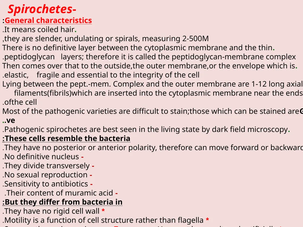
General characteristics:
.It means coiled hair.
they are slender, undulating or spirals, measuring 2-500M,
.There is no definitive layer between the cytoplasmic membrane and the thin peptidoglycan layers; therefore it is called the peptidoglycan-membrane complex.
.Then comes over that to the outside,the outer membrane,or the envelope which is elastic, fragile and essential to the integrity of the cell.
. Lying between the pept.-mem. Complex and the outer membrane are 1-12 long axial filaments(fibrils)which are inserted into the cytoplasmic membrane near the ends ofthe cell.
.Most of the pathogenic varieties are difficult to stain;those which can be stained areG –ve..
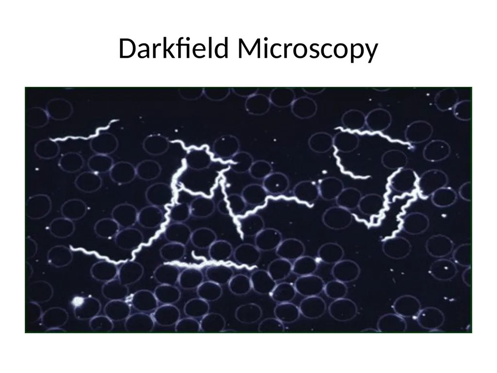
.Pathogenic spirochetes are best seen in the living state by dark field microscopy.
These cells resemble the bacteria:
- They have no posterior or anterior polarity, therefore can move forward or backward.
- No definitive nucleus.
- They divide transversely.
- No sexual reproduction.
- Sensitivity to antibiotics.
- Their content of muramic acid.
But they differ from bacteria in:
* They have no rigid cell wall.
* Motility is a function of cell structure rather than flagella.
* Some pathogenic species, e.g. Treponema. Have not been cultured artificially.<br>
04
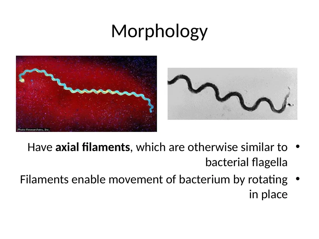
Morphology Have axial filaments, which are otherwise similar to bacterial flagella
Filaments enable movement of bacterium by rotating in place<br>
Filaments enable movement of bacterium by rotating in place<br>
05
Darkfield Microscopy<br>
06
Classification:
The order spirochetales has been divided into two families:
Spirochetaceae, include 3 non pathogenic free living genera:
1- Spirocheta.
2- Saprospira.
3- Cristispira .
Treponemataceae which include3genera,each contains few specie pathogenic for human Treponema.
Borrelia .
Leptospira . Treponema
Include 3 species:
1- Treponema palladium, the causative agent of syphilis.
2- Trep. Pertenue, the causative agent of Yaws.
3- Trep. Carateum, the causative agents of Pinta.
Treponema pallidum:
* Very delicate thin organism, measuring 6-14 X 0.15-0.2M.
* Cannot be stained by Gram stain, but by Giemsa solution 1:10.
* Very sensitive to drying, O2, soap, mercuric ointment, water and to several common bacterial agents.
* Very sensitive to temperature, killed at 390C for 5 hrs. At 410C for 2 hrs. And at 41.50C for 1hr. fever induction by malaria was used to cure the disease.
* The antigenic structure, studies revealed the presence of proteins, polysaccharide.And two different lipids, some are group specific, others are species specific<br>
07
Pathogenicity:
-The organism is transmitted from spirochete- containing lesions of the skin and mucous membranes,such as: genitalia,mouth and rectum of infected person to others by intimate contact.Also from syphilitic pregnant woman to fetus,and rarely through blood transfusion.
-The organism penetrates the mucous membranes and multiplies at the site of infection and in to 2-10 weeks leads to the formation of the primary syphilis, characterized by the formation of primary inflammatory lesion known as chancre, which is a superficial ulcer with clean firm base.
- Chancre heals spontaneously, and the organisms invade the regional lymph nodes forming satellite buboes, and eventually reach the blood stream where they establish systemic infections.
- 2-12 weeks after the appearance of the primary lesion, a secondary syphilis appears characterized by a generalized skin rash appearance (mainly on the palm and soles), and also moist lesions appear on the genitalia where it is called condylomata lata , which are rich in organisms.
- Lesions may develop also in bones, liver, kidney, CNS, mouth, and other organs.
- Primary and secondary syphilis are rich in organisms, and are very infectious.<br>
08
*** Syphilitic infection may remain subclinical, and the patient may pass through the primary or the secondary(or both)without symptoms or signs and develop tertiary lesions. 1/3 of these primary and secondary syphilis will heal spontaneously that even serologic test for syphilis appears negative.
1/3 remain latent, Early latent and Late latent.
Early latent: can last for 1 or 2years after the secondary stage, then the symptoms of the secondary syphilis reappear and are infectious.
Late latent: last for many years with no symptoms, only antibodies could be detected in serum.
1/3 of the untreated patients develop tertiary syphilis,several years after the appearance of the primary syphilis. This is characterized by the development of granulomatous lesions (gummas) in the skin, bones, liver, and degenerative changes in CNS (paresis, tabes), syphilitic cardiovascular lesions, particularly aortitis and aortic valve insufficiency. In this stage the organisms are very rare.
Congenital syphilis: a pregnant syphilitic woman can pass the Treponema to the fetus during, about, the 10th week of gestation, which leads to miscarriage or death of the fetus or born dead. Others are born alive but develop the signs of congenital syphilis in childhood, such as: interstitial keratitis, Hutchinson’s teeth, saddle nose, Periostitis and a variety of CNS anomalies.
Experimental disease: rabbits can be experimentally infected, with the human treponemes, in the skin, eyes, and testes. The animal develops chancre rich in spirochetes and organisms persist in lymph nodes, spleen and bone marrow.<br>
09
Laboratory diagnosis:
I- Microscopic examination:
samples taken from primary or secondary lesions are examined only by dark field microscopy or by direct florescent antibody.
II- Serologic tests (serological tests for syphilis STS):
A- Non-treponemal antigen tests:(these are non specific serological tests)the antigen used is the cardiolipin (diphosphotidyl glycerol) purified from alcoholic extract of beef heart with lecithin and cholesterol as sensitizers to react with regain, which is a mixture of IgM & IgG antibodies.It is found in patient’s serum after2-3 weeks of infection,and in spinal fluid after 4-8weeks of infection. Two tests are performed:
1.VDRL“venereal disease research laboratories”which is rapid slide flocculation test used for screening.
2.Wasserman test: which is a confirmatory complement fixation test.
These tests are positive in most cases of primary and secondary syphilisThe titer of these non specific antibodies decreases with effective treatment; in contrast to the specific antibodies which are positive for life. False positive may be shown in varies diseases such as, malaria, measles, infectious mononucleosis, L.E. , R.A. , leprosy, hepatitis B and some microbial infections.
B-Treponemal antigen tests (these are more specific test):
1- Florescent treponemal antibody-absorbed test (FTA-ABS):
Killed T.pallidum + patient’s serum + labeled antihuman Gamma globulin.<br>
10
2-TP.CFT:
Spirochetes extracted from syphilomas of rabbits from specific antigens for CT test.
3-TP passive heamagglutination test “TPHA” :
Formalized tanned sheep RBCs are conjugated with the antigens of a lysate of TP. When anti- treponemal Ab. Are added, clumping occur.
4-TP immunization test “TPI” :
Treponemes obtained from rabbit’s syphilitic lesions are mixed with patient’s serum and fresh guinea pig serum and incubated for 18 hrs.At 350C. Immobilization of 50%of the organisms is considered +ve
Treatment:
Penicillin is effective in all stages of syphilis. No resistance to penicillin has been observed.<br>
11
Borrelia
* The causative agent of relapsing fever.
* They are large, motile, retractile, 10-30M long X 0.3-0.7M wide, possess 6 or more fibrils
* Easy stained Gram –ve.
* Some cause diseases, other are normal commensals on mucous membranes.
* Some species can cause relapsing fever, such as:
1-Borrelia recurrentis :
European relapsing fever, transmitted from person to person by the body louse pediculus humans (variant corpora’s).
2-Borrelia duttoni, Borrelia hermsii :
African relapsing fever transmitted by the ticks Ornithodorus moubata between animals and animals to human.
The animal reservoirs are the rodents, where the organisms are passed trasovarially.<br>
12
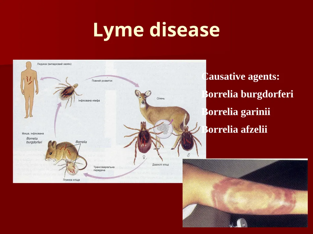
Lyme disease Causative agents:
Borrelia burgdorferi
Borrelia garinii
Borrelia afzelii<br>
Borrelia burgdorferi
Borrelia garinii
Borrelia afzelii<br>
13
Leptospira
* Leptospira interrogans is the causative agent of leptospirosis.
Leptospira are tightly coiled, 6-20 X 0.1 M
* It infects the kidneys of several types of animals, such as: rodents, dogs, pigs, goats, cattle, frogs, tortoises…etc. where they excrete the organisms with urine, which contaminates water and soil.
* Human infection results when organisms are ingested or pass through the skin or mucous membranes.
* The organism may infect several organs such as: liver |(jaundice: weil’s disease), kidneys ( uremia), lung (hemorrhage) ,and CNS (aseptic meningitis).
Diagnosis :
Depends on history of exposure,clinical signs, and marked rise in IgM antibodies
*It is rarely isolated from blood or urine.<br>
14
THANKS<br>