Maintaining the partogram Sri Lanka College of Obstetricians Gynaecologists Scientific Activities Research Committee Partogram: Is a graphic representation of the events of labour All the observations made on the mother and the fetus
"Maintaining the partogram Sri Lanka College of" is the property of its rightful owner. Permission is granted to
download and print the materials on this website for personal, non-commercial use only, and to display it
on your personal computer provided you do not modify the materials and that you retain all copyright
notices contained in the materials. By downloading content from our website, you accept the terms of this
agreement.
Presentation Transcript
01
Maintaining the partogram Sri Lanka College of Obstetricians & Gynaecologists
Scientific Activities & Research Committee<br>
02
Partogram: Is a graphic representation of the events of labour
All the observations made on the mother and the fetus are plotted in one sheet
Helps in early recognition of abnormal labour and fetal distress.
Now let us look at the partogram in detail.<br>
03
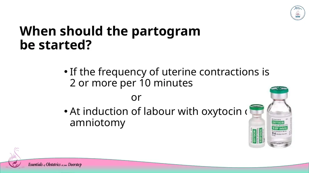
When should the partogram be started? If the frequency of uterine contractions is 2 or more per 10 minutes
or
At induction of labour with oxytocin or by amniotomy<br>
04
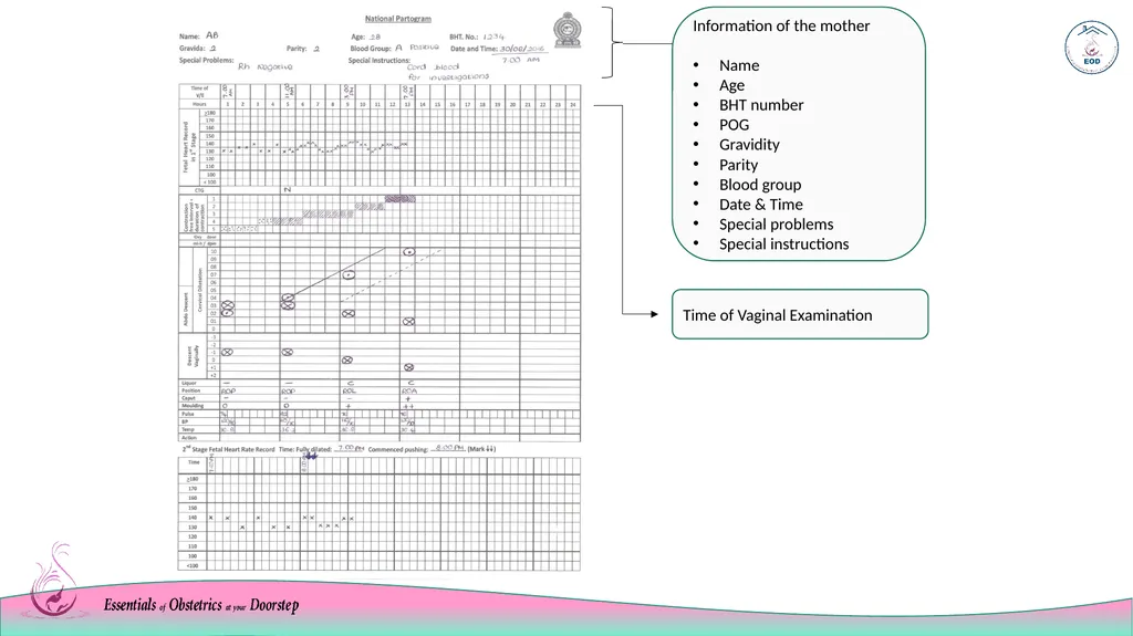
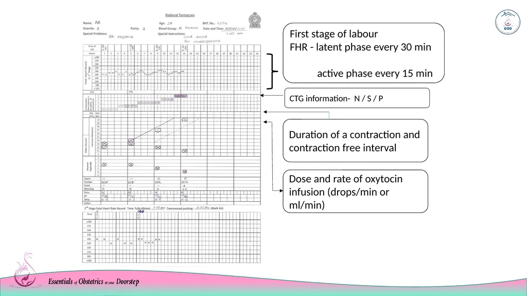
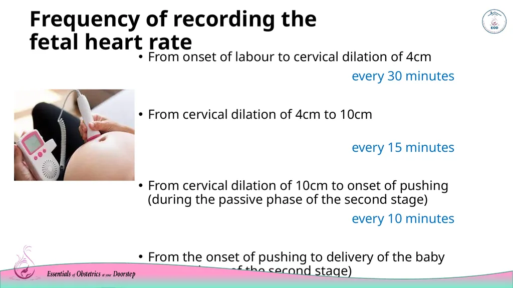
Frequency of recording the fetal heart rate From onset of labour to cervical dilation of 4cm
every 30 minutes
From cervical dilation of 4cm to 10cm
every 15 minutes
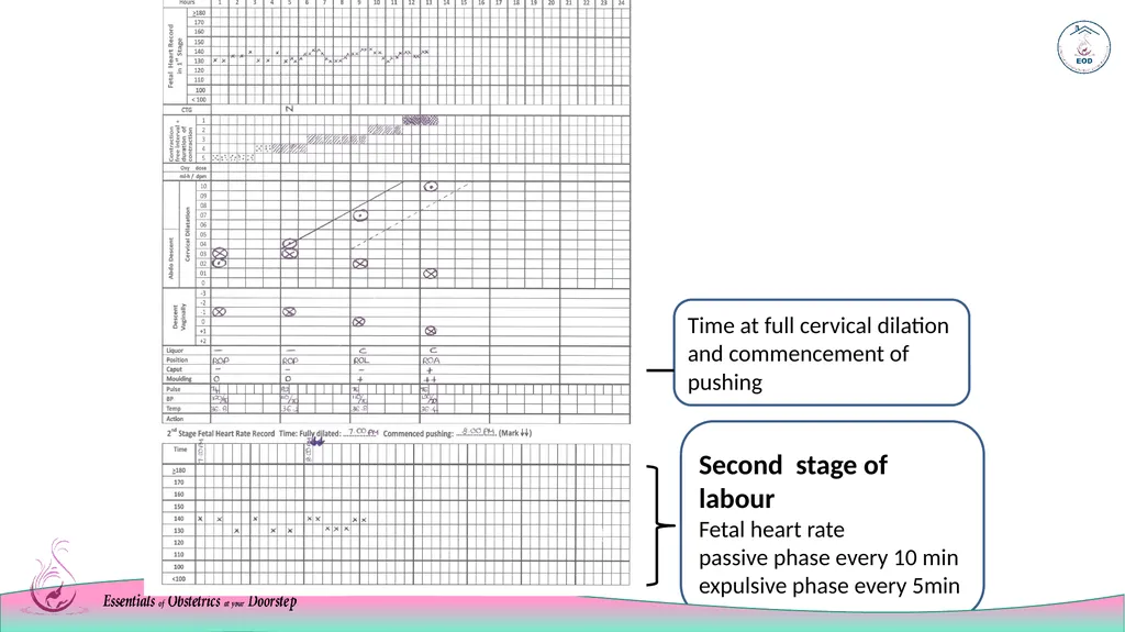
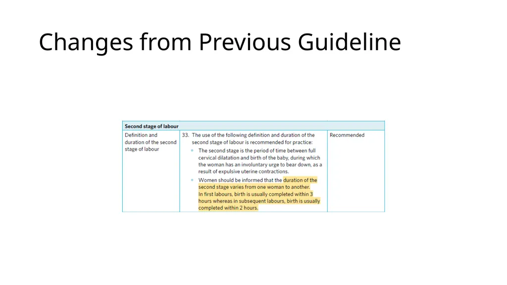
From cervical dilation of 10cm to onset of pushing (during the passive phase of the second stage)
every 10 minutes
From the onset of pushing to delivery of the baby (active phase of the second stage)
every 5 minutes<br>
05
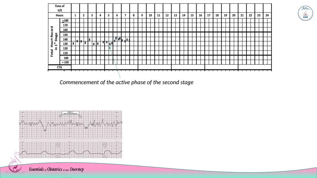
example of a FHR recording If a CTG is carried out, findings to be documented as; N (normal) S (suspicious) P (pathological) Commencement of the active phase of the second stage<br>
06
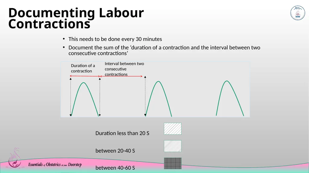
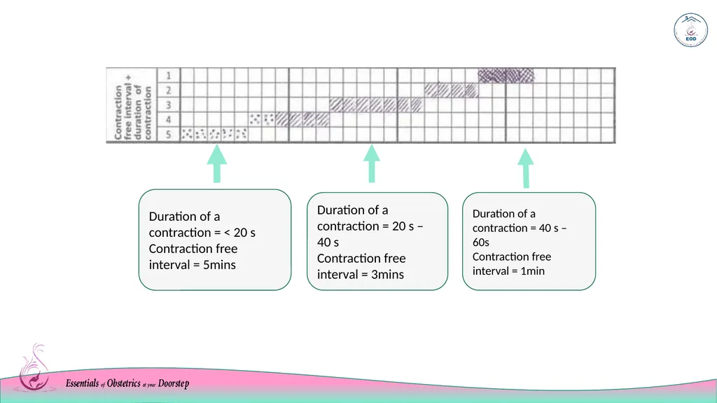
Documenting Labour Contractions This needs to be done every 30 minutes
Document the sum of the ‘duration of a contraction and the interval between two consecutive contractions’
<
Duration less than 20 S
between 20-40 S
between 40-60 S Duration of a contraction Interval between two consecutive contractions<br>
07
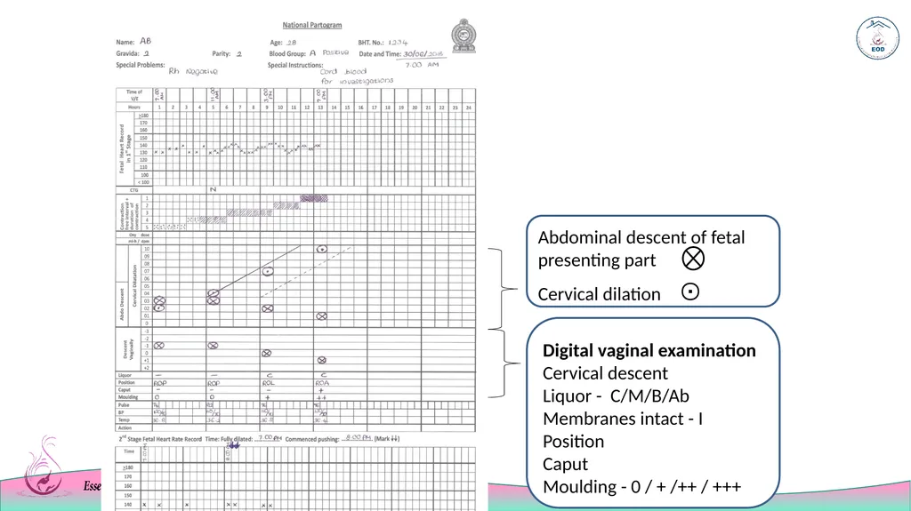
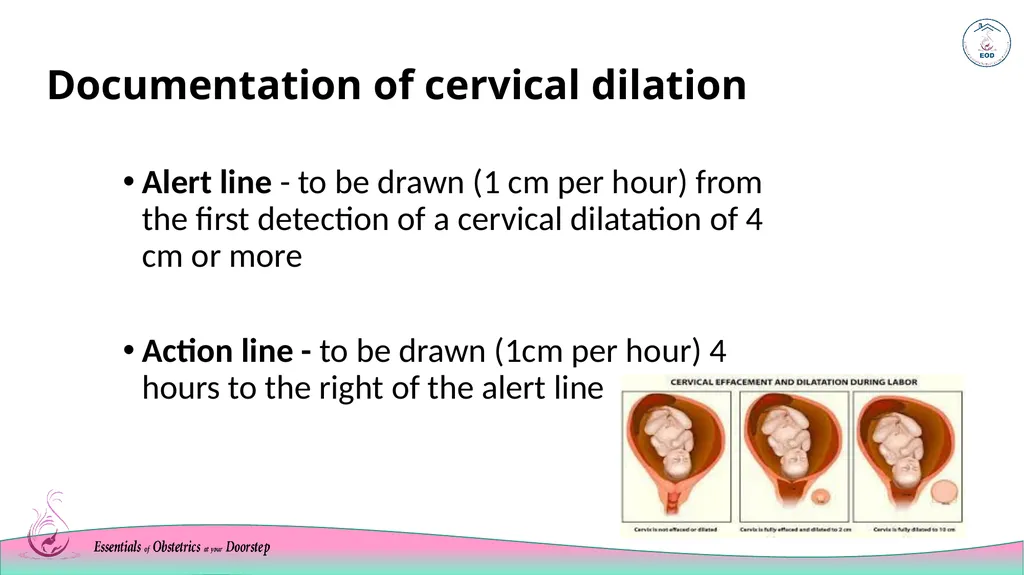
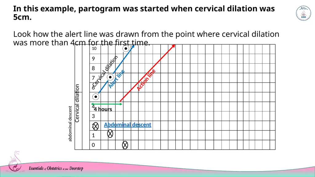
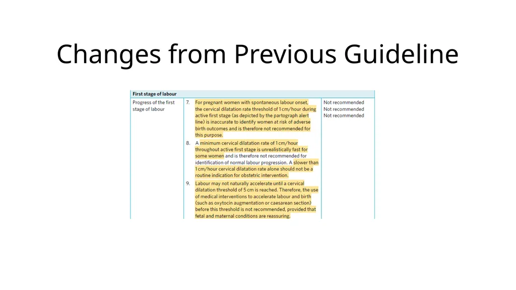
Documentation of cervical dilation Alert line - to be drawn (1 cm per hour) from the first detection of a cervical dilatation of 4 cm or more
Action line - to be drawn (1cm per hour) 4 hours to the right of the alert line<br>
08
Cervical dilation abdominal descent In this example, partogram was started when cervical dilation was 5cm. Look how the alert line was drawn from the point where cervical dilation was more than 4cm for the first time.<br>
09
Descent of the fetal head on abdominal palpation<br>
10
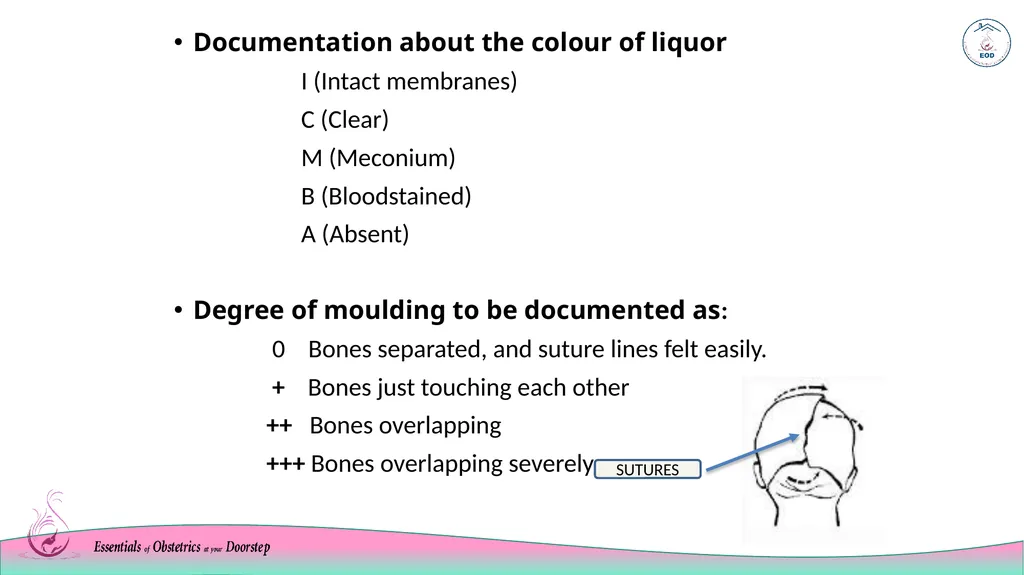
Documentation about the colour of liquor
I (Intact membranes)
C (Clear)
M (Meconium)
B (Bloodstained)
A (Absent)
Degree of moulding to be documented as:
0 Bones separated, and suture lines felt easily.
+ Bones just touching each other
++ Bones overlapping
+++ Bones overlapping severely suTURES<br>
11
Maintaining the partogram during the second stage of labour<br>
12
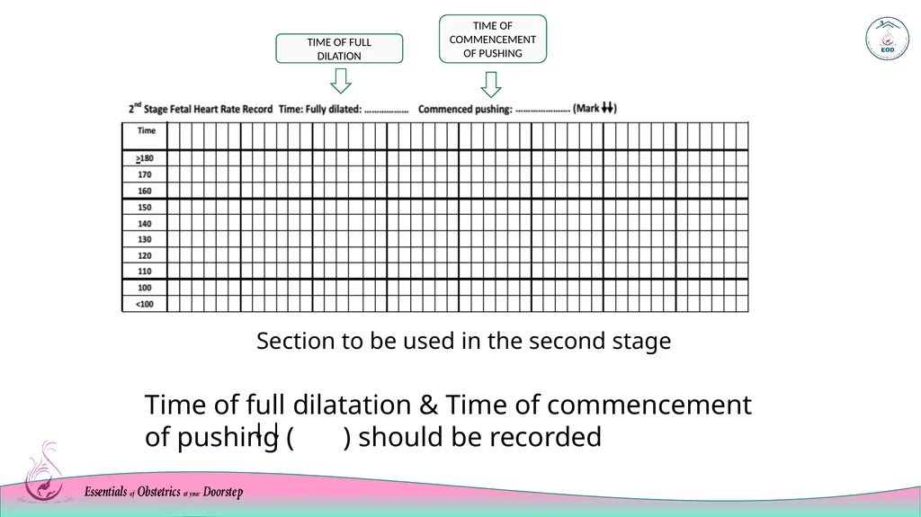
Section to be used in the second stage Time of full dilatation & Time of commencement of pushing ( ) should be recorded<br>
13
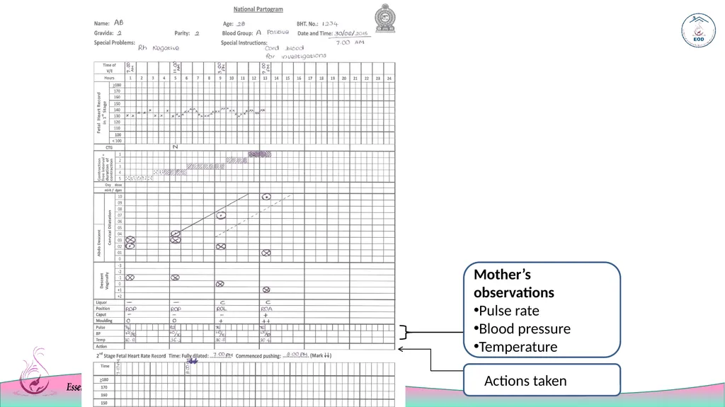
During the intrapartum period (during both the first and second stages) document the observations of the mother as follows ,
Pulse rate – every 30 minutes
Blood pressure and temperature – every 4hrs The second stage of labour<br>
14
Monitoring the mother during the third stage of labour<br>
15
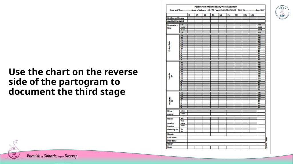
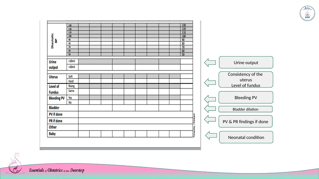
Use the chart on the reverse side of the partogram to document the third stage<br>
16
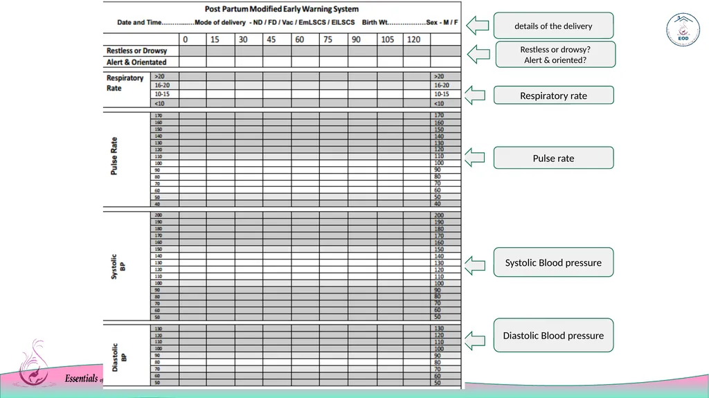
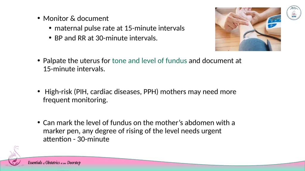
Monitor & document
maternal pulse rate at 15-minute intervals
BP and RR at 30-minute intervals.
Palpate the uterus for tone and level of fundus and document at 15-minute intervals.
High-risk (PIH, cardiac diseases, PPH) mothers may need more frequent monitoring.
Can mark the level of fundus on the mother’s abdomen with a marker pen, any degree of rising of the level needs urgent attention - 30-minute<br>
17
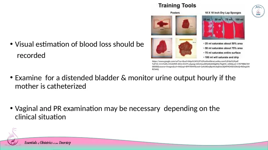
Visual estimation of blood loss should be
recorded
Examine for a distended bladder & monitor urine output hourly if the mother is catheterized
Vaginal and PR examination may be necessary depending on the clinical situation https://www.google.com/url?sa=i&url=https%3A%2F%2Fonlinelibrary.wiley.com%2Fdoi%2Fpdf%2F10.1111%2Fj.15526909.2012.01375.x&psig=AOvVaw2ENxWaNWgKNn7kigM4_U0O&ust=1707988676900000&source=images&cd=vfe&opi=89978449&ved=2ahUKEwj8pJrkv6qEAxU6jWMGHSD1D6sQr4kDegUIARCNAQ<br>
18
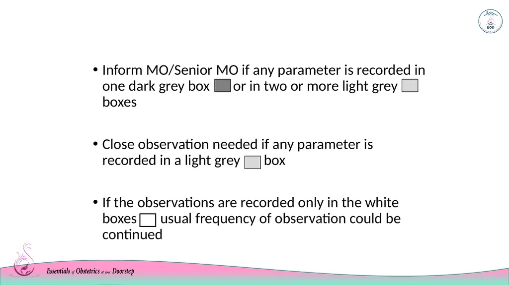
Inform MO/Senior MO if any parameter is recorded in one dark grey box or in two or more light grey boxes
Close observation needed if any parameter is recorded in a light grey box
If the observations are recorded only in the white boxes usual frequency of observation could be continued<br>
19
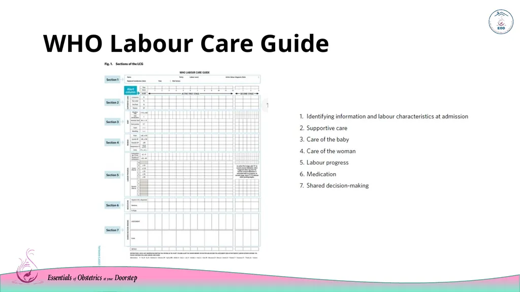
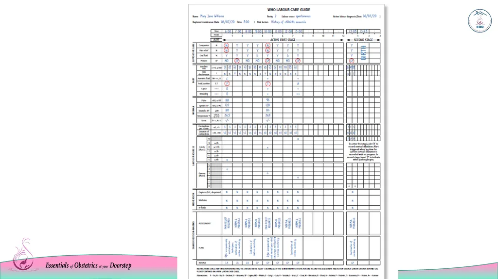
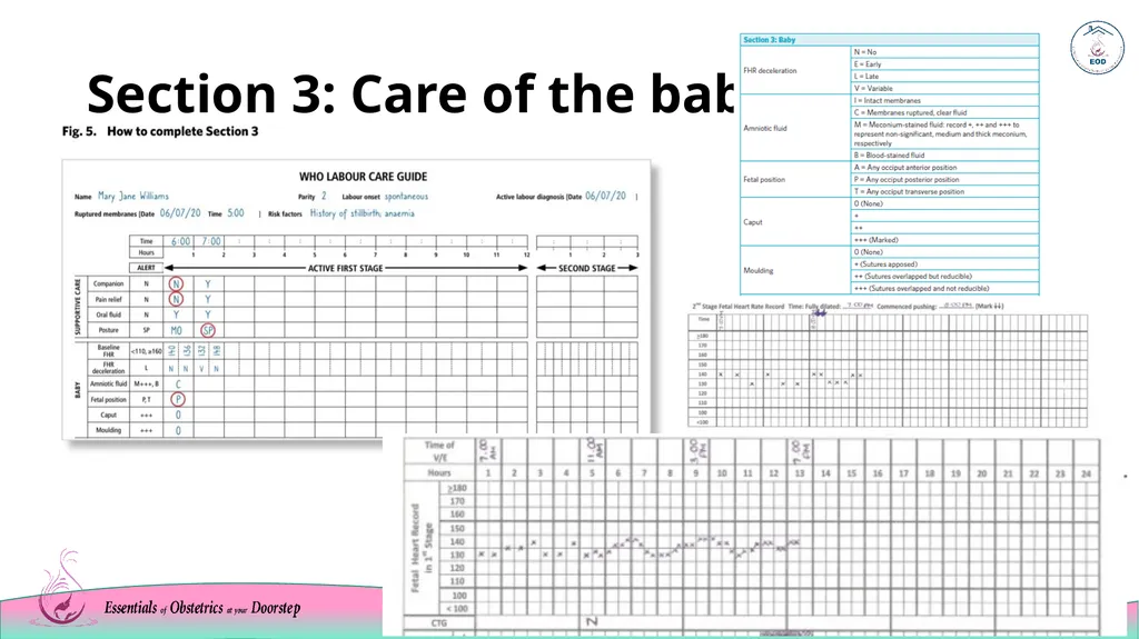
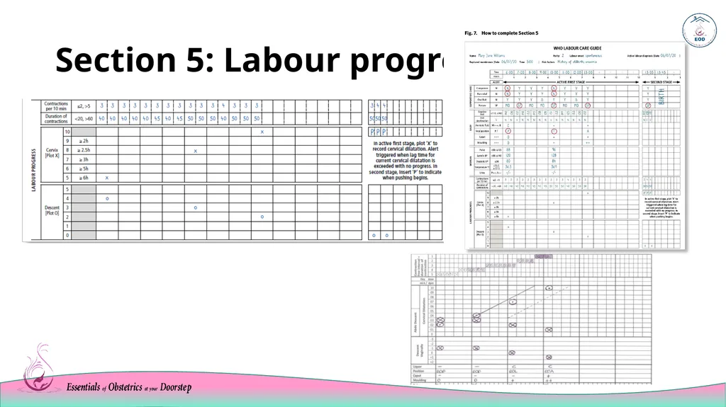
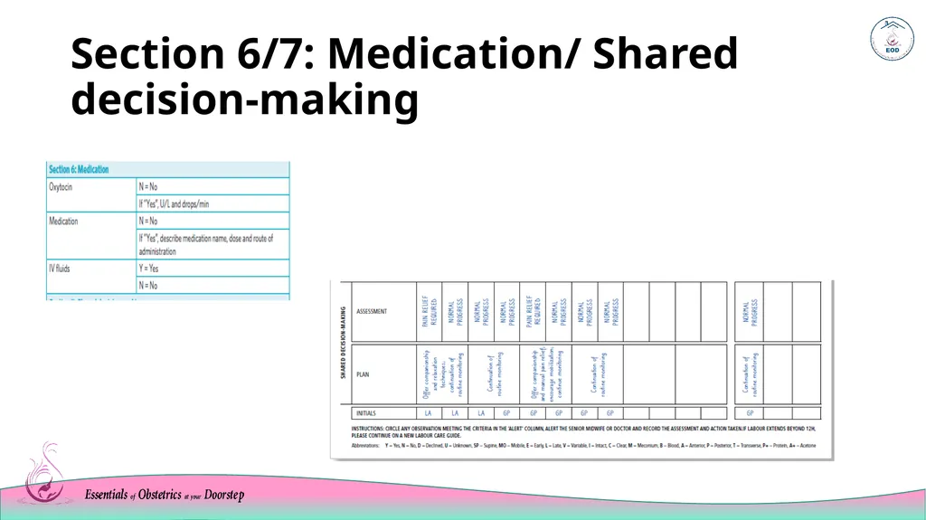
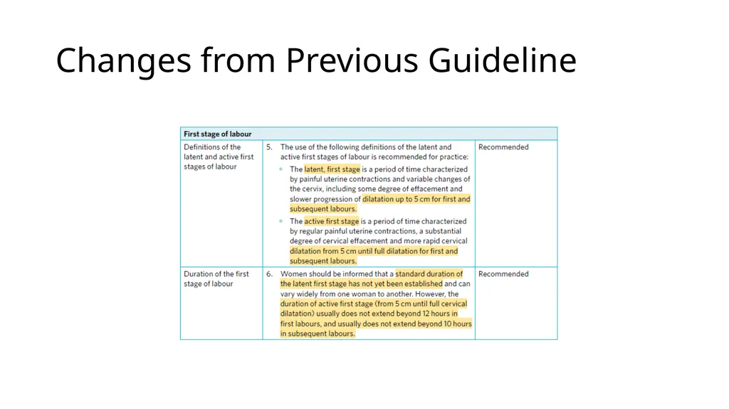
WHO Labour Care Guide<br>
20
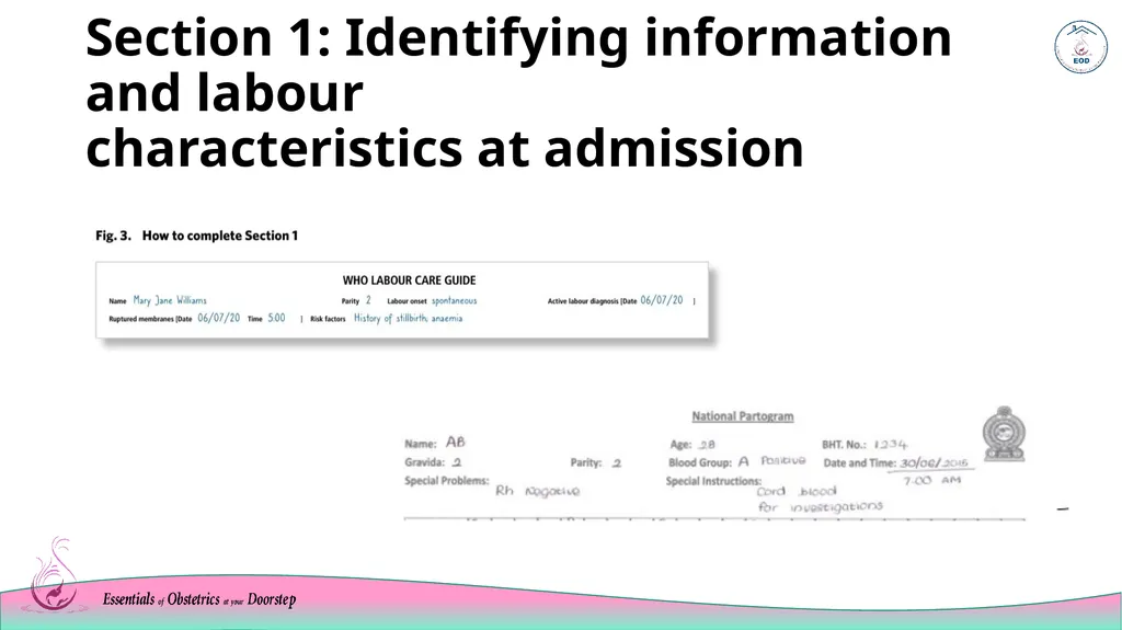
Section 1: Identifying information and labour characteristics at admission<br>