
Medicare Preventive Services for Bone Mass




Description: Medicare Preventive Services for Bone Mass Measurement May 8, 2024 Disclaimer We prepared this education as a tool to assist the provider community. Medicare rules change often. They are in the relevant laws, regulations and rulings on the
Related Topics
Download Presentation
"Medicare Preventive Services for Bone Mass" is the property of its rightful owner. Permission is granted to download and print the materials on this website for personal, non-commercial use only, and to display it on your personal computer provided you do not modify the materials and that you retain all copyright notices contained in the materials. By downloading content from our website, you accept the terms of this agreement.
Presentation Transcript
slide1. Medicare Preventive Services for Bone Mass Measurement May 8, 2024<br>
slide2. Disclaimer We prepared this education as a tool to assist the provider community. Medicare rules change often. They are in the relevant laws, regulations and rulings on the Centers for Medicare & Medicaid Services (CMS) website.
We will provide responses to questions based on the facts given, but the Medicare rules will determine final coverage.
CMS prohibits recording of the presentation for profit-making purposes.<br>
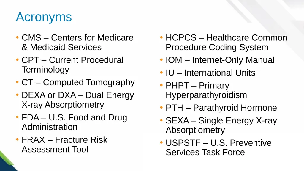
slide3. Acronyms CMS – Centers for Medicare & Medicaid Services
CPT – Current Procedural Terminology
CT – Computed Tomography
DEXA or DXA – Dual Energy X-ray Absorptiometry
FDA – U.S. Food and Drug Administration
FRAX – Fracture Risk Assessment Tool HCPCS – Healthcare Common Procedure Coding System
IOM – Internet-Only Manual
IU – International Units
PHPT – Primary Hyperparathyroidism
PTH – Parathyroid Hormone
SEXA – Single Energy X-ray Absorptiometry
USPSTF – U.S. Preventive Services Task Force<br>
slide4. Agenda This webinar will cover
Background information about osteoporosis and primary hyperparathyroidism
Bone density tests
Treatment options and preventive strategies
Medicare Part B coverage criteria for Bone Mass Measurement<br>
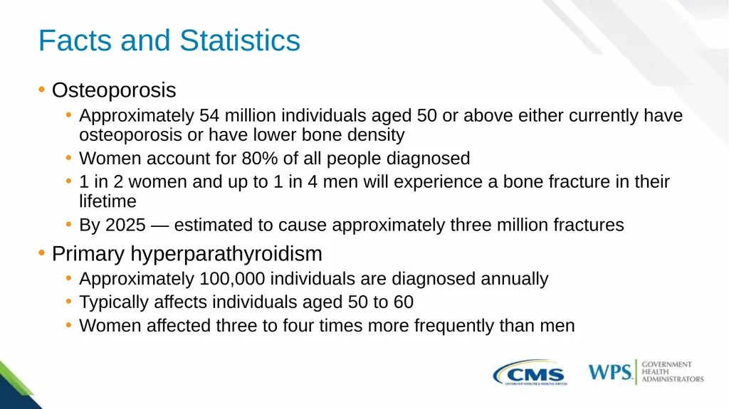
slide5. Facts and Statistics Osteoporosis
Approximately 54 million individuals aged 50 or above either currently have osteoporosis or have lower bone density
Women account for 80% of all people diagnosed
1 in 2 women and up to 1 in 4 men will experience a bone fracture in their lifetime
By 2025 — estimated to cause approximately three million fractures
Primary hyperparathyroidism
Approximately 100,000 individuals are diagnosed annually
Typically affects individuals aged 50 to 60
Women affected three to four times more frequently than men<br>
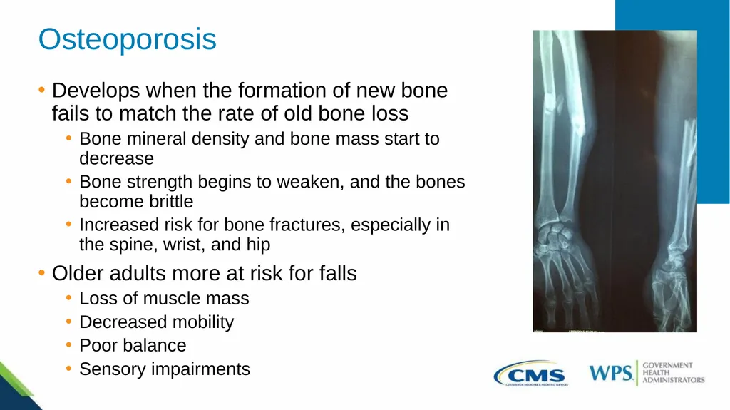
slide6. Osteoporosis Develops when the formation of new bone fails to match the rate of old bone loss
Bone mineral density and bone mass start to decrease
Bone strength begins to weaken, and the bones become brittle
Increased risk for bone fractures, especially in the spine, wrist, and hip
Older adults more at risk for falls
Loss of muscle mass
Decreased mobility
Poor balance
Sensory impairments<br>
slide7. Nonmodifiable Risk Factors Sex
Women are at greater risk
Age
Likelihood increases as you age
Race
Higher in non-Hispanic White and Asian women and men
Body size
People with smaller body frames more at risk
Family history
Having a parent with a history of osteoporosis or hip fracture<br>
slide8. Modifiable Risk Factors Low body weight
Changes in hormone levels
Certain pre-existing conditions
Taking certain medications
Lack of physical activity and exercise
Diet
Alcohol use
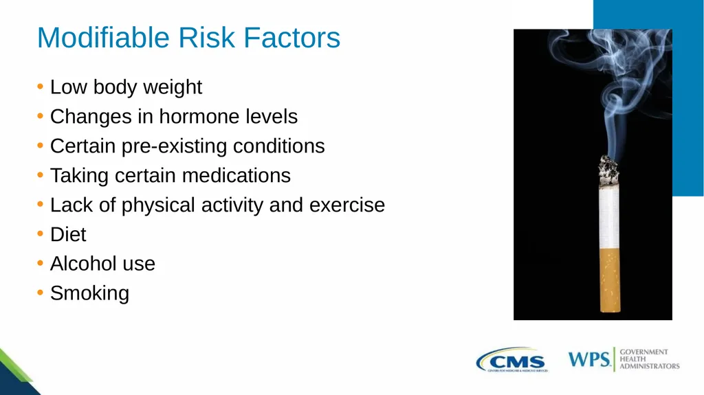
Smoking<br>
slide9. Signs, Symptoms, and Complications Signs and Symptoms
Asymptomatic - “silent” disease
Fractures during everyday activities
If signs and symptoms are present, include
Back pain from broken or collapsed vertebrae
Kyphosis
Gradual loss of height
Fractures from activities that would normally not cause a break in a healthy bone
Complications
Fractures
Femur
Vertebrae<br>
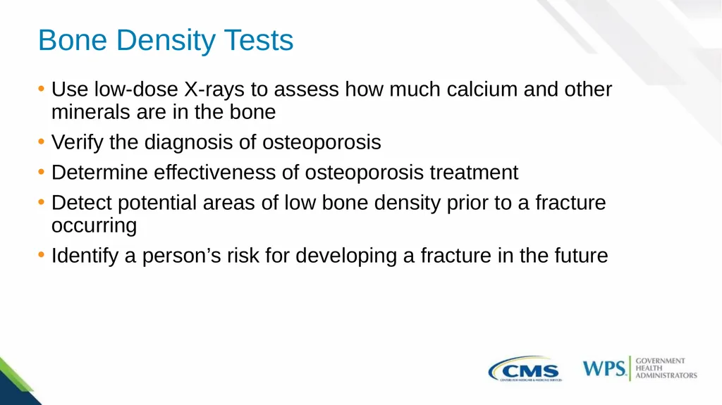
slide10. Bone Density Tests Use low-dose X-rays to assess how much calcium and other minerals are in the bone
Verify the diagnosis of osteoporosis
Determine effectiveness of osteoporosis treatment
Detect potential areas of low bone density prior to a fracture occurring
Identify a person’s risk for developing a fracture in the future<br>
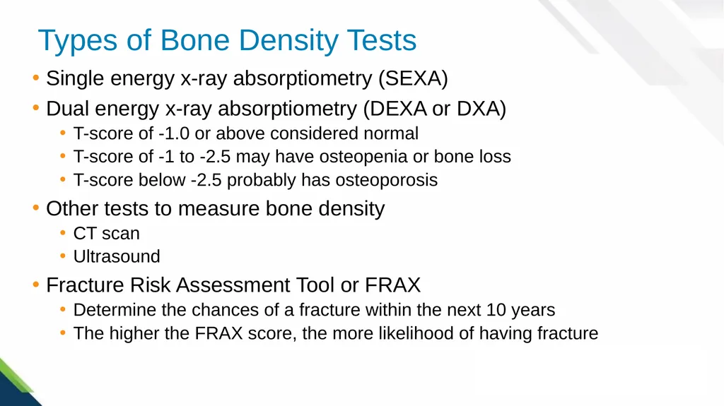
slide11. Types of Bone Density Tests Single energy x-ray absorptiometry (SEXA)
Dual energy x-ray absorptiometry (DEXA or DXA)
T-score of -1.0 or above considered normal
T-score of -1 to -2.5 may have osteopenia or bone loss
T-score below -2.5 probably has osteoporosis
Other tests to measure bone density
CT scan
Ultrasound
Fracture Risk Assessment Tool or FRAX
Determine the chances of a fracture within the next 10 years
The higher the FRAX score, the more likelihood of having fracture<br>
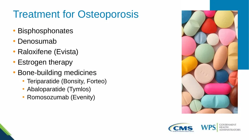
slide12. Treatment for Osteoporosis Bisphosphonates
Denosumab
Raloxifene (Evista)
Estrogen therapy
Bone-building medicines
Teriparatide (Bonsity, Forteo)
Abaloparatide (Tymlos)
Romosozumab (Evenity)<br>
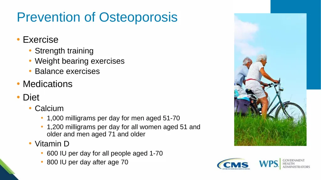
slide13. Prevention of Osteoporosis Exercise
Strength training
Weight bearing exercises
Balance exercises
Medications
Diet
Calcium
1,000 milligrams per day for men aged 51-70
1,200 milligrams per day for all women aged 51 and older and men aged 71 and older
Vitamin D
600 IU per day for all people aged 1-70
800 IU per day after age 70<br>
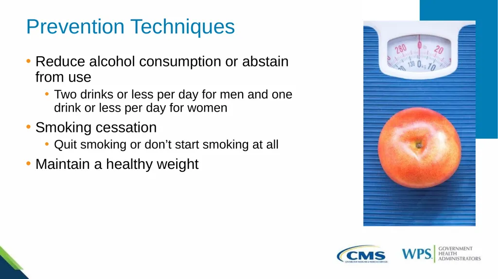
slide14. Prevention Techniques Reduce alcohol consumption or abstain from use
Two drinks or less per day for men and one drink or less per day for women
Smoking cessation
Quit smoking or don’t start smoking at all
Maintain a healthy weight<br>
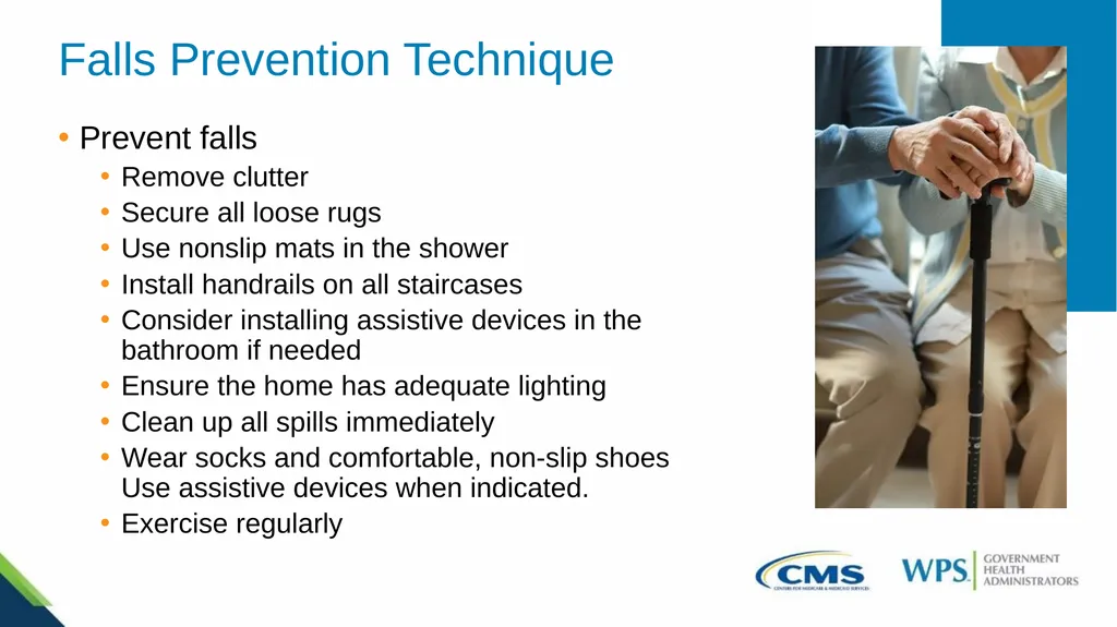
slide15. Falls Prevention Technique Prevent falls
Remove clutter
Secure all loose rugs
Use nonslip mats in the shower
Install handrails on all staircases
Consider installing assistive devices in the bathroom if needed
Ensure the home has adequate lighting
Clean up all spills immediately
Wear socks and comfortable, non-slip shoes Use assistive devices when indicated.
Exercise regularly<br>
slide16. Primary Hyperparathyroidism Occurs when one or more of the parathyroid glands becomes overactive
Leads to an excessive production of parathyroid hormone
Elevated calcium levels in the blood (hypercalcemia)
Causes thinning and weakening of the bone
Typically detected in its early stages during a routine blood test<br>
slide17. Causes of Primary Hyperparathyroidism Results from an abnormal growth of one or more of the parathyroid glands
Adenoma of one or more of the parathyroid glands
Most common cause
Accounts for around 85% of all cases
Enlargement or hyperplasia of two or more parathyroid glands
Accounts for around 15% of all cases of primary hyperparathyroidism
Individuals with a rare inherited disease that affects the parathyroid glands have a higher probability of involvement of more than one gland
Parathyroid cancer
Very rare- accounts for less than 1% of all cases<br>
slide18. Risk Factors Post-menopausal women
Individuals who have taken lithium
People who have experienced an extended period of significant calcium or vitamin D deficiency
Those who have undergone radiation therapy for cancer that involved exposure to the neck
Those who have a rare inherited disease that affects the parathyroid glands, such as multiple endocrine neoplasia, type 1 or familial hypocalciuric hypercalcemia<br>
slide19. Signs and Symptoms Fragile and brittle bones that are prone to breaking
Kidney stones
Muscle weakness
Bone and joint pain
Abdominal pain
Nausea and vomiting
Loss of appetite Polydipsia or excessive thirst
Polyuria or excessive urination
Weakness and fatigue
Depression
Confusion
Constipation<br>
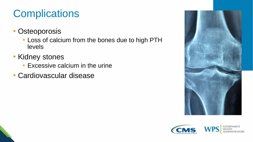
slide20. Complications Osteoporosis
Loss of calcium from the bones due to high PTH levels
Kidney stones
Excessive calcium in the urine
Cardiovascular disease<br>
slide21. Diagnosis and Tests Laboratory tests for diagnosis
Blood tests to check for high levels of calcium and PTH
Blood tests to check for low levels of Vitamin D
24-hour urine test to check kidney function and amount of calcium in urine
Tests to determine if any complications are present
Bone mineral density test such as a DEXA scan to check for osteoporosis
Imaging tests of the kidneys to check for kidney stones and any kidney issues
Tests to determine parathyroid gland involvement
CT scan
Ultrasound<br>
slide22. Treatment Parathyroidectomy
Most common treatment
Monitoring
Asymptomatic
Blood calcium levels are only slightly elevated
Kidney function and bone density in the normal range
Medication
Cinacalcet
Lowers blood calcium levels by reducing the amount of PTH made<br>
slide23. Prevention of Complications Eat a healthy diet and drink plenty of fluids
Avoid medications that increase calcium levels
Restriction of calcium intake is not recommended
Staying physically active and exercising regularly
Not smoking<br>
slide24. Eligibility Beneficiaries with Medicare Part B who meet at least one of the following criteria
Women who are identified by their physician or qualified nonphysician practitioner as being estrogen deficient and being at risk for clinical osteoporosis
Individuals with vertebral abnormalities
Those getting (or expecting to get) glucocorticoid therapy for more than three months
People with primary hyperparathyroidism
Individuals being monitored to assess response to FDA-approved osteoporosis drug therapy<br>
slide25. Frequency and Cost Frequency
Once every two years
May be performed more frequently if medically necessary
Cost
No copayment, coinsurance, or deductible<br>
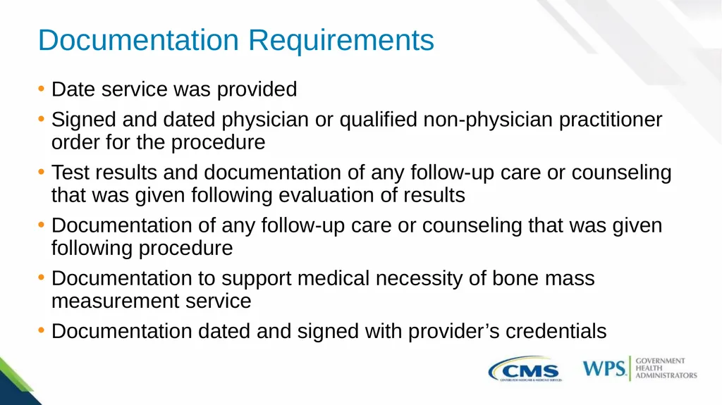
slide26. Documentation Requirements Date service was provided
Signed and dated physician or qualified non-physician practitioner order for the procedure
Test results and documentation of any follow-up care or counseling that was given following evaluation of results
Documentation of any follow-up care or counseling that was given following procedure
Documentation to support medical necessity of bone mass measurement service
Documentation dated and signed with provider’s credentials<br>
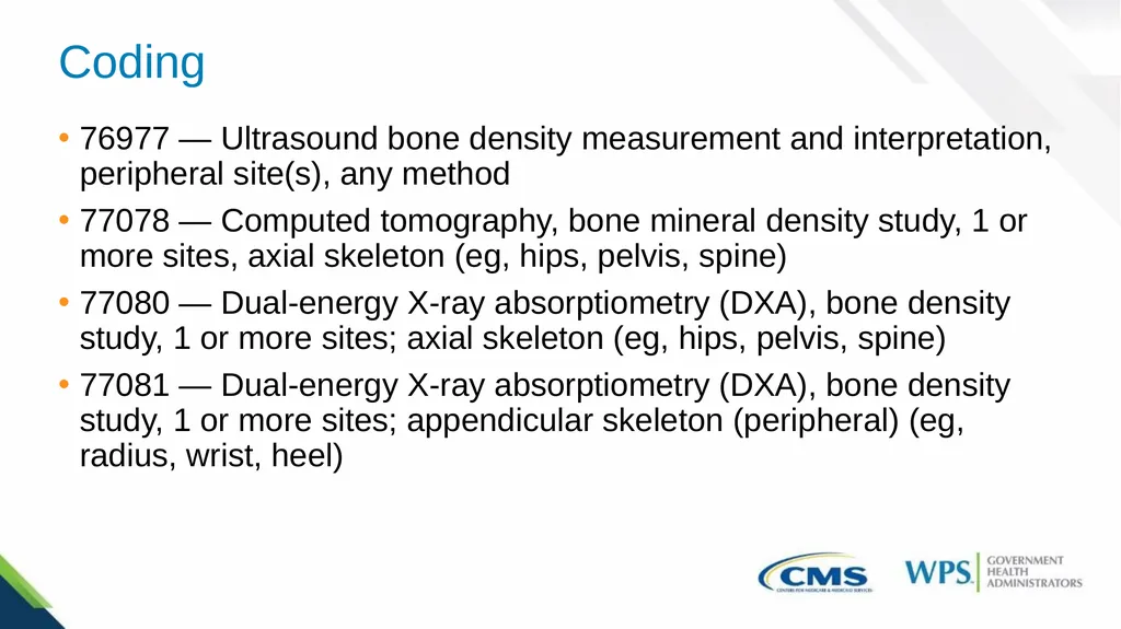
slide27. Coding 76977 — Ultrasound bone density measurement and interpretation, peripheral site(s), any method
77078 — Computed tomography, bone mineral density study, 1 or more sites, axial skeleton (eg, hips, pelvis, spine)
77080 — Dual-energy X-ray absorptiometry (DXA), bone density study, 1 or more sites; axial skeleton (eg, hips, pelvis, spine)
77081 — Dual-energy X-ray absorptiometry (DXA), bone density study, 1 or more sites; appendicular skeleton (peripheral) (eg, radius, wrist, heel)<br>
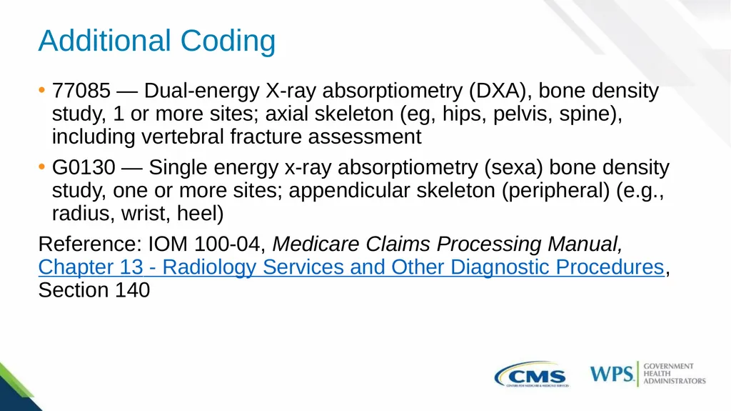
slide28. Additional Coding 77085 — Dual-energy X-ray absorptiometry (DXA), bone density study, 1 or more sites; axial skeleton (eg, hips, pelvis, spine), including vertebral fracture assessment
G0130 — Single energy x-ray absorptiometry (sexa) bone density study, one or more sites; appendicular skeleton (peripheral) (e.g., radius, wrist, heel)
Reference: IOM 100-04, Medicare Claims Processing Manual, Chapter 13 - Radiology Services and Other Diagnostic Procedures, Section 140<br>
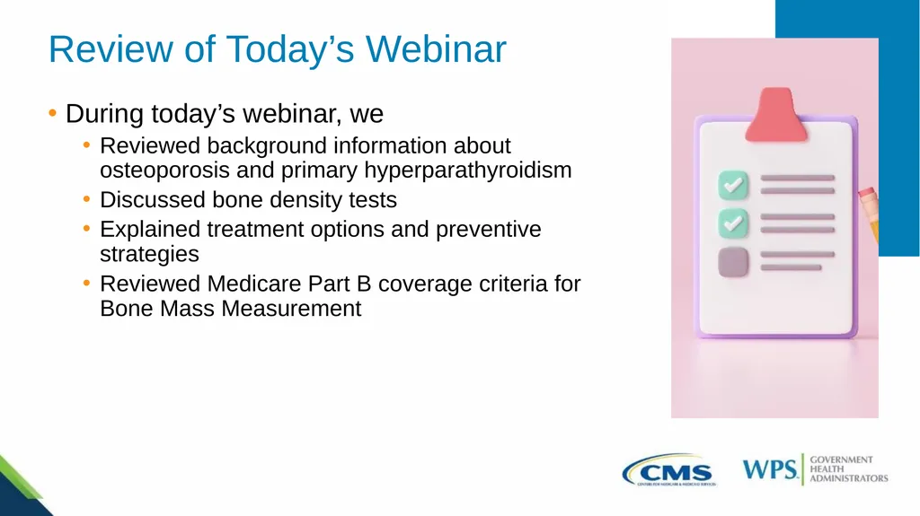
slide29. Review of Today’s Webinar During today’s webinar, we
Reviewed background information about osteoporosis and primary hyperparathyroidism
Discussed bone density tests
Explained treatment options and preventive strategies
Reviewed Medicare Part B coverage criteria for Bone Mass Measurement<br>
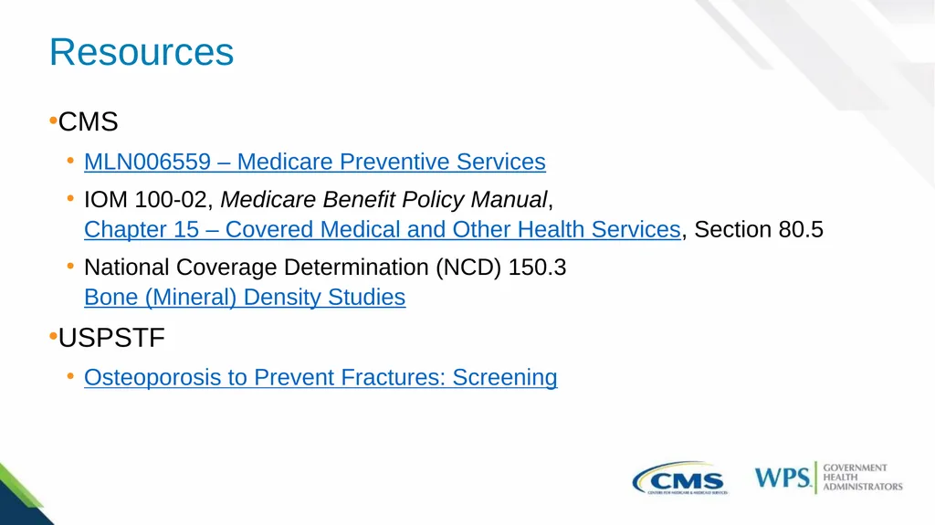
slide30. Resources CMS
MLN006559 – Medicare Preventive Services
IOM 100-02, Medicare Benefit Policy Manual, Chapter 15 – Covered Medical and Other Health Services, Section 80.5
National Coverage Determination (NCD) 150.3 Bone (Mineral) Density Studies
USPSTF
Osteoporosis to Prevent Fractures: Screening<br>
slide31. Questions<br>
slide32. Thank you for attending<br>
slide2. Disclaimer We prepared this education as a tool to assist the provider community. Medicare rules change often. They are in the relevant laws, regulations and rulings on the Centers for Medicare & Medicaid Services (CMS) website.
We will provide responses to questions based on the facts given, but the Medicare rules will determine final coverage.
CMS prohibits recording of the presentation for profit-making purposes.<br>
slide3. Acronyms CMS – Centers for Medicare & Medicaid Services
CPT – Current Procedural Terminology
CT – Computed Tomography
DEXA or DXA – Dual Energy X-ray Absorptiometry
FDA – U.S. Food and Drug Administration
FRAX – Fracture Risk Assessment Tool HCPCS – Healthcare Common Procedure Coding System
IOM – Internet-Only Manual
IU – International Units
PHPT – Primary Hyperparathyroidism
PTH – Parathyroid Hormone
SEXA – Single Energy X-ray Absorptiometry
USPSTF – U.S. Preventive Services Task Force<br>
slide4. Agenda This webinar will cover
Background information about osteoporosis and primary hyperparathyroidism
Bone density tests
Treatment options and preventive strategies
Medicare Part B coverage criteria for Bone Mass Measurement<br>
slide5. Facts and Statistics Osteoporosis
Approximately 54 million individuals aged 50 or above either currently have osteoporosis or have lower bone density
Women account for 80% of all people diagnosed
1 in 2 women and up to 1 in 4 men will experience a bone fracture in their lifetime
By 2025 — estimated to cause approximately three million fractures
Primary hyperparathyroidism
Approximately 100,000 individuals are diagnosed annually
Typically affects individuals aged 50 to 60
Women affected three to four times more frequently than men<br>
slide6. Osteoporosis Develops when the formation of new bone fails to match the rate of old bone loss
Bone mineral density and bone mass start to decrease
Bone strength begins to weaken, and the bones become brittle
Increased risk for bone fractures, especially in the spine, wrist, and hip
Older adults more at risk for falls
Loss of muscle mass
Decreased mobility
Poor balance
Sensory impairments<br>
slide7. Nonmodifiable Risk Factors Sex
Women are at greater risk
Age
Likelihood increases as you age
Race
Higher in non-Hispanic White and Asian women and men
Body size
People with smaller body frames more at risk
Family history
Having a parent with a history of osteoporosis or hip fracture<br>
slide8. Modifiable Risk Factors Low body weight
Changes in hormone levels
Certain pre-existing conditions
Taking certain medications
Lack of physical activity and exercise
Diet
Alcohol use
Smoking<br>
slide9. Signs, Symptoms, and Complications Signs and Symptoms
Asymptomatic - “silent” disease
Fractures during everyday activities
If signs and symptoms are present, include
Back pain from broken or collapsed vertebrae
Kyphosis
Gradual loss of height
Fractures from activities that would normally not cause a break in a healthy bone
Complications
Fractures
Femur
Vertebrae<br>
slide10. Bone Density Tests Use low-dose X-rays to assess how much calcium and other minerals are in the bone
Verify the diagnosis of osteoporosis
Determine effectiveness of osteoporosis treatment
Detect potential areas of low bone density prior to a fracture occurring
Identify a person’s risk for developing a fracture in the future<br>
slide11. Types of Bone Density Tests Single energy x-ray absorptiometry (SEXA)
Dual energy x-ray absorptiometry (DEXA or DXA)
T-score of -1.0 or above considered normal
T-score of -1 to -2.5 may have osteopenia or bone loss
T-score below -2.5 probably has osteoporosis
Other tests to measure bone density
CT scan
Ultrasound
Fracture Risk Assessment Tool or FRAX
Determine the chances of a fracture within the next 10 years
The higher the FRAX score, the more likelihood of having fracture<br>
slide12. Treatment for Osteoporosis Bisphosphonates
Denosumab
Raloxifene (Evista)
Estrogen therapy
Bone-building medicines
Teriparatide (Bonsity, Forteo)
Abaloparatide (Tymlos)
Romosozumab (Evenity)<br>
slide13. Prevention of Osteoporosis Exercise
Strength training
Weight bearing exercises
Balance exercises
Medications
Diet
Calcium
1,000 milligrams per day for men aged 51-70
1,200 milligrams per day for all women aged 51 and older and men aged 71 and older
Vitamin D
600 IU per day for all people aged 1-70
800 IU per day after age 70<br>
slide14. Prevention Techniques Reduce alcohol consumption or abstain from use
Two drinks or less per day for men and one drink or less per day for women
Smoking cessation
Quit smoking or don’t start smoking at all
Maintain a healthy weight<br>
slide15. Falls Prevention Technique Prevent falls
Remove clutter
Secure all loose rugs
Use nonslip mats in the shower
Install handrails on all staircases
Consider installing assistive devices in the bathroom if needed
Ensure the home has adequate lighting
Clean up all spills immediately
Wear socks and comfortable, non-slip shoes Use assistive devices when indicated.
Exercise regularly<br>
slide16. Primary Hyperparathyroidism Occurs when one or more of the parathyroid glands becomes overactive
Leads to an excessive production of parathyroid hormone
Elevated calcium levels in the blood (hypercalcemia)
Causes thinning and weakening of the bone
Typically detected in its early stages during a routine blood test<br>
slide17. Causes of Primary Hyperparathyroidism Results from an abnormal growth of one or more of the parathyroid glands
Adenoma of one or more of the parathyroid glands
Most common cause
Accounts for around 85% of all cases
Enlargement or hyperplasia of two or more parathyroid glands
Accounts for around 15% of all cases of primary hyperparathyroidism
Individuals with a rare inherited disease that affects the parathyroid glands have a higher probability of involvement of more than one gland
Parathyroid cancer
Very rare- accounts for less than 1% of all cases<br>
slide18. Risk Factors Post-menopausal women
Individuals who have taken lithium
People who have experienced an extended period of significant calcium or vitamin D deficiency
Those who have undergone radiation therapy for cancer that involved exposure to the neck
Those who have a rare inherited disease that affects the parathyroid glands, such as multiple endocrine neoplasia, type 1 or familial hypocalciuric hypercalcemia<br>
slide19. Signs and Symptoms Fragile and brittle bones that are prone to breaking
Kidney stones
Muscle weakness
Bone and joint pain
Abdominal pain
Nausea and vomiting
Loss of appetite Polydipsia or excessive thirst
Polyuria or excessive urination
Weakness and fatigue
Depression
Confusion
Constipation<br>
slide20. Complications Osteoporosis
Loss of calcium from the bones due to high PTH levels
Kidney stones
Excessive calcium in the urine
Cardiovascular disease<br>
slide21. Diagnosis and Tests Laboratory tests for diagnosis
Blood tests to check for high levels of calcium and PTH
Blood tests to check for low levels of Vitamin D
24-hour urine test to check kidney function and amount of calcium in urine
Tests to determine if any complications are present
Bone mineral density test such as a DEXA scan to check for osteoporosis
Imaging tests of the kidneys to check for kidney stones and any kidney issues
Tests to determine parathyroid gland involvement
CT scan
Ultrasound<br>
slide22. Treatment Parathyroidectomy
Most common treatment
Monitoring
Asymptomatic
Blood calcium levels are only slightly elevated
Kidney function and bone density in the normal range
Medication
Cinacalcet
Lowers blood calcium levels by reducing the amount of PTH made<br>
slide23. Prevention of Complications Eat a healthy diet and drink plenty of fluids
Avoid medications that increase calcium levels
Restriction of calcium intake is not recommended
Staying physically active and exercising regularly
Not smoking<br>
slide24. Eligibility Beneficiaries with Medicare Part B who meet at least one of the following criteria
Women who are identified by their physician or qualified nonphysician practitioner as being estrogen deficient and being at risk for clinical osteoporosis
Individuals with vertebral abnormalities
Those getting (or expecting to get) glucocorticoid therapy for more than three months
People with primary hyperparathyroidism
Individuals being monitored to assess response to FDA-approved osteoporosis drug therapy<br>
slide25. Frequency and Cost Frequency
Once every two years
May be performed more frequently if medically necessary
Cost
No copayment, coinsurance, or deductible<br>
slide26. Documentation Requirements Date service was provided
Signed and dated physician or qualified non-physician practitioner order for the procedure
Test results and documentation of any follow-up care or counseling that was given following evaluation of results
Documentation of any follow-up care or counseling that was given following procedure
Documentation to support medical necessity of bone mass measurement service
Documentation dated and signed with provider’s credentials<br>
slide27. Coding 76977 — Ultrasound bone density measurement and interpretation, peripheral site(s), any method
77078 — Computed tomography, bone mineral density study, 1 or more sites, axial skeleton (eg, hips, pelvis, spine)
77080 — Dual-energy X-ray absorptiometry (DXA), bone density study, 1 or more sites; axial skeleton (eg, hips, pelvis, spine)
77081 — Dual-energy X-ray absorptiometry (DXA), bone density study, 1 or more sites; appendicular skeleton (peripheral) (eg, radius, wrist, heel)<br>
slide28. Additional Coding 77085 — Dual-energy X-ray absorptiometry (DXA), bone density study, 1 or more sites; axial skeleton (eg, hips, pelvis, spine), including vertebral fracture assessment
G0130 — Single energy x-ray absorptiometry (sexa) bone density study, one or more sites; appendicular skeleton (peripheral) (e.g., radius, wrist, heel)
Reference: IOM 100-04, Medicare Claims Processing Manual, Chapter 13 - Radiology Services and Other Diagnostic Procedures, Section 140<br>
slide29. Review of Today’s Webinar During today’s webinar, we
Reviewed background information about osteoporosis and primary hyperparathyroidism
Discussed bone density tests
Explained treatment options and preventive strategies
Reviewed Medicare Part B coverage criteria for Bone Mass Measurement<br>
slide30. Resources CMS
MLN006559 – Medicare Preventive Services
IOM 100-02, Medicare Benefit Policy Manual, Chapter 15 – Covered Medical and Other Health Services, Section 80.5
National Coverage Determination (NCD) 150.3 Bone (Mineral) Density Studies
USPSTF
Osteoporosis to Prevent Fractures: Screening<br>
slide31. Questions<br>
slide32. Thank you for attending<br>