04
2. Depolarization Overshoot Smaller fibers and
Many Central nervous system (CNS) neurons
The potential merely approaches the zero level
Does not overshoot to the positive level 4<br>
05
2. Depolarization Overshoot During overshoot
Direction of electrical gradient
For Na+ is reversed 5<br>
06
2. Depolarization Overshoot Because
Membrane potential is reversed
Limits Na+ influx
Voltage gated K+ channels Open 6<br>
07
Rising membrane potential Within
Fraction of a millisecond
Causes 7<br>
08
Rising membrane potential Beginning of Closure of
Sodium channels
Opening of
Potassium channel
Action potential terminates 8<br>
09
3. Repolarization stage of action potential Within few 10,000ths of a second
Na+ channels begin to close
After membrane becomes
Highly permeable to K+ ions 9<br>
10
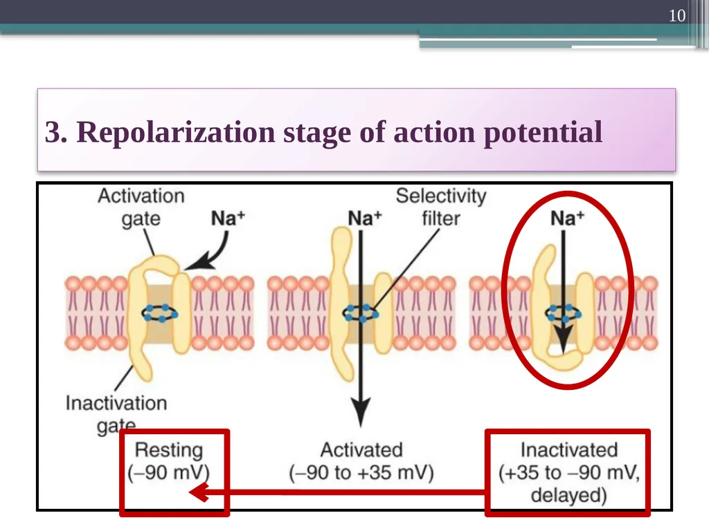
3. Repolarization stage of action potential 10<br>
11
3. Repolarization stage of action potential The K+ channels open
More than normally
Rapid diffusion of K+ ions to the exterior
(Higher Concentration to Lower Concentration) 11<br>
12
3. Repolarization stage of action potential Re-established
The normal negative resting membrane potential (RMP: -90 mV ) called
Repolarization 12<br>
13
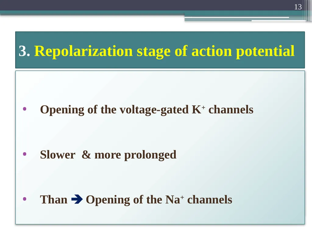
3. Repolarization stage of action potential Opening of the voltage-gated K+ channels
Slower & more prolonged
Than Opening of the Na+ channels 13<br>
14
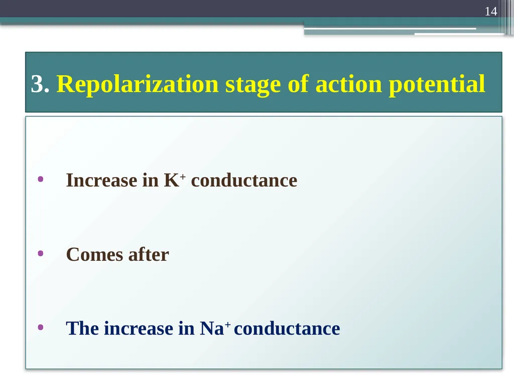
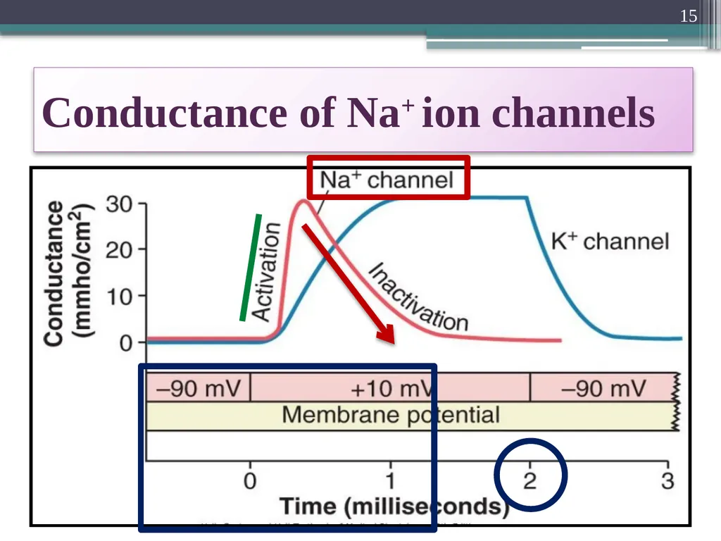
3. Repolarization stage of action potential Increase in K+ conductance
Comes after
The increase in Na+ conductance 14<br>
15
Conductance of Na+ ion channels 15<br>
16
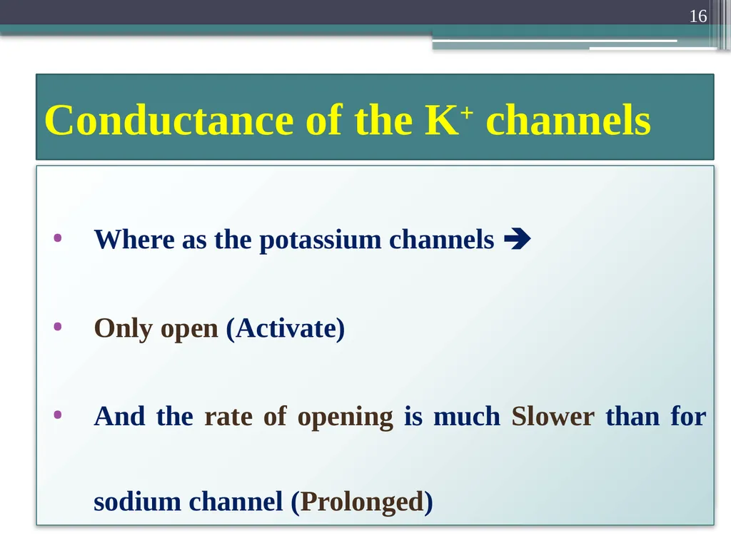
Conductance of the K+ channels Where as the potassium channels
Only open (Activate)
And the rate of opening is much Slower than for sodium channel (Prolonged) 16<br>
17
During the Resting stage:
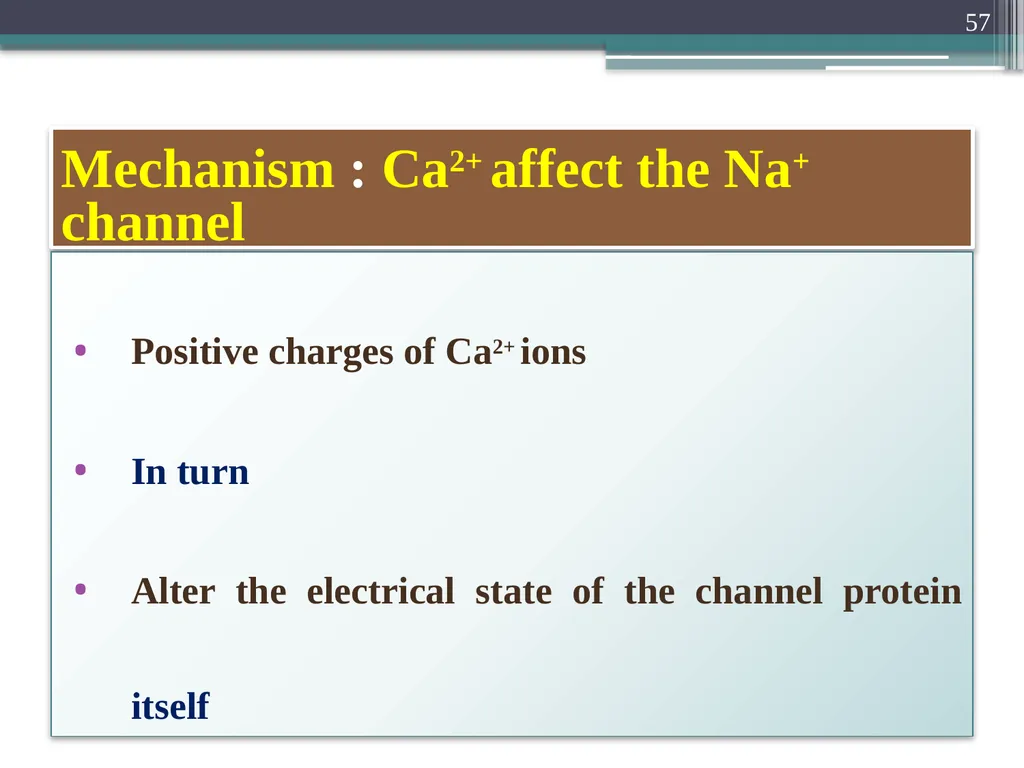
The Gate of the potassium channel is
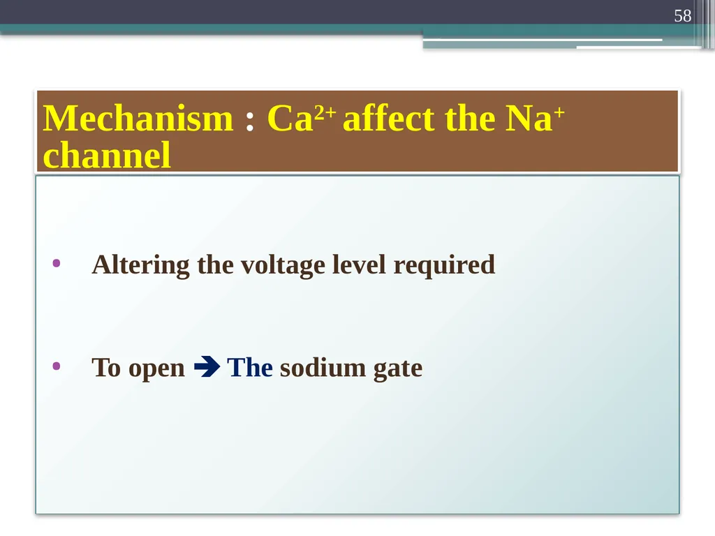
Closed Voltage-Gated Potassium Channel 17<br>
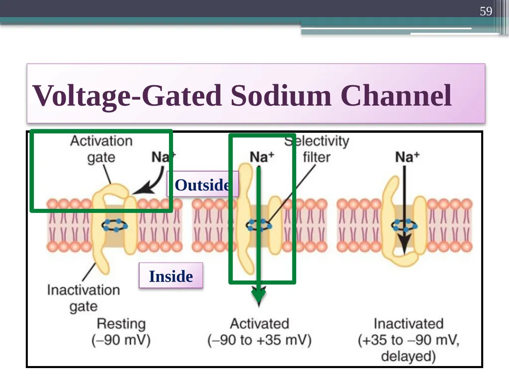
18
Potassium ions are
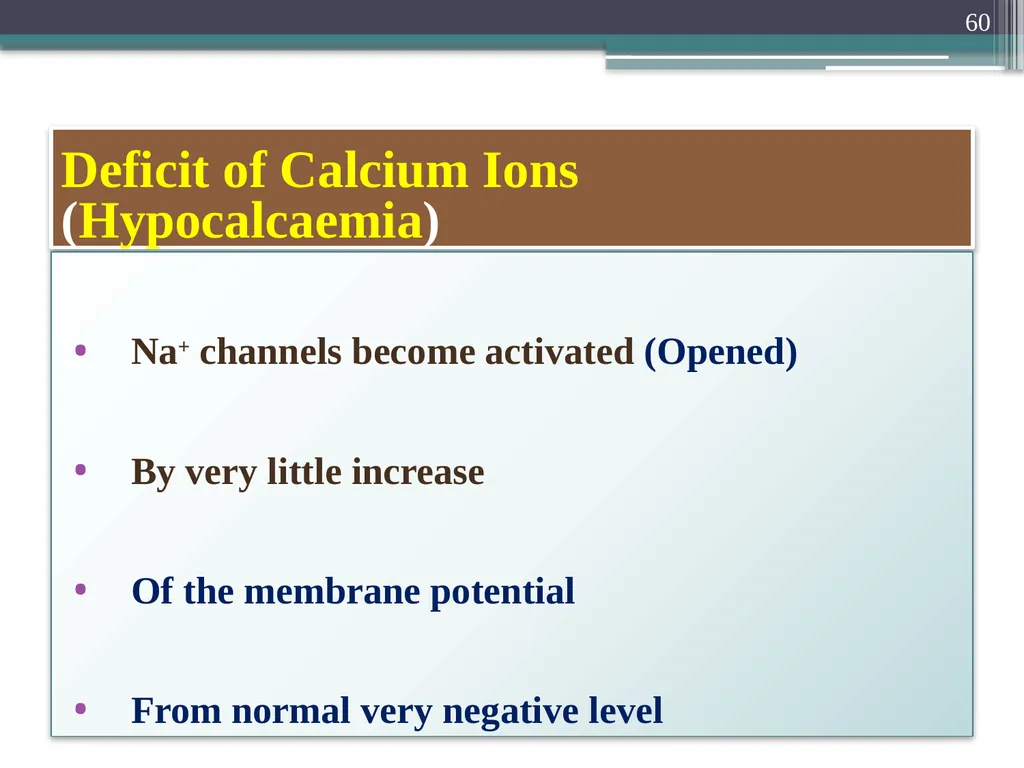
Prevented from passing through this channel
To the exterior Voltage-Gated Potassium Channel 18<br>
19
When membrane potential rises
From -90mV Towards Zero
Voltage change
Cause slow conformational opening of the gate
Allows increased potassium diffusion outward Voltage-Gated Potassium Channel 19<br>
20
K+ channels open Just at the same time
Na+ channels Beginning to close
Due to Inactivation Slowness of the K+ Channels 20<br>
21
3. Repolarization stage of action potential The net movement of positively charge
Out of the cell
Due to K+ efflux Completes
The process of repolarization 21<br>
22
Stages of Nerve Action Potential Resting stage
Depolarization stage and Overshoot
Repolarization stage
After-hyperpolarization 22<br>
23
4. “Positive” After potential Membrane potential becomes more negative
Than Original RMP (- 90 mV)
For few milliseconds
After action potential Over 23<br>
24
4. “Positive” After potential “Positive” after potential is Misnomer
Because positive afterpotential
Is even more negative
Than resting membrane potential (RMP =-90mV) 24<br>
25
4. “Positive” After potential Reason for calling it “Positive” Historically
The first potential measurement
Were made on
The outside of the nerve fiber membrane
Was Positive 25<br>
26
4. “Positive” After potential Than The inside
When measured on the outside
This potential causes a positive record
Rather than a negative one 26<br>
27
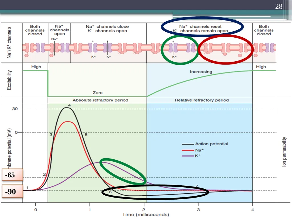
4. “Positive” After potential Cause of the positive afterpotential Mainly
Many potassium channels
Remain open for several milliseconds
After complete repolarization of the membrane 27<br>
29
4. After-hyperpolarization The slow return of the K+ channels
To the closed state explain
After-hyperpolarization
F/b return
To the resting membrane potential 29<br>
30
5. End of action potential Voltage-gated K+ channels
Bring the action potential To the end
Cause closer of their gates through
Negative feedback process 30<br>
31
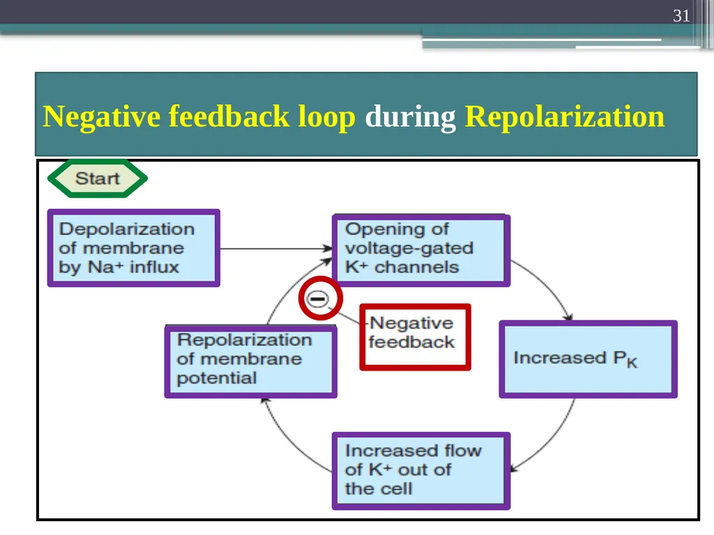
Negative feedback loop during Repolarization 31<br>
32
Resting Membrane Potential in Neurons About -70mV
Close to the equilibrium potential for K+
Because there are more open K+ channels
Than Na+ channels at rest
Membrane permeability to K+ is greater at rest 32<br>
33
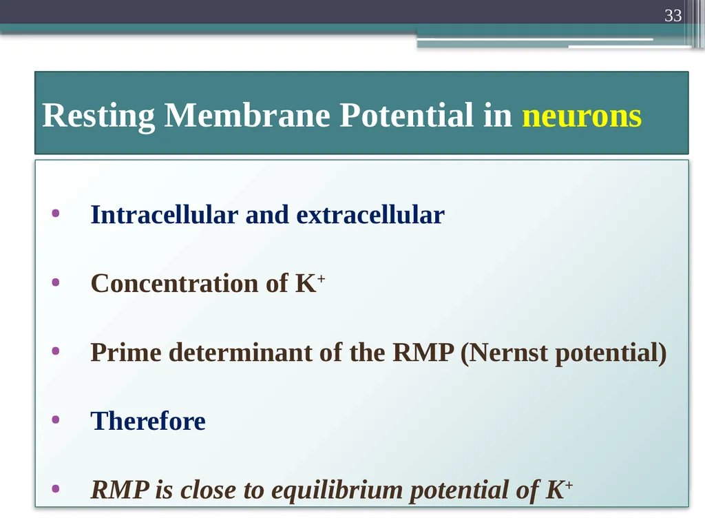
Resting Membrane Potential in neurons Intracellular and extracellular
Concentration of K+
Prime determinant of the RMP (Nernst potential)
Therefore
RMP is close to equilibrium potential of K+ 33<br>
34
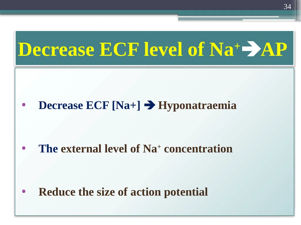
Decrease ECF level of Na+AP Decrease ECF [Na+] Hyponatraemia
The external level of Na+ concentration
Reduce the size of action potential 34<br>
35
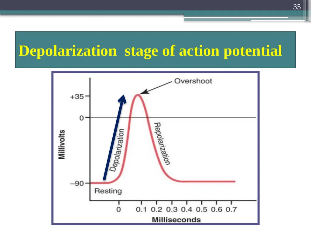
Depolarization stage of action potential 35<br>
36
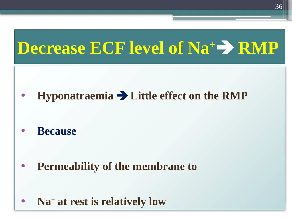
Decrease ECF level of Na+ RMP Hyponatraemia Little effect on the RMP
Because
Permeability of the membrane to
Na+ at rest is relatively low 36<br>
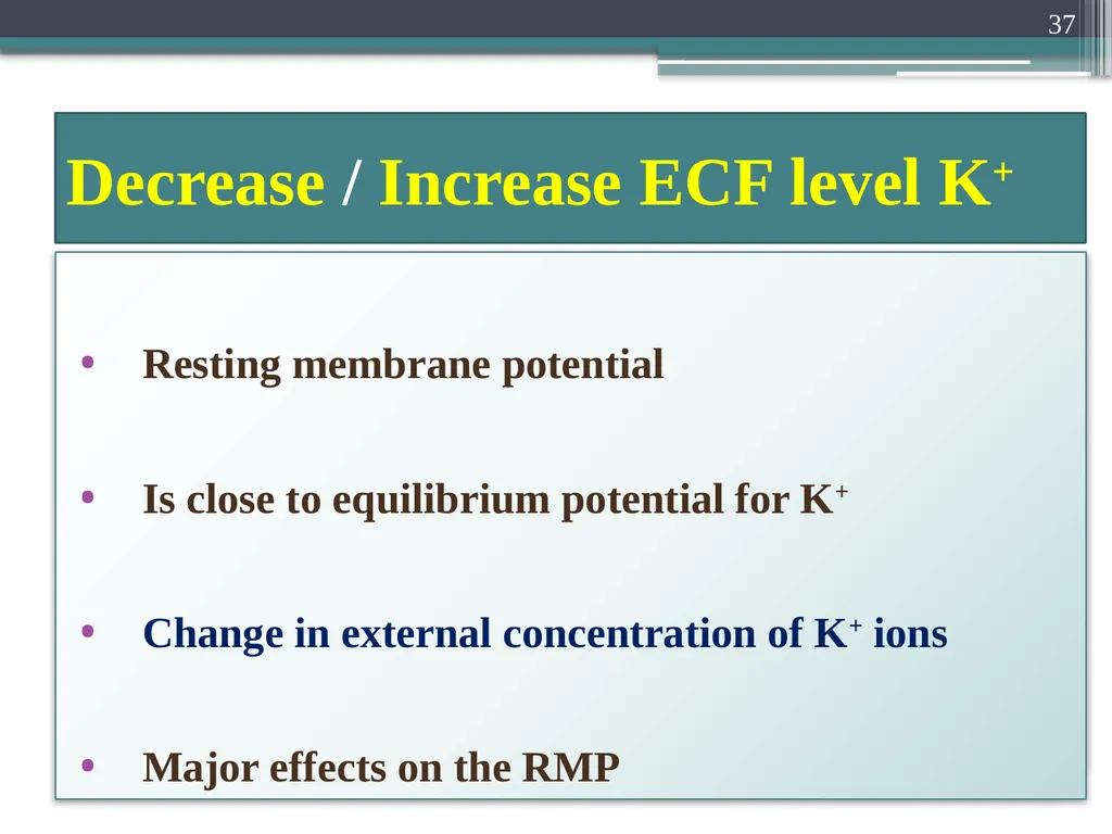
37
Decrease / Increase ECF level K+ Resting membrane potential
Is close to equilibrium potential for K+
Change in external concentration of K+ ions
Major effects on the RMP 37<br>
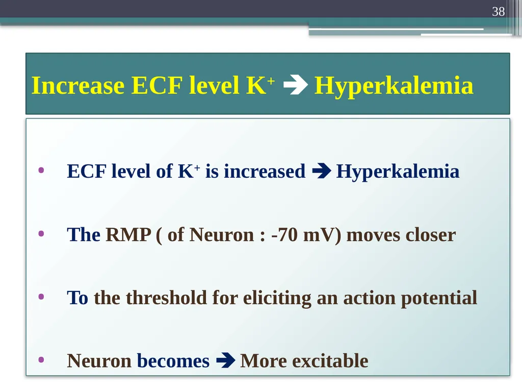
38
Increase ECF level K+ Hyperkalemia ECF level of K+ is increased Hyperkalemia
The RMP ( of Neuron : -70 mV) moves closer
To the threshold for eliciting an action potential
Neuron becomes More excitable 38<br>
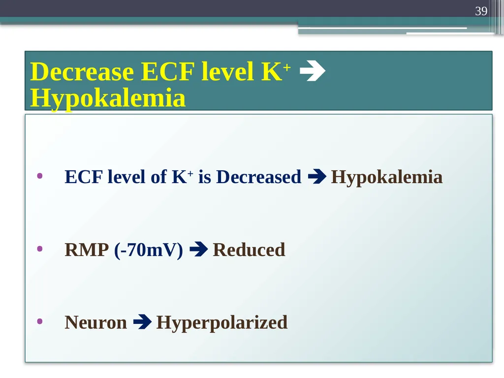
39
Decrease ECF level K+ Hypokalemia ECF level of K+ is Decreased Hypokalemia
RMP (-70mV) Reduced
Neuron Hyperpolarized 39<br>
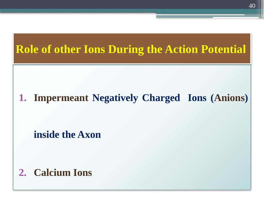
40
Role of other Ions During the Action Potential Impermeant Negatively Charged Ions (Anions) inside the Axon
Calcium Ions 40<br>
41
Impermeant Anions inside the axon Inside the axon
Many negatively charged ions (Anions)
That can not go through the membrane channels 41<br>
42
Impermeant Anions inside the axon Includes
Anions of the Protein molecules
Anions of many Organic phosphate compounds
Anions of Sulfate compounds 42<br>
43
Impermeant Anions inside the axon Because these ions
Cannot leave the interior of the axon 43<br>
44
Impermeant Anions inside the axon Excess of these impermeant anions
Deficit of positive ions inside the membrane 44<br>
45
Impermeant Anions inside the axon Responsible
For the negative charge inside the fiber
When there is deficit of positive charged K+
And other positive ions 45<br>
46
Calcium Ions Membranes of almost all cells of the body
Have Ca2+ pump
Similar to Na+ pump 46<br>
47
Calcium ions serves Along with or Instead of Na+
In some cells
To cause most of action potential 47<br>
48
Calcium pump Like
Sodium (Na+) pump
Pumps Ca2+ ions 48<br>
49
Calcium pump Ca2+ From the interior
To the exterior of the cell membrane
Or
To endoplasmic reticulum (ER) 49<br>
50
Calcium ions gradient Of 10,000 folds due to it
Internal cell concentration of calcium ions of
10-7 molar
External concentration of 10-3 molar 50<br>
51
Voltage gated Ca2+ Channels Slightly permeable to Na+ ions also
When channels open Both Ca2+ and Na+ ions
Flow exterior of the fiber 51<br>
52
Ca2+-Na+ Channels Slow channels Slow to activation
Require 10-20 times a long for activation
As the Sodium channels Fast channels 52<br>
53
Ca2+ channels Numerous Cardiac muscle
Smooth muscle 53<br>
54
Some types of Smooth Muscle Fast sodium channels
Hardly present 54<br>
55
Some types of Smooth Muscle Action potential caused Entirely by
Activation of the slow calcium channels 55<br>
56
Mechanism : Ca2+ affect the Na+ channel Ca2+ ions bind
To the exterior surfaces of
The Na+ channel protein molecules 56<br>
57
Mechanism : Ca2+ affect the Na+ channel Positive charges of Ca2+ ions
In turn
Alter the electrical state of the channel protein itself 57<br>
58
Mechanism : Ca2+ affect the Na+ channel Altering the voltage level required
To open The sodium gate 58<br>
59
Voltage-Gated Sodium Channel Inside Outside 59<br>
60
Deficit of Calcium Ions (Hypocalcaemia) Na+ channels become activated (Opened)
By very little increase
Of the membrane potential
From normal very negative level 60<br>
61
Calcium Ions falls 50% below normal Spontaneous discharge in peripheral nerves 61<br>
62
Calcium Ions falls 50% below normal Often causing muscle “Tetany”
Lethal Death
Tetanic contraction of the respiratory muscles 62<br>
63
Attend Your Roll Call 63<br>