
Obstetrics and Neonatal Care CHAPTER 35: Focused
PC
Published · 126 slides · 0 views

1 / 1






Description
Obstetrics and Neonatal Care CHAPTER 35: Focused Lecture National EMS Education Standard Competencies (1 of 9) Special Patient Populations Applies a fundamental knowledge of growth, development, and aging and assessment findings to provide
Related Topics
Share
Embed code
Download this presentation From Below
"Obstetrics and Neonatal Care CHAPTER 35: Focused" is the property of its rightful owner. Permission is granted to download and print the materials on this website for personal, non-commercial use only, and to display it on your personal computer provided you do not modify the materials and that you retain all copyright notices contained in the materials. By downloading content from our website, you accept the terms of this agreement.
Presentation Transcript
01
Obstetrics and Neonatal Care CHAPTER 35: Focused Lecture<br>
02
National EMS Education Standard Competencies (1 of 9) Special Patient Populations
Applies a fundamental knowledge of growth, development, and aging and assessment findings to provide basic and selected advanced emergency care and transportation for a patient with special needs.<br>
Applies a fundamental knowledge of growth, development, and aging and assessment findings to provide basic and selected advanced emergency care and transportation for a patient with special needs.<br>
03
National EMS Education Standard Competencies (2 of 9) Obstetrics
Recognition and management of
Normal delivery
Vaginal bleeding in the pregnant patient
Anatomy and physiology of normal pregnancy<br>
Recognition and management of
Normal delivery
Vaginal bleeding in the pregnant patient
Anatomy and physiology of normal pregnancy<br>
04
National EMS Education Standard Competencies (3 of 9) Obstetrics
Recognition and management of
Pathophysiology of complications of pregnancy
Assessment of the pregnant patient<br>
Recognition and management of
Pathophysiology of complications of pregnancy
Assessment of the pregnant patient<br>
05
National EMS Education Standard Competencies (4 of 9) Management of:
Normal delivery
Abnormal delivery
Nuchal cord
Prolapsed cord
Breech delivery<br>
Normal delivery
Abnormal delivery
Nuchal cord
Prolapsed cord
Breech delivery<br>
06
National EMS Education Standard Competencies (5 of 9) Management of:
Third trimester bleeding
Placenta previa
Abruptio placenta
Spontaneous abortion/miscarriage
Ectopic pregnancy
Preeclampsia/eclampsia<br>
Third trimester bleeding
Placenta previa
Abruptio placenta
Spontaneous abortion/miscarriage
Ectopic pregnancy
Preeclampsia/eclampsia<br>
07
National EMS Education Standard Competencies (6 of 9) Neonatal care
Assessment and management
Newborn
Neonatal resuscitation<br>
Assessment and management
Newborn
Neonatal resuscitation<br>
08
National EMS Education Standard Competencies (7 of 9) Trauma
Applies fundamental knowledge to provide basic and selected advanced emergency care and transportation based on assessment findings for an acutely injured patient.<br>
Applies fundamental knowledge to provide basic and selected advanced emergency care and transportation based on assessment findings for an acutely injured patient.<br>
09
National EMS Education Standard Competencies (8 of 9) Special Considerations in Trauma
Recognition and management of trauma in
Pregnant patient
Pediatric patient
Geriatric patient<br>
Recognition and management of trauma in
Pregnant patient
Pediatric patient
Geriatric patient<br>
10
National EMS Education Standard Competencies (9 of 9) Special Considerations in Trauma
Pathophysiology, assessment, and management of trauma in the
Pregnant patient
Pediatric patient
Geriatric patient
Cognitively impaired patient<br>
Pathophysiology, assessment, and management of trauma in the
Pregnant patient
Pediatric patient
Geriatric patient
Cognitively impaired patient<br>
11
Introduction Most births require little or no medical intervention beyond basic interventions, such as suctioning, drying, and warming the baby.
Others may be life threatening to both the woman and baby.<br>
Others may be life threatening to both the woman and baby.<br>
12
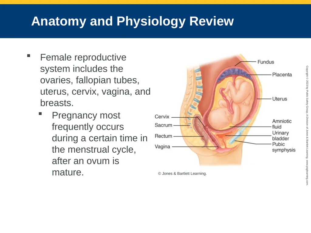
Anatomy and Physiology Review Female reproductive system includes the ovaries, fallopian tubes, uterus, cervix, vagina, and breasts.
Pregnancy most frequently occurs during a certain time in the menstrual cycle, after an ovum is mature. © Jones & Bartlett Learning.<br>
Pregnancy most frequently occurs during a certain time in the menstrual cycle, after an ovum is mature. © Jones & Bartlett Learning.<br>
13
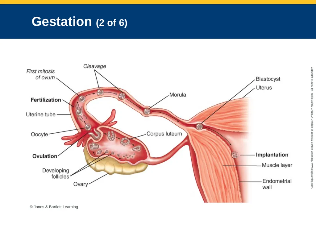
Gestation (1 of 6) Process of fetal development following fertilization of an egg
Fertilization usually in the distal third of the fallopian tube.
From time of fertilization to end of ninth week, the developing embryoblast is an embryo.
From 10th week forward, it is a fetus.<br>
Fertilization usually in the distal third of the fallopian tube.
From time of fertilization to end of ninth week, the developing embryoblast is an embryo.
From 10th week forward, it is a fetus.<br>
14
Gestation (2 of 6) © Jones & Bartlett Learning.<br>
15
Gestation (3 of 6) Around the fourth week of pregnancy, the placenta develops and serves the following functions:
Early liver for the fetus
Respiratory gas exchange
Transport of nutrients to fetal circulation
Excretion of wastes
Transfer of heat
Hormone production
Formation of barrier against harmful substances<br>
Early liver for the fetus
Respiratory gas exchange
Transport of nutrients to fetal circulation
Excretion of wastes
Transfer of heat
Hormone production
Formation of barrier against harmful substances<br>
16
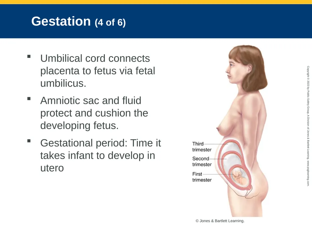
Gestation (4 of 6) Umbilical cord connects placenta to fetus via fetal umbilicus.
Amniotic sac and fluid protect and cushion the developing fetus.
Gestational period: Time it takes infant to develop in utero © Jones & Bartlett Learning.<br>
Amniotic sac and fluid protect and cushion the developing fetus.
Gestational period: Time it takes infant to develop in utero © Jones & Bartlett Learning.<br>
17
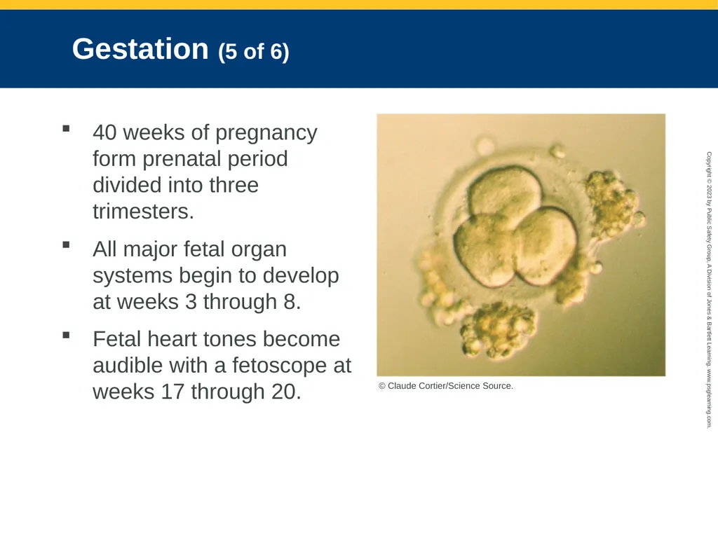
Gestation (5 of 6) 40 weeks of pregnancy form prenatal period divided into three trimesters.
All major fetal organ systems begin to develop at weeks 3 through 8.
Fetal heart tones become audible with a fetoscope at weeks 17 through 20. © Claude Cortier/Science Source.<br>
All major fetal organ systems begin to develop at weeks 3 through 8.
Fetal heart tones become audible with a fetoscope at weeks 17 through 20. © Claude Cortier/Science Source.<br>
18
Gestation (6 of 6) Female and male genitalia may be distinguished by ultrasonography at week 18.
Neonate: Baby during the first 28 days of life
Infant: Baby between the ages of 1 month and 1 year<br>
Neonate: Baby during the first 28 days of life
Infant: Baby between the ages of 1 month and 1 year<br>
19
Normal Maternal Changes of Pregnancy (1 of 2) During pregnancy, many other body systems undergo changes.
Total blood volume increases by about 50% by 40 weeks and red blood cells increase in number by about 30%.
Cardiac output increases by 30% to 50%.<br>
Total blood volume increases by about 50% by 40 weeks and red blood cells increase in number by about 30%.
Cardiac output increases by 30% to 50%.<br>
20
Normal Maternal Changes of Pregnancy (2 of 2) Increase in red blood cells heightens pregnant woman’s need for iron.
Respiratory minute volume increases by about 40% by full term.
Decreased gastrointestinal function could affect the absorption of medications.
Pregnant women often have edema in their lower extremities.<br>
Respiratory minute volume increases by about 40% by full term.
Decreased gastrointestinal function could affect the absorption of medications.
Pregnant women often have edema in their lower extremities.<br>
21
Pathophysiology During Pregnancy Complications can occur that threaten the health or life of both the woman and fetus.
Vaginal bleeding is a common early pregnancy emergency.<br>
Vaginal bleeding is a common early pregnancy emergency.<br>
22
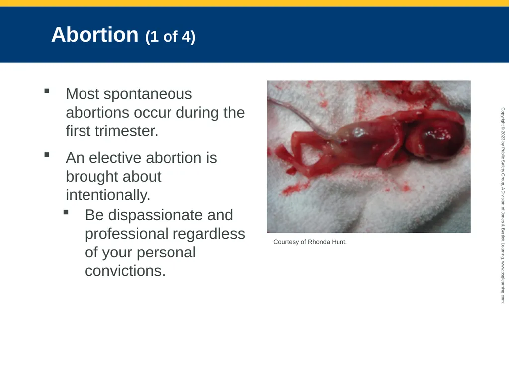
Abortion (1 of 4) Most spontaneous abortions occur during the first trimester.
An elective abortion is brought about intentionally.
Be dispassionate and professional regardless of your personal convictions. Courtesy of Rhonda Hunt.<br>
An elective abortion is brought about intentionally.
Be dispassionate and professional regardless of your personal convictions. Courtesy of Rhonda Hunt.<br>
23
Abortion (2 of 4) Threatened abortion: An abortion that is impending or potentially occurring
Characterized by vaginal bleeding during the first half of pregnancy
Can progress to a miscarriage or abortion, or it may subside
Treatment is usually complete bed rest.<br>
Characterized by vaginal bleeding during the first half of pregnancy
Can progress to a miscarriage or abortion, or it may subside
Treatment is usually complete bed rest.<br>
24
Abortion (3 of 4) Imminent abortion: An impending or threatened spontaneous abortion that cannot be prevented
Maintain blood pressure and prevent hypovolemia.
Treatment consists of:
Administering 250-mL boluses of normal saline.
100% supplemental oxygen to maintain an SpO2 level of greater than 94%
Providing emotional support with rapid transport<br>
Maintain blood pressure and prevent hypovolemia.
Treatment consists of:
Administering 250-mL boluses of normal saline.
100% supplemental oxygen to maintain an SpO2 level of greater than 94%
Providing emotional support with rapid transport<br>
25
Abortion (4 of 4) An incomplete abortion occurs when part of the products of conception are expelled but some remain in the uterus.
Vaginal bleeding will be present.
Be alert for signs and symptoms of shock, and start an IV line of normal saline.
If products of conception are protruding from the vagina, consult medical control for instructions.<br>
Vaginal bleeding will be present.
Be alert for signs and symptoms of shock, and start an IV line of normal saline.
If products of conception are protruding from the vagina, consult medical control for instructions.<br>
26
Ectopic Pregnancy Implantation and growth of the embryo outside of the uterus
Woman usually feels lower abdominal pain and cramping.
Can be a life-threatening emergency
Embryo will not survive and must be removed surgically to save the woman.<br>
Woman usually feels lower abdominal pain and cramping.
Can be a life-threatening emergency
Embryo will not survive and must be removed surgically to save the woman.<br>
27
Third-Trimester Bleeding Vaginal bleeding is a serious sign at any stage of pregnancy.
Complications of bleeding increase as the gestation progresses.
Third-trimester bleeding presents the most dangerous hemorrhage in terms of risk to the health of the mother.<br>
Complications of bleeding increase as the gestation progresses.
Third-trimester bleeding presents the most dangerous hemorrhage in terms of risk to the health of the mother.<br>
28
Bleeding and the Placenta (1 of 3) Major causes of substantial hemorrhage before delivery are abruptio placentae and placenta previa<br>
29
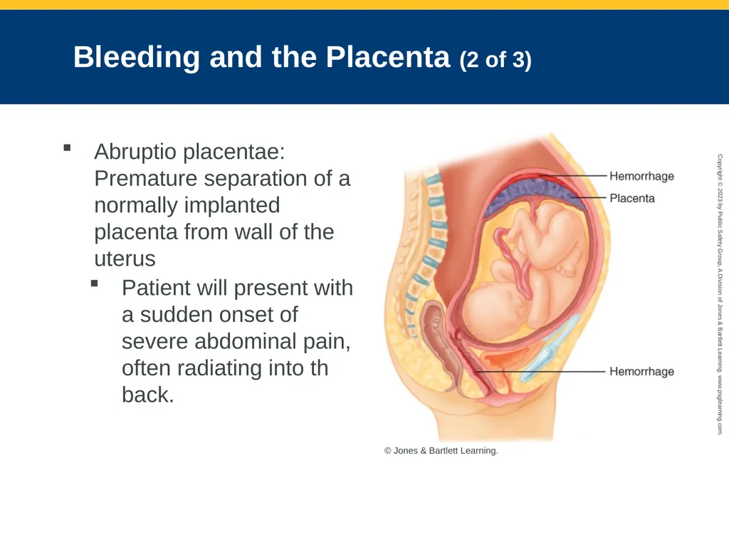
Bleeding and the Placenta (2 of 3) Abruptio placentae: Premature separation of a normally implanted placenta from wall of the uterus
Patient will present with a sudden onset of severe abdominal pain, often radiating into th back. © Jones & Bartlett Learning.<br>
Patient will present with a sudden onset of severe abdominal pain, often radiating into th back. © Jones & Bartlett Learning.<br>
30
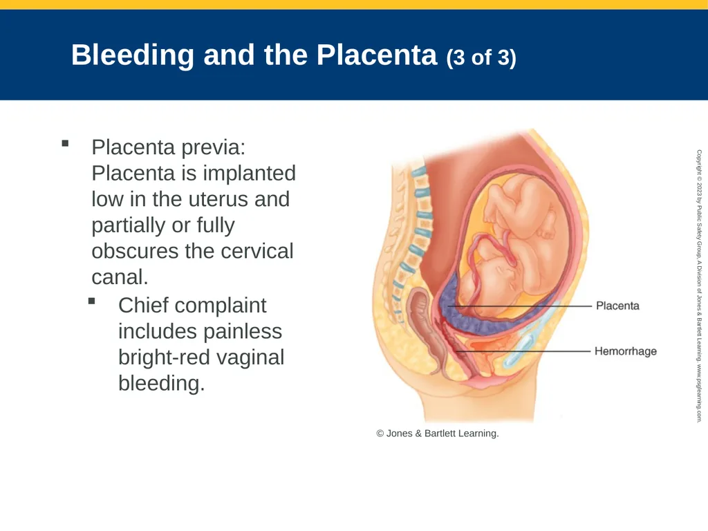
Bleeding and the Placenta (3 of 3) Placenta previa: Placenta is implanted low in the uterus and partially or fully obscures the cervical canal.
Chief complaint includes painless bright-red vaginal bleeding. © Jones & Bartlett Learning.<br>
Chief complaint includes painless bright-red vaginal bleeding. © Jones & Bartlett Learning.<br>
31
Hypertensive Disorders (1 of 4) Chronic hypertension: Blood pressure equal to or greater than 130/80 mm Hg
Diastolic pressures higher than 110 mm Hg place the patient at an increased risk for stroke and other cardiovascular dangers.<br>
Diastolic pressures higher than 110 mm Hg place the patient at an increased risk for stroke and other cardiovascular dangers.<br>
32
Hypertensive Disorders (2 of 4) Gestational hypertension develops after the 20th week of pregnancy in women with previously normal blood pressures
Resolves spontaneously in postpartum period
Preeclampsia: Increase in blood pressure after the 20th week of gestation
Accompanied by protein in the urine and edema<br>
Resolves spontaneously in postpartum period
Preeclampsia: Increase in blood pressure after the 20th week of gestation
Accompanied by protein in the urine and edema<br>
33
Hypertensive Disorders (3 of 4) Symptoms: Hypertension, severe headache, nausea and vomiting, agitation, rapid weight gain, and visual disturbances
Eclampsia: Seizure in a pregnant woman who has preeclampsia and no other cause for the seizure
Postpartum eclampsia can occur up to 4 weeks following birth.<br>
Eclampsia: Seizure in a pregnant woman who has preeclampsia and no other cause for the seizure
Postpartum eclampsia can occur up to 4 weeks following birth.<br>
34
Hypertensive Disorders (4 of 4) Supine hypotensive syndrome: Occurs when a pregnant patient has hypotension when lying supine
Results from compression of the vena cava by the weight of the gravid uterus
In most women, symptoms never develop.<br>
Results from compression of the vena cava by the weight of the gravid uterus
In most women, symptoms never develop.<br>
35
Effects of Substance Abuse (1 of 2) Substances pass through the placental barrier and enter the fetal circulation.
Effects on the fetus can include:
Birth defects
Addiction
Prematurity
Low birth weight
Severe respiratory depression<br>
Effects on the fetus can include:
Birth defects
Addiction
Prematurity
Low birth weight
Severe respiratory depression<br>
36
Effects of Substance Abuse (2 of 2) Fetal alcohol syndrome (FAS): A condition seen in infants born to women who have abused alcohol
Physical characteristics of newborns include a small head, abnormal facial features, low birth weight, and deformities of the extremities.<br>
Physical characteristics of newborns include a small head, abnormal facial features, low birth weight, and deformities of the extremities.<br>
37
Isoimmunization (Rh Sensitization)
(1 of 2) Rh factor: Protein found on the red blood cells of most people
When a woman who is Rh negative becomes pregnant by a man who is Rh positive and the fetus is Rh positive, fetal blood can pass into the woman’s circulation and produce maternal antibody to the factor.<br>
When a woman who is Rh negative becomes pregnant by a man who is Rh positive and the fetus is Rh positive, fetal blood can pass into the woman’s circulation and produce maternal antibody to the factor.<br>
38
Isoimmunization (Rh Sensitization)
(2 of 2) In subsequent pregnancies, antibody will aggressively cross placental barrier to attack fetal red blood cells
Woman’s body identifies them as foreign proteins.
Can result in death for the fetus or hemolytic disease<br>
Woman’s body identifies them as foreign proteins.
Can result in death for the fetus or hemolytic disease<br>
39
Gestational Diabetes (1 of 2) Inability to process carbohydrates during pregnancy
Patient may be asymptomatic or exhibit the same signs observed in patients with diabetes mellitus
Polyuria, polydipsia, and polyphagia<br>
Patient may be asymptomatic or exhibit the same signs observed in patients with diabetes mellitus
Polyuria, polydipsia, and polyphagia<br>
40
Gestational Diabetes (2 of 2) Treatment is limited to meal planning, blood glucose testing, and insulin use.
Gestational diabetes predisposes patient to hyperglycemia or hypoglycemia.
Fetus may grow to a larger-than-average size and not fit through the birth canal.
Usually resolves following delivery<br>
Gestational diabetes predisposes patient to hyperglycemia or hypoglycemia.
Fetus may grow to a larger-than-average size and not fit through the birth canal.
Usually resolves following delivery<br>
41
Hyperemesis Gravidarum Persistent nausea and vomiting during pregnancy
Results in dehydration and malnutrition
Suspected causes include increased hormone levels, stress, and changes to the gastrointestinal system.
Treatment includes administering 100% oxygen via nonrebreathing mask, checking blood glucose levels, providing an IV line, and transport.<br>
Results in dehydration and malnutrition
Suspected causes include increased hormone levels, stress, and changes to the gastrointestinal system.
Treatment includes administering 100% oxygen via nonrebreathing mask, checking blood glucose levels, providing an IV line, and transport.<br>
42
Premature Rupture of Membranes Amniotic sac ruptures or “opens” more than 1 hour before labor.
If the pregnancy is not yet at term, there is a risk of infection.
Provide emotional support to the patient and transport to the hospital.<br>
If the pregnancy is not yet at term, there is a risk of infection.
Provide emotional support to the patient and transport to the hospital.<br>
43
Effects of Physical Abuse Violence often begins or worsens during pregnancy.
Increased risk of spontaneous abortion, premature delivery, and low birth weight
Suspect abuse when story of how injury happened does not make sense.
Support ABCs, control bleeding, stabilize extremity injuries, and treat for shock.
Mandatory reporting responsibility<br>
Increased risk of spontaneous abortion, premature delivery, and low birth weight
Suspect abuse when story of how injury happened does not make sense.
Support ABCs, control bleeding, stabilize extremity injuries, and treat for shock.
Mandatory reporting responsibility<br>
44
Special Considerations for Trauma and Pregnancy (1 of 4) The woman and her fetus are vulnerable to trauma.
Severe hemorrhaging may occur from injuries to the pregnant uterus.
Pregnant women have an increased risk of falls.
Aggressively treat a pregnant woman with an MOI that indicates shock.<br>
Severe hemorrhaging may occur from injuries to the pregnant uterus.
Pregnant women have an increased risk of falls.
Aggressively treat a pregnant woman with an MOI that indicates shock.<br>
45
Special Considerations for Trauma and Pregnancy (2 of 4) Assessment and treatment of pregnant trauma patient
A pregnant woman must be evaluated in the ED.
Management of pregnant women with abdominal trauma is the same as for nonpregnant patients.
Airway, breathing, and circulation remain the highest priorities.<br>
A pregnant woman must be evaluated in the ED.
Management of pregnant women with abdominal trauma is the same as for nonpregnant patients.
Airway, breathing, and circulation remain the highest priorities.<br>
46
Special Considerations for Trauma and Pregnancy (3 of 4) Transport patient on her left side or with her right hip elevated about 6 inches.
Treatment includes:
Maintain an open airway
Administer high-flow oxygen
Ensure adequate ventilation
Assess circulation
Provide IV fluids
Transport considerations<br>
Treatment includes:
Maintain an open airway
Administer high-flow oxygen
Ensure adequate ventilation
Assess circulation
Provide IV fluids
Transport considerations<br>
47
Special Considerations for Trauma and Pregnancy (4 of 4) Maternal cardiac arrest
Provide CPR, beginning with chest compressions.
Position the patient supine while another rescuer provides manual left uterine displacement.
Good CPR and ventilatory support may keep the fetus viable until the cesarean section can be performed.<br>
Provide CPR, beginning with chest compressions.
Position the patient supine while another rescuer provides manual left uterine displacement.
Good CPR and ventilatory support may keep the fetus viable until the cesarean section can be performed.<br>
48
Cultural Value Considerations Different cultures may view pregnancy differently in terms of social, psychological, and emotional issues.
Your responsibility is to the patient and is limited to providing care and transport.
Respect these differences and honor requests from the patients.<br>
Your responsibility is to the patient and is limited to providing care and transport.
Respect these differences and honor requests from the patients.<br>
49
Teenage Pregnancy Pregnant teenagers may or may not know that they are pregnant or may be in denial.
Respect privacy and need for independence.
If possible, perform your assessment and obtain the patient’s history away from her parents.<br>
Respect privacy and need for independence.
If possible, perform your assessment and obtain the patient’s history away from her parents.<br>
50
Patient Assessment (1 of 12) Terms unique to pregnancy:
Gravida: Number of times a woman has been pregnant
Para: Number of times a pregnant woman has delivered a viable newborn
Primigravida: Woman who is pregnant for first time
Primipara: Woman who has had one delivery<br>
Gravida: Number of times a woman has been pregnant
Para: Number of times a pregnant woman has delivered a viable newborn
Primigravida: Woman who is pregnant for first time
Primipara: Woman who has had one delivery<br>
51
Patient Assessment (2 of 12) Multigravida: Woman who has been pregnant two or more times
Nullipara: Woman who has never delivered a viable newborn
Multipara: Woman who has delivered two or more viable newborns
Grand multipara: Woman who has delivered five or more viable newborns<br>
Nullipara: Woman who has never delivered a viable newborn
Multipara: Woman who has delivered two or more viable newborns
Grand multipara: Woman who has delivered five or more viable newborns<br>
52
Patient Assessment (3 of 12) Scene size-up
Safety is the top priority.
Take standard precautions.
Determine the MOI or NOI.<br>
Safety is the top priority.
Take standard precautions.
Determine the MOI or NOI.<br>
53
Patient Assessment (4 of 12) Primary survey
Assess ABCDEs and manage life threats.
Confirm whether the infant will be delivered in the next few minutes.
Evaluate trauma or other medical problems first and then assess the effect of these problems on the fetus.
Use the AVPU scale to determine level of consciousness<br>
Assess ABCDEs and manage life threats.
Confirm whether the infant will be delivered in the next few minutes.
Evaluate trauma or other medical problems first and then assess the effect of these problems on the fetus.
Use the AVPU scale to determine level of consciousness<br>
54
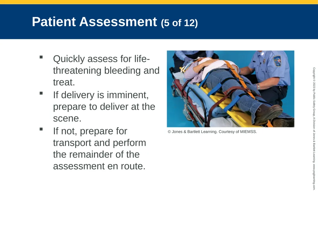
Patient Assessment (5 of 12) Quickly assess for life-threatening bleeding and treat.
If delivery is imminent, prepare to deliver at the scene.
If not, prepare for transport and perform the remainder of the assessment en route. © Jones & Bartlett Learning. Courtesy of MIEMSS.<br>
If delivery is imminent, prepare to deliver at the scene.
If not, prepare for transport and perform the remainder of the assessment en route. © Jones & Bartlett Learning. Courtesy of MIEMSS.<br>
55
Patient Assessment (6 of 12) History taking
Determine patient’s chief complaint, and elaborate using OPQRST-I mnemonic.
Ask about gravida and para.
Ask about the length of gestation and estimated due date.
Ask about fetal movement.
Identify potential complications prior to the delivery.
Ask whether she has experienced complications with any pregnancies.<br>
Determine patient’s chief complaint, and elaborate using OPQRST-I mnemonic.
Ask about gravida and para.
Ask about the length of gestation and estimated due date.
Ask about fetal movement.
Identify potential complications prior to the delivery.
Ask whether she has experienced complications with any pregnancies.<br>
56
Patient Assessment (7 of 12) Consider delivering a newborn at the scene when:
Delivery is expected within a few minutes.
Some type of catastrophe makes it impossible to reach the hospital.
No transportation is available.<br>
Delivery is expected within a few minutes.
Some type of catastrophe makes it impossible to reach the hospital.
No transportation is available.<br>
57
Patient Assessment (8 of 12) To determine whether delivery may occur within a few minutes, look for crowning and ask:
How many weeks pregnant are you?
Is this your first baby?
Are you having contractions? How far apart are the contractions? How long do the contractions last?
Do you feel the urge to move your bowels?
Have you had any spotting or bleeding? If so, what color was it and how much?<br>
How many weeks pregnant are you?
Is this your first baby?
Are you having contractions? How far apart are the contractions? How long do the contractions last?
Do you feel the urge to move your bowels?
Have you had any spotting or bleeding? If so, what color was it and how much?<br>
58
Patient Assessment (9 of 12) Do not allow the pregnant woman to get up to go to the bathroom.
Reassure her that the sensation of needing to move her bowels is normal and that it means she is about to deliver.<br>
Reassure her that the sensation of needing to move her bowels is normal and that it means she is about to deliver.<br>
59
Patient Assessment (10 of 12) Secondary assessment
Based on patient’s chief complaint
Includes a complete set of vital signs
Obtain fetal heart tones and heart rate.
Full-term delivery usually occurs when the contractions are 1 to 2 minutes apart and last from 30 to 60 seconds.<br>
Based on patient’s chief complaint
Includes a complete set of vital signs
Obtain fetal heart tones and heart rate.
Full-term delivery usually occurs when the contractions are 1 to 2 minutes apart and last from 30 to 60 seconds.<br>
60
Patient Assessment (11 of 12) Braxton-Hicks contractions
Irregularly spaced uterine contractions that are more uncomfortable than painful
Patient needs to be transported to the ED.
Physical examination should focus on contractions and possible delivery.<br>
Irregularly spaced uterine contractions that are more uncomfortable than painful
Patient needs to be transported to the ED.
Physical examination should focus on contractions and possible delivery.<br>
61
Patient Assessment (12 of 12) Reassessment
Repeat primary survey with a focus on ABCs and vaginal bleeding.
Obtain another set of vital signs and compare results with earlier.
Recheck interventions and treatments.
If your assessment determines that delivery is imminent, notify staff at the receiving hospital.<br>
Repeat primary survey with a focus on ABCs and vaginal bleeding.
Obtain another set of vital signs and compare results with earlier.
Recheck interventions and treatments.
If your assessment determines that delivery is imminent, notify staff at the receiving hospital.<br>
62
Assessment and Management of Nondelivery Emergencies (1 of 5) Spontaneous abortion
Characterized by vaginal bleeding and abdominal cramping during first half of pregnancy
In second half of pregnancy, patient may present with severe abdominal pain, substantial vaginal bleeding, and cervical dilation.
No specific treatment<br>
Characterized by vaginal bleeding and abdominal cramping during first half of pregnancy
In second half of pregnancy, patient may present with severe abdominal pain, substantial vaginal bleeding, and cervical dilation.
No specific treatment<br>
63
Assessment and Management of Nondelivery Emergencies (2 of 5) Third-trimester bleeding
Determine as much as possible about the nature of the bleeding.
When did the bleeding start?
What activity was the woman engaged in?
How much blood has been lost?
Is she experiencing abdominal pain? If so, what is the nature of the pain? Is it sharp, cramping, dull, achy?
Use OPQRST-I mnemonic.
Look for positive Grey Turner sign or Cullen sign.<br>
Determine as much as possible about the nature of the bleeding.
When did the bleeding start?
What activity was the woman engaged in?
How much blood has been lost?
Is she experiencing abdominal pain? If so, what is the nature of the pain? Is it sharp, cramping, dull, achy?
Use OPQRST-I mnemonic.
Look for positive Grey Turner sign or Cullen sign.<br>
64
Assessment and Management of Nondelivery Emergencies (3 of 5) Ectopic pregnancy
Patient is in severe pain, possibly hypovolemic shock.
Treatment is focused on supporting the ABCs.
Preeclampsia
Characterized by:
Headache, anxiety
Swelling in the hands, face, and feet
Nausea/vomiting<br>
Patient is in severe pain, possibly hypovolemic shock.
Treatment is focused on supporting the ABCs.
Preeclampsia
Characterized by:
Headache, anxiety
Swelling in the hands, face, and feet
Nausea/vomiting<br>
65
Assessment and Management of Nondelivery Emergencies (4 of 5) Severe preeclampsia:
Pulmonary edema/shortness of breath
Confusion or other altered level of consciousness
Visual disturbances
Upper abdominal pain
Myoclonus
Treatment is mostly supportive.<br>
Pulmonary edema/shortness of breath
Confusion or other altered level of consciousness
Visual disturbances
Upper abdominal pain
Myoclonus
Treatment is mostly supportive.<br>
66
Assessment and Management of Nondelivery Emergencies (5 of 5) Supine hypotensive syndrome
Most easily treated by placing the woman onto her left side
If the patient must be immobilized on the long backboard, place blanket rolls or something similar under the right side of the board.
Gestational diabetes
Management should include high-flow oxygen, IV fluids, and administration of dextrose if indicated.<br>
Most easily treated by placing the woman onto her left side
If the patient must be immobilized on the long backboard, place blanket rolls or something similar under the right side of the board.
Gestational diabetes
Management should include high-flow oxygen, IV fluids, and administration of dextrose if indicated.<br>
67
Normal Childbirth (1 of 15) Stages of labor
Onset of labor starts with contractions of the uterus.
Lightening refers to movement of the baby moving down into the pelvis prior to birth.<br>
Onset of labor starts with contractions of the uterus.
Lightening refers to movement of the baby moving down into the pelvis prior to birth.<br>
68
Normal Childbirth (2 of 15) Three stages of labor:
First stage: Begins with onset of contractions and ends when cervix is fully effaced and dilated
Second stage: From full cervical effacement and dilation until fetus is delivered
Third stage: Begins with delivery of the newborn and ends with delivery of the placenta<br>
First stage: Begins with onset of contractions and ends when cervix is fully effaced and dilated
Second stage: From full cervical effacement and dilation until fetus is delivered
Third stage: Begins with delivery of the newborn and ends with delivery of the placenta<br>
69
Normal Childbirth (3 of 15) Preparing for delivery
Prepare your equipment and position the patient for the delivery.
Your part is to help, guide, and support the newborn as it is born.
If delivery must occur during transport, stop the vehicle and have your partner or partners assist with the delivery.<br>
Prepare your equipment and position the patient for the delivery.
Your part is to help, guide, and support the newborn as it is born.
If delivery must occur during transport, stop the vehicle and have your partner or partners assist with the delivery.<br>
70
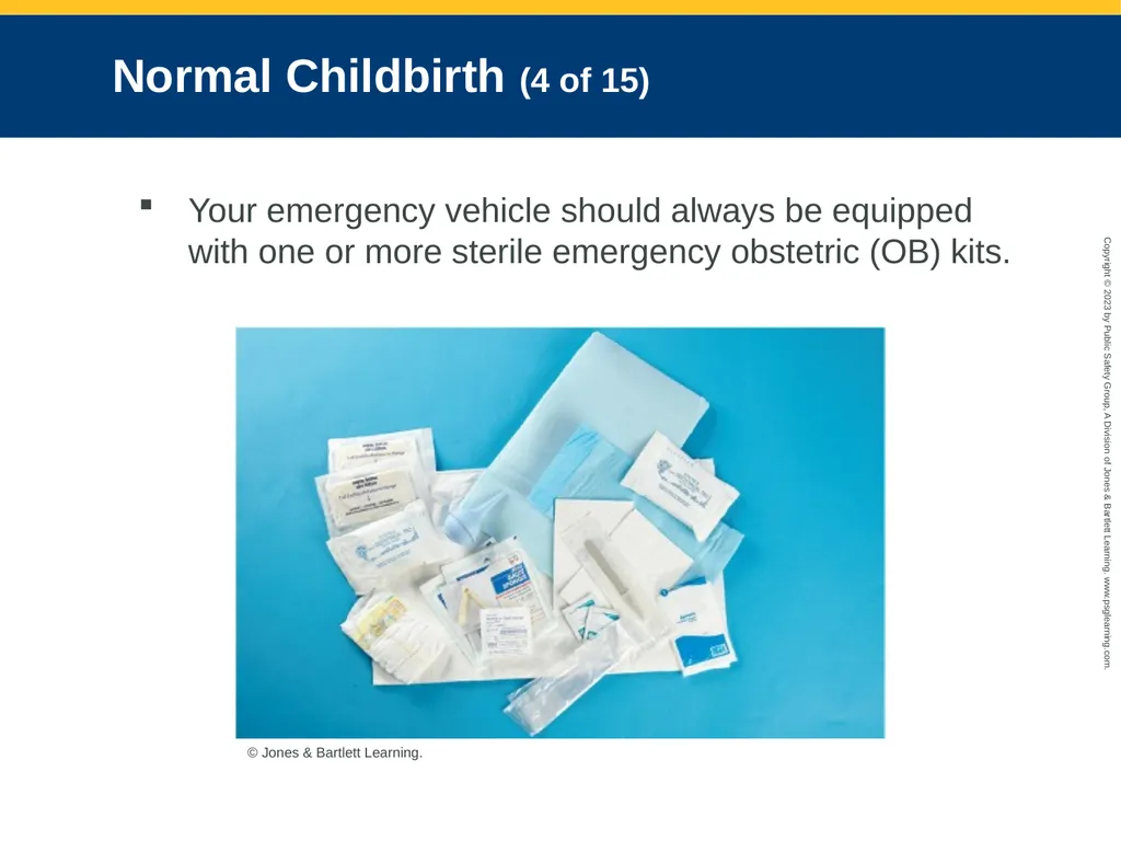
Normal Childbirth (4 of 15) Your emergency vehicle should always be equipped with one or more sterile emergency obstetric (OB) kits. © Jones & Bartlett Learning.<br>
71
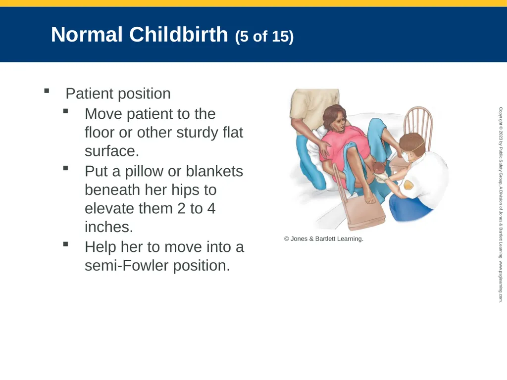
Normal Childbirth (5 of 15) Patient position
Move patient to the floor or other sturdy flat surface.
Put a pillow or blankets beneath her hips to elevate them 2 to 4 inches.
Help her to move into a semi-Fowler position. © Jones & Bartlett Learning.<br>
Move patient to the floor or other sturdy flat surface.
Put a pillow or blankets beneath her hips to elevate them 2 to 4 inches.
Help her to move into a semi-Fowler position. © Jones & Bartlett Learning.<br>
72
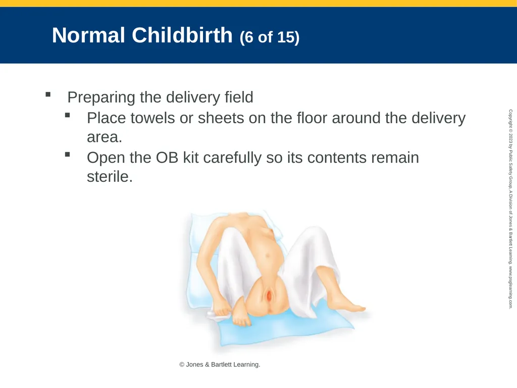
Normal Childbirth (6 of 15) Preparing the delivery field
Place towels or sheets on the floor around the delivery area.
Open the OB kit carefully so its contents remain sterile. © Jones & Bartlett Learning.<br>
Place towels or sheets on the floor around the delivery area.
Open the OB kit carefully so its contents remain sterile. © Jones & Bartlett Learning.<br>
73
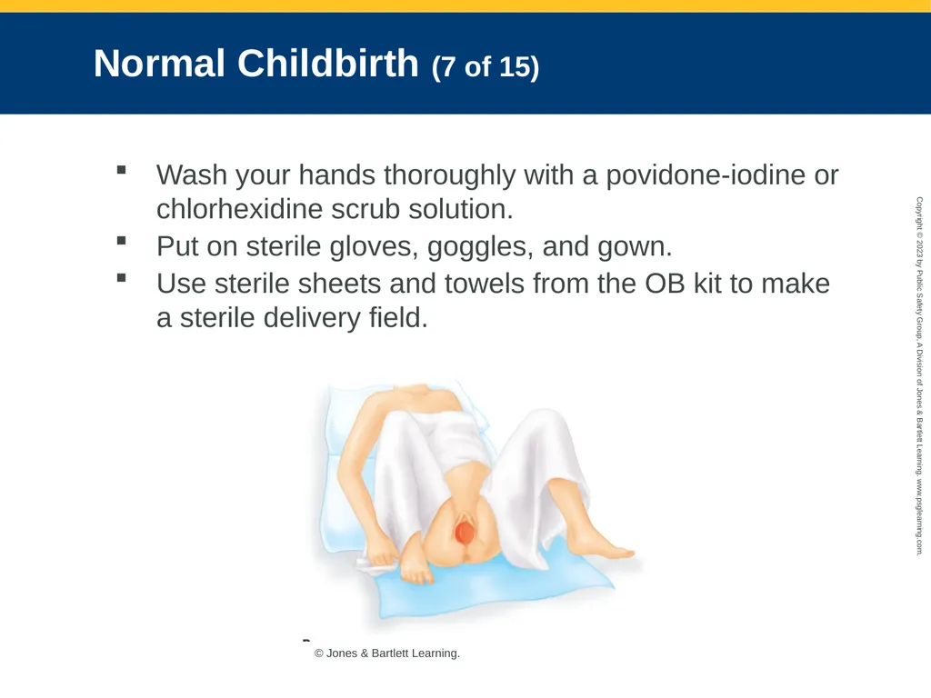
Normal Childbirth (7 of 15) Wash your hands thoroughly with a povidone-iodine or chlorhexidine scrub solution.
Put on sterile gloves, goggles, and gown.
Use sterile sheets and towels from the OB kit to make a sterile delivery field. © Jones & Bartlett Learning.<br>
Put on sterile gloves, goggles, and gown.
Use sterile sheets and towels from the OB kit to make a sterile delivery field. © Jones & Bartlett Learning.<br>
74
Normal Childbirth (8 of 15) Delivering the newborn
Your partner should be at the patient’s head to comfort, soothe, and reassure her during the delivery.
Continually assess the patient for crowning.
Do not allow an abrupt delivery to occur.
Position yourself so that you can see the perineal area at all times.<br>
Your partner should be at the patient’s head to comfort, soothe, and reassure her during the delivery.
Continually assess the patient for crowning.
Do not allow an abrupt delivery to occur.
Position yourself so that you can see the perineal area at all times.<br>
75
Normal Childbirth (9 of 15) Between contractions, encourage the patient to rest and breathe deeply through her mouth.
Wrap the newborn so that only the face is exposed, and that the top of the head is covered.
After suctioning, keep the newborn at the same level as the woman’s vagina until the umbilical cord is cut.<br>
Wrap the newborn so that only the face is exposed, and that the top of the head is covered.
After suctioning, keep the newborn at the same level as the woman’s vagina until the umbilical cord is cut.<br>
76
Normal Childbirth (10 of 15) Apgar scoring
Evaluates adequacy of a newborn’s vital functions immediately after birth
Five parameters: Heart rate, respiratory effort, muscle tone, reflex irritability, and color
Each is given a score from 0 to 2 at 1 minute and again at 5 minutes after birth.
A score in the 4 to 6 range will require resuscitation.<br>
Evaluates adequacy of a newborn’s vital functions immediately after birth
Five parameters: Heart rate, respiratory effort, muscle tone, reflex irritability, and color
Each is given a score from 0 to 2 at 1 minute and again at 5 minutes after birth.
A score in the 4 to 6 range will require resuscitation.<br>
77
Normal Childbirth (11 of 15) Cutting the umbilical cord
Clamping of the cord should be delayed 30 seconds after delivery, unless the newborn requires resuscitation.
Handle the umbilical cord with care.
After the cord has stopped pulsating, tie or clamp it 4 inches from the newborn’s navel, and place the second tie 2 inches from the first.
Cut the cord between the two ties or clamps.
Wrap the newborn in a dry blanket.<br>
Clamping of the cord should be delayed 30 seconds after delivery, unless the newborn requires resuscitation.
Handle the umbilical cord with care.
After the cord has stopped pulsating, tie or clamp it 4 inches from the newborn’s navel, and place the second tie 2 inches from the first.
Cut the cord between the two ties or clamps.
Wrap the newborn in a dry blanket.<br>
78
Normal Childbirth (12 of 15) Delivery of the placenta
Usually placenta delivers within a few minutes of the birth (may take as long as 30 minutes)
Never pull on the end of the umbilical cord to speed delivery of the placenta.
Wrap the entire placenta and umbilical cord in a towel and place them in a plastic bag.<br>
Usually placenta delivers within a few minutes of the birth (may take as long as 30 minutes)
Never pull on the end of the umbilical cord to speed delivery of the placenta.
Wrap the entire placenta and umbilical cord in a towel and place them in a plastic bag.<br>
79
Normal Childbirth (13 of 15) Postpartum care
Place a sterile pad or sanitary napkin over the vagina and straighten the woman’s legs.
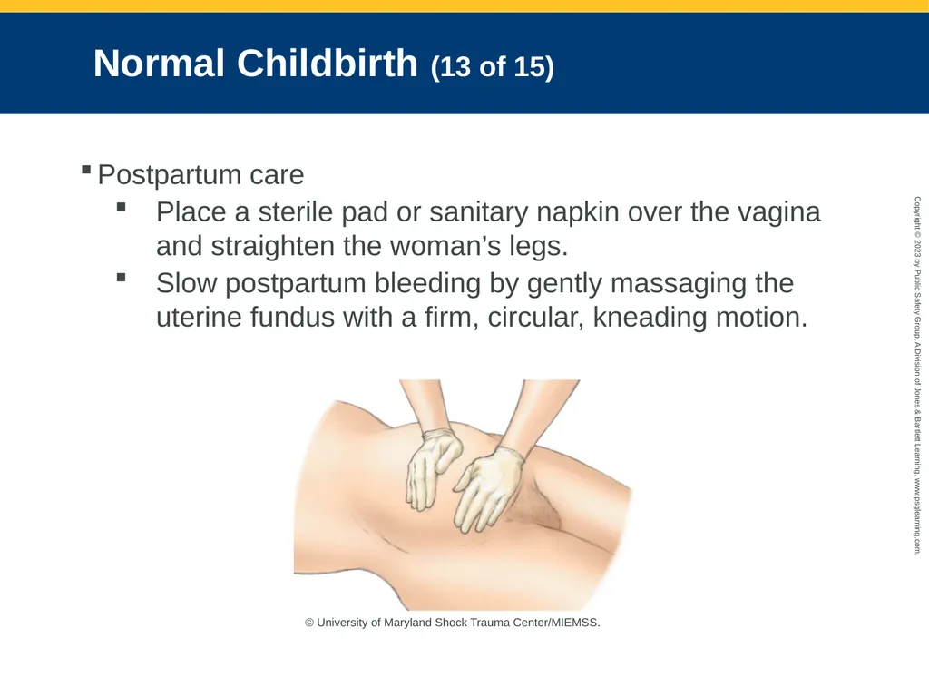
Slow postpartum bleeding by gently massaging the uterine fundus with a firm, circular, kneading motion. © University of Maryland Shock Trauma Center/MIEMSS.<br>
Place a sterile pad or sanitary napkin over the vagina and straighten the woman’s legs.
Slow postpartum bleeding by gently massaging the uterine fundus with a firm, circular, kneading motion. © University of Maryland Shock Trauma Center/MIEMSS.<br>
80
Normal Childbirth (14 of 15) Massaging the uterus and having the newborn nurse will help contract the uterus and slow bleeding.
Record time of birth.
After delivery of the newborn, obtain the woman’s vital signs.
Monitor closely for postpartum hemorrhage and shock, seizure activity, or respiratory difficulty.<br>
Record time of birth.
After delivery of the newborn, obtain the woman’s vital signs.
Monitor closely for postpartum hemorrhage and shock, seizure activity, or respiratory difficulty.<br>
81
Normal Childbirth (15 of 15) Postpartum hemorrhage
Blood loss exceeds 1 pint during the first 24 hours after giving birth
Emergency situations:
Placenta has not delivered after 30 minutes.
More than 500 mL of bleeding occurs before delivery of the placenta.
Substantial bleeding occurs after placenta delivery.
Provide rapid transport.<br>
Blood loss exceeds 1 pint during the first 24 hours after giving birth
Emergency situations:
Placenta has not delivered after 30 minutes.
More than 500 mL of bleeding occurs before delivery of the placenta.
Substantial bleeding occurs after placenta delivery.
Provide rapid transport.<br>
82
Complications of Labor (1 of 5) Definitive treatment for many abnormal labor or delivery problems is a cesarean section.
Preterm labor
Labor that begins after the 20th week of gestation but before the 37th week
Best treatment option is a bolus of an isotonic crystalloid solution.<br>
Preterm labor
Labor that begins after the 20th week of gestation but before the 37th week
Best treatment option is a bolus of an isotonic crystalloid solution.<br>
83
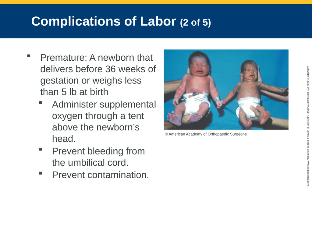
Complications of Labor (2 of 5) Premature: A newborn that delivers before 36 weeks of gestation or weighs less than 5 lb at birth
Administer supplemental oxygen through a tent above the newborn’s head.
Prevent bleeding from the umbilical cord.
Prevent contamination. © American Academy of Orthopaedic Surgeons.<br>
Administer supplemental oxygen through a tent above the newborn’s head.
Prevent bleeding from the umbilical cord.
Prevent contamination. © American Academy of Orthopaedic Surgeons.<br>
84
Complications of Labor (3 of 5) Postterm pregnancy
Pregnancy that is longer than 41 completed weeks of gestation.
Risks to the mother
More difficult labor and delivery
Increased likelihood of cesarean section
Greater risk of perineal tears and infection
Be prepared to resuscitate the newborn<br>
Pregnancy that is longer than 41 completed weeks of gestation.
Risks to the mother
More difficult labor and delivery
Increased likelihood of cesarean section
Greater risk of perineal tears and infection
Be prepared to resuscitate the newborn<br>
85
Complications of Labor (4 of 5) Fetal distress
May be caused by hypoxia, nuchal cord, trauma, abruptio placentae, fetal developmental disabilities, and a prolapsed cord
Rely on the information provided by the woman on fetus movement.
Provide support and rapid transport.<br>
May be caused by hypoxia, nuchal cord, trauma, abruptio placentae, fetal developmental disabilities, and a prolapsed cord
Rely on the information provided by the woman on fetus movement.
Provide support and rapid transport.<br>
86
Complications of Labor (5 of 5) Uterine rupture
At greatest risk are women who have had several children and those with a scar on the uterus.
A woman may have initially had very strong and painful contractions, but contractions slackened off and she has severe abdominal pain.
Substantial vaginal bleeding may or may not be obvious.<br>
At greatest risk are women who have had several children and those with a scar on the uterus.
A woman may have initially had very strong and painful contractions, but contractions slackened off and she has severe abdominal pain.
Substantial vaginal bleeding may or may not be obvious.<br>
87
High-Risk Pregnancy Conditions (1 of 6) Meconium staining
In cases of fetal distress, or the stress of labor and delivery, fetus may expel the meconium into the amniotic fluid.
May result in chemical pneumonia in newborn
Be vigilant regarding the need for suctioning.
Call early for paramedic backup.<br>
In cases of fetal distress, or the stress of labor and delivery, fetus may expel the meconium into the amniotic fluid.
May result in chemical pneumonia in newborn
Be vigilant regarding the need for suctioning.
Call early for paramedic backup.<br>
88
High-Risk Pregnancy Conditions (2 of 6) Multiple gestation
Always keep a spare OB kit in your equipment.
Consider possibility of twins if the uterus is still large and firm after delivery of the first newborn.<br>
Always keep a spare OB kit in your equipment.
Consider possibility of twins if the uterus is still large and firm after delivery of the first newborn.<br>
89
High-Risk Pregnancy Conditions (3 of 6) If there is more than one fetus:
Repeat earlier preparations for delivery.
When the first newborn is born, clamp and cut the cord before the second newborn is delivered.
Keep the newborns warm, well oxygenated, and in as sterile an environment as possible.
Identify first newborn delivered as “Baby A”.
Record time of birth of each twin separately.<br>
Repeat earlier preparations for delivery.
When the first newborn is born, clamp and cut the cord before the second newborn is delivered.
Keep the newborns warm, well oxygenated, and in as sterile an environment as possible.
Identify first newborn delivered as “Baby A”.
Record time of birth of each twin separately.<br>
90
High-Risk Pregnancy Conditions (4 of 6) Cephalopelvic disproportion
Head of the fetus is larger than the pelvis.
Cesarean section will be required.
May cause massive hemorrhage, along with other postpartum complications<br>
Head of the fetus is larger than the pelvis.
Cesarean section will be required.
May cause massive hemorrhage, along with other postpartum complications<br>
91
High-Risk Pregnancy Conditions (5 of 6) Intrauterine fetal death
Complications of labor and delivery can result in death of fetus.
Grieving parents will require all your professionalism and support skills.
Do not attempt to resuscitate an obviously dead newborn.
You must attempt to resuscitate normal-appearing newborns.<br>
Complications of labor and delivery can result in death of fetus.
Grieving parents will require all your professionalism and support skills.
Do not attempt to resuscitate an obviously dead newborn.
You must attempt to resuscitate normal-appearing newborns.<br>
92
High-Risk Pregnancy Conditions (6 of 6) Amniotic fluid embolism
Extremely rare
Risk factors: Maternal age greater than 35 years, eclampsia, abruptio placentae, placenta previa, uterine rupture, fetal distress
Signs and symptoms include a sudden onset of respiratory distress and hypotension.
Treatment focuses on supporting vital systems and providing rapid transport.<br>
Extremely rare
Risk factors: Maternal age greater than 35 years, eclampsia, abruptio placentae, placenta previa, uterine rupture, fetal distress
Signs and symptoms include a sudden onset of respiratory distress and hypotension.
Treatment focuses on supporting vital systems and providing rapid transport.<br>
93
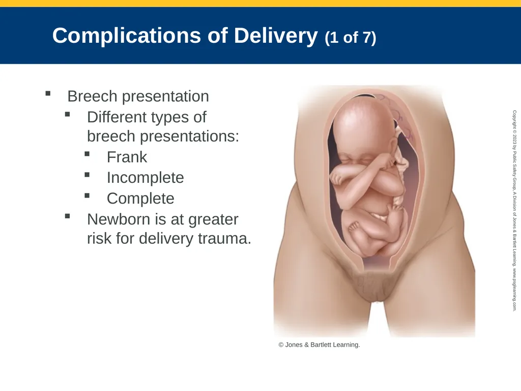
Complications of Delivery (1 of 7) Breech presentation
Different types of breech presentations:
Frank
Incomplete
Complete
Newborn is at greater risk for delivery trauma. © Jones & Bartlett Learning.<br>
Different types of breech presentations:
Frank
Incomplete
Complete
Newborn is at greater risk for delivery trauma. © Jones & Bartlett Learning.<br>
94
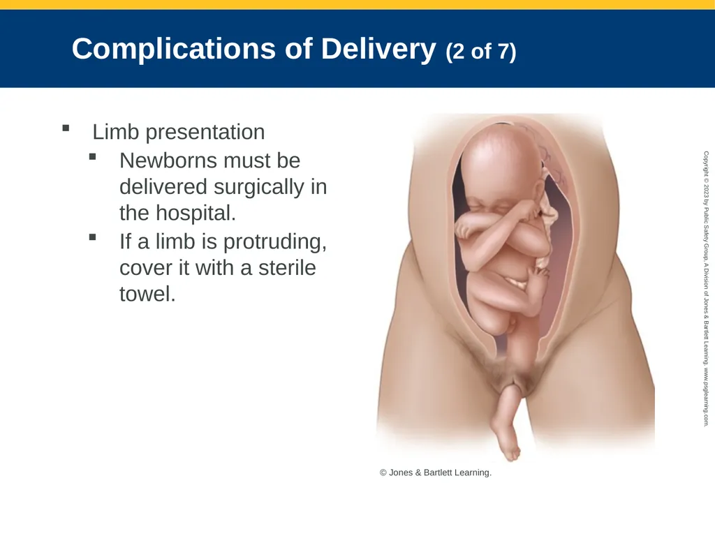
Complications of Delivery (2 of 7) Limb presentation
Newborns must be delivered surgically in the hospital.
If a limb is protruding, cover it with a sterile towel. © Jones & Bartlett Learning.<br>
Newborns must be delivered surgically in the hospital.
If a limb is protruding, cover it with a sterile towel. © Jones & Bartlett Learning.<br>
95
Complications of Delivery (3 of 7) Place the patient on her back with head down and hips elevated.
Prevent further trauma to the newborn that could result from the patient’s continued pushing.
Transport rapidly.<br>
Prevent further trauma to the newborn that could result from the patient’s continued pushing.
Transport rapidly.<br>
96
Complications of Delivery (4 of 7) Shoulder dystocia
Women with diabetes, large fetuses, and postterm fetuses are at increased risk.
Occurs after head has delivered and shoulder cannot get past the woman’s symphysis pubis
Major concern for the newborn is damage to the brachial nerve plexus.
McRoberts maneuver<br>
Women with diabetes, large fetuses, and postterm fetuses are at increased risk.
Occurs after head has delivered and shoulder cannot get past the woman’s symphysis pubis
Major concern for the newborn is damage to the brachial nerve plexus.
McRoberts maneuver<br>
97
Complications of Delivery (5 of 7) Nuchal cord
Wound tightly around the newborn’s neck could cause asphyxiation
Slip your finger under the cord and gently attempt to slip it over newborn’s shoulder and head.
If unsuccessful, cut the cord before delivery can continue.<br>
Wound tightly around the newborn’s neck could cause asphyxiation
Slip your finger under the cord and gently attempt to slip it over newborn’s shoulder and head.
If unsuccessful, cut the cord before delivery can continue.<br>
98
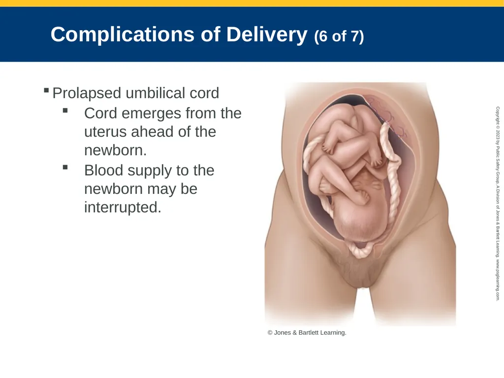
Complications of Delivery (6 of 7) Prolapsed umbilical cord
Cord emerges from the uterus ahead of the newborn.
Blood supply to the newborn may be interrupted. © Jones & Bartlett Learning.<br>
Cord emerges from the uterus ahead of the newborn.
Blood supply to the newborn may be interrupted. © Jones & Bartlett Learning.<br>
99
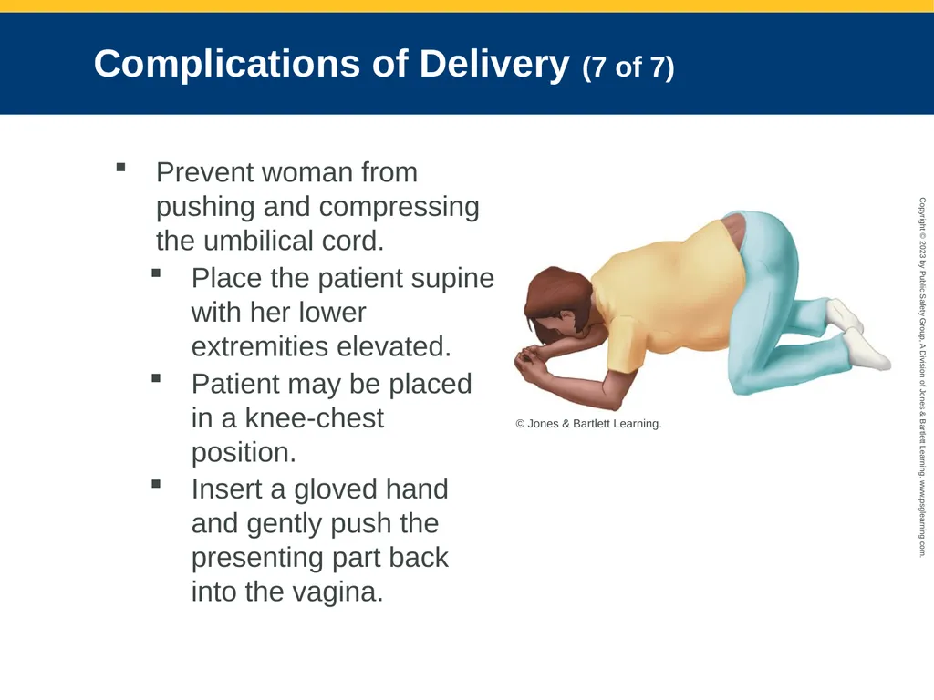
Complications of Delivery (7 of 7) Prevent woman from pushing and compressing the umbilical cord.
Place the patient supine with her lower extremities elevated.
Patient may be placed in a knee-chest position.
Insert a gloved hand and gently push the presenting part back into the vagina. © Jones & Bartlett Learning.<br>
Place the patient supine with her lower extremities elevated.
Patient may be placed in a knee-chest position.
Insert a gloved hand and gently push the presenting part back into the vagina. © Jones & Bartlett Learning.<br>
100
Postpartum Complications (1 of 5) Excessive bleeding
Bleeding that exceeds approximately 1,000 mL is considered high risk for maternal mortality and morbidity
Usually caused by uterus not fully contracting
Cover vagina with sterile pad.
Save any blood-soaked pads or tissue.
Administer oxygen as needed, monitor vital signs frequently, and provide rapid transport.<br>
Bleeding that exceeds approximately 1,000 mL is considered high risk for maternal mortality and morbidity
Usually caused by uterus not fully contracting
Cover vagina with sterile pad.
Save any blood-soaked pads or tissue.
Administer oxygen as needed, monitor vital signs frequently, and provide rapid transport.<br>
101
Postpartum Complications (2 of 5) Uterine inversion
Placenta fails to detach properly and adheres to uterine wall when it is expelled.
Can result from placing excessive pressure on the uterus during fundal massage or from exerting excess traction on the umbilical cord
Make one attempt at replacement by using the palm of the hand to try to push it back inside the body.<br>
Placenta fails to detach properly and adheres to uterine wall when it is expelled.
Can result from placing excessive pressure on the uterus during fundal massage or from exerting excess traction on the umbilical cord
Make one attempt at replacement by using the palm of the hand to try to push it back inside the body.<br>
102
Postpartum Complications (3 of 5) Never use your fingers to try to replace a prolapsed uterus.
Administer 100% supplemental oxygen via a nonrebreathing mask.
Start two IV lines with normal saline, and titrate fluid administration based on the vital signs.
Treat for shock.<br>
Administer 100% supplemental oxygen via a nonrebreathing mask.
Start two IV lines with normal saline, and titrate fluid administration based on the vital signs.
Treat for shock.<br>
103
Postpartum Complications (4 of 5) Pulmonary embolism
May form from several sources
Suspect if woman experiences sudden dyspnea, tachycardia, or hypotension in postpartum state
Management: High-flow oxygen and rapid transport<br>
May form from several sources
Suspect if woman experiences sudden dyspnea, tachycardia, or hypotension in postpartum state
Management: High-flow oxygen and rapid transport<br>
104
Postpartum Complications (5 of 5) Spina bifida
Portion of spinal cord or meninges protrudes outside of the vertebrae
Cover the open area of the spinal cord with a sterile, moist dressing immediately after birth.
Maintenance of body temperature is important when applying moist dressings.<br>
Portion of spinal cord or meninges protrudes outside of the vertebrae
Cover the open area of the spinal cord with a sterile, moist dressing immediately after birth.
Maintenance of body temperature is important when applying moist dressings.<br>
105
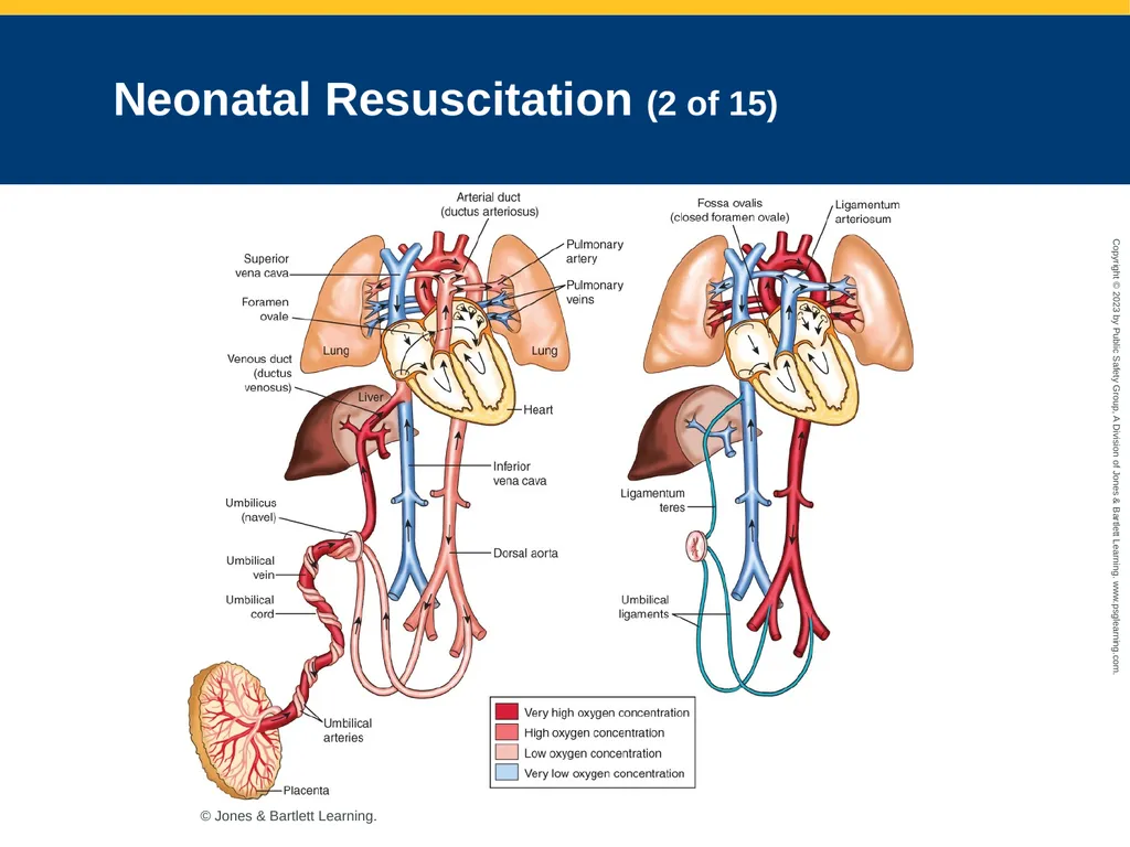
Neonatal Resuscitation (1 of 15) Physiology
In utero, a fetus receives its oxygen from the placenta.
Fetal transition: As fetus is delivered, rapid series of events occurs to enable newborn to breathe.
Following fetal transition, neonate breathes and oxygenates the blood.<br>
In utero, a fetus receives its oxygen from the placenta.
Fetal transition: As fetus is delivered, rapid series of events occurs to enable newborn to breathe.
Following fetal transition, neonate breathes and oxygenates the blood.<br>
106
Neonatal Resuscitation (2 of 15) © Jones & Bartlett Learning.<br>
107
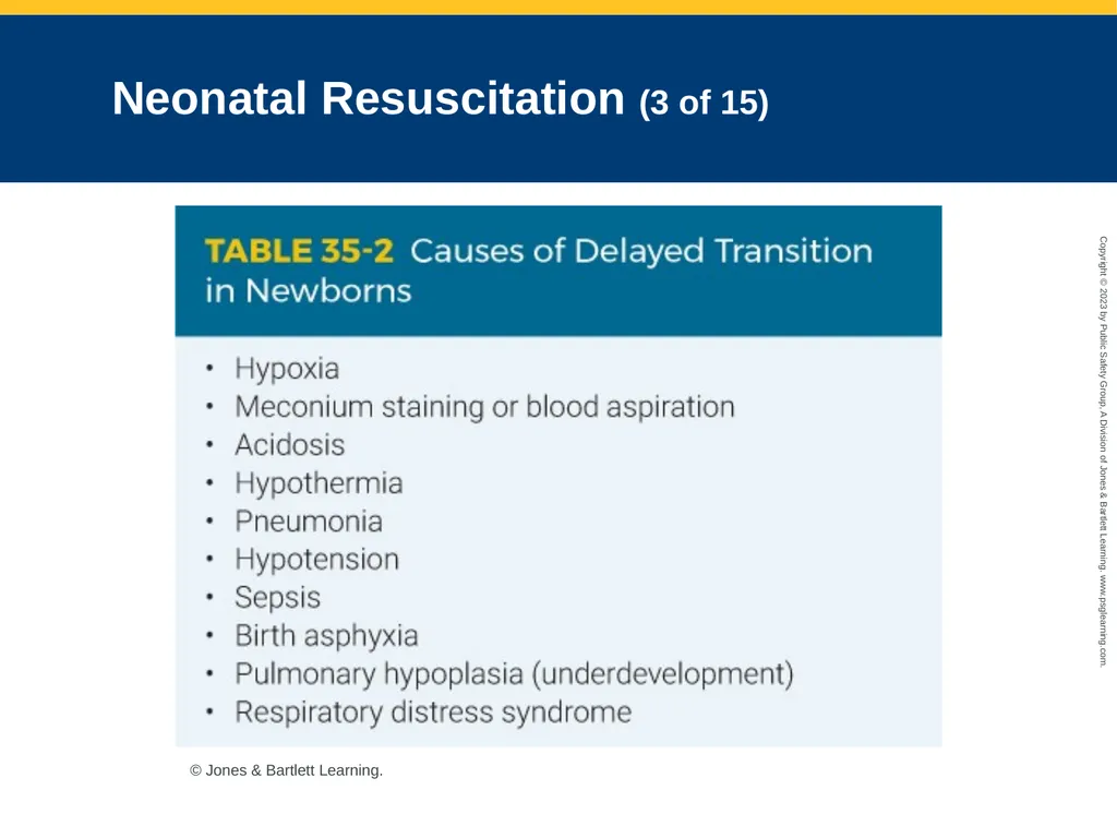
Neonatal Resuscitation (3 of 15) © Jones & Bartlett Learning.<br>
108
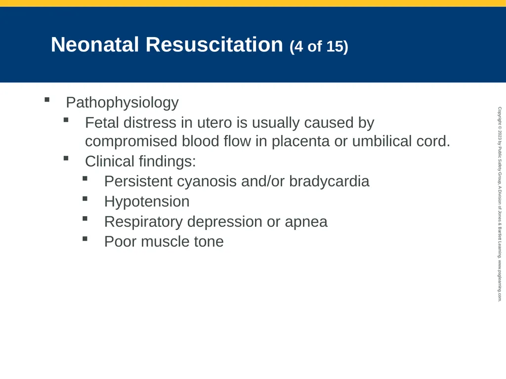
Neonatal Resuscitation (4 of 15) Pathophysiology
Fetal distress in utero is usually caused by compromised blood flow in placenta or umbilical cord.
Clinical findings:
Persistent cyanosis and/or bradycardia
Hypotension
Respiratory depression or apnea
Poor muscle tone<br>
Fetal distress in utero is usually caused by compromised blood flow in placenta or umbilical cord.
Clinical findings:
Persistent cyanosis and/or bradycardia
Hypotension
Respiratory depression or apnea
Poor muscle tone<br>
109
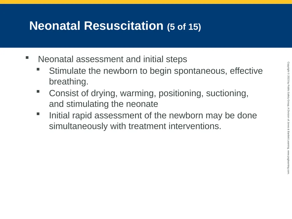
Neonatal Resuscitation (5 of 15) Neonatal assessment and initial steps
Stimulate the newborn to begin spontaneous, effective breathing.
Consist of drying, warming, positioning, suctioning, and stimulating the neonate
Initial rapid assessment of the newborn may be done simultaneously with treatment interventions.<br>
Stimulate the newborn to begin spontaneous, effective breathing.
Consist of drying, warming, positioning, suctioning, and stimulating the neonate
Initial rapid assessment of the newborn may be done simultaneously with treatment interventions.<br>
110
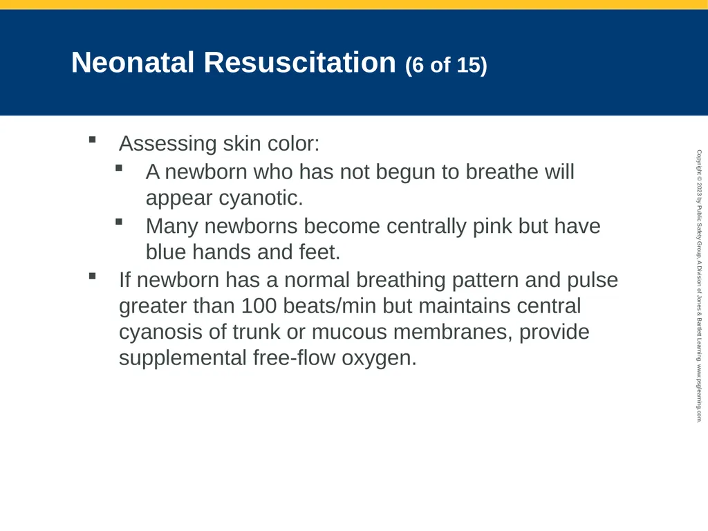
Neonatal Resuscitation (6 of 15) Assessing skin color:
A newborn who has not begun to breathe will appear cyanotic.
Many newborns become centrally pink but have blue hands and feet.
If newborn has a normal breathing pattern and pulse greater than 100 beats/min but maintains central cyanosis of trunk or mucous membranes, provide supplemental free-flow oxygen.<br>
A newborn who has not begun to breathe will appear cyanotic.
Many newborns become centrally pink but have blue hands and feet.
If newborn has a normal breathing pattern and pulse greater than 100 beats/min but maintains central cyanosis of trunk or mucous membranes, provide supplemental free-flow oxygen.<br>
111
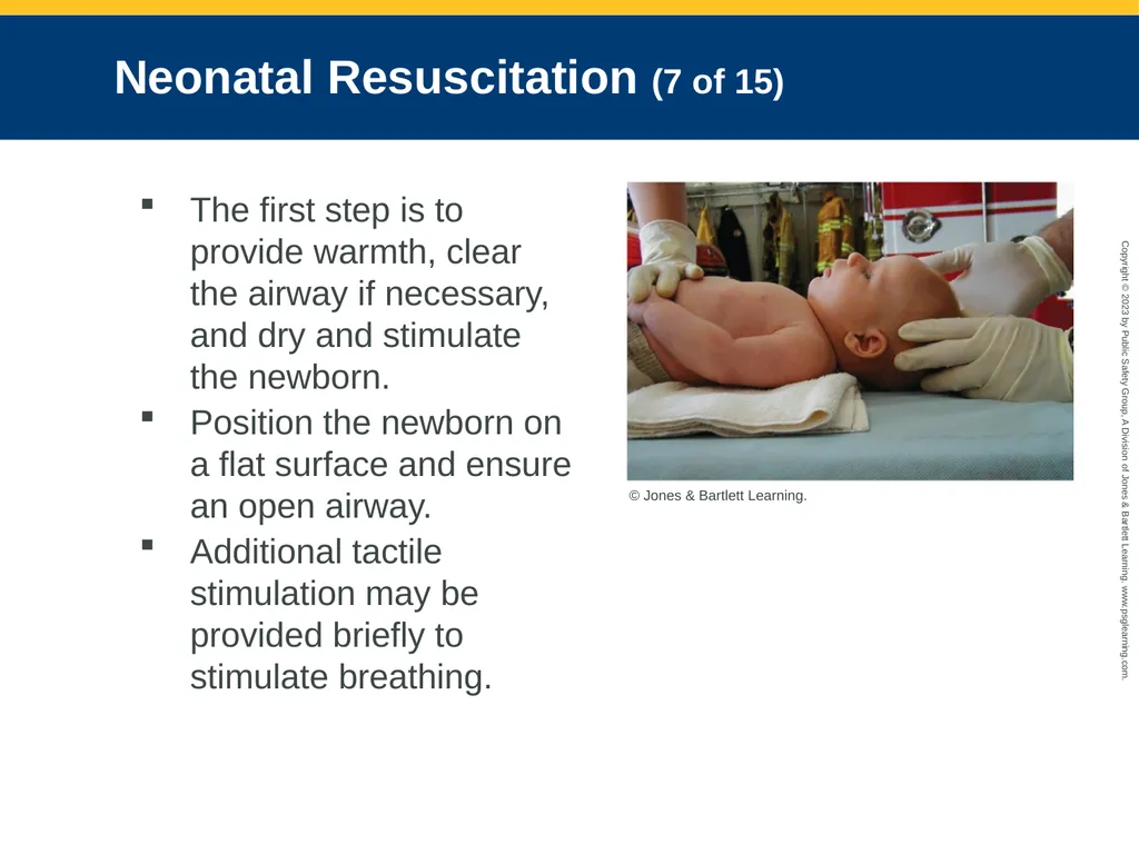
Neonatal Resuscitation (7 of 15) The first step is to provide warmth, clear the airway if necessary, and dry and stimulate the newborn.
Position the newborn on a flat surface and ensure an open airway.
Additional tactile stimulation may be provided briefly to stimulate breathing. © Jones & Bartlett Learning.<br>
Position the newborn on a flat surface and ensure an open airway.
Additional tactile stimulation may be provided briefly to stimulate breathing. © Jones & Bartlett Learning.<br>
112
Neonatal Resuscitation (8 of 15) Neonatal resuscitation consists of:
Initial steps in stabilization
Positive-pressure ventilation (PPV) with oxygen saturation monitoring
Chest compressions if heart rate is below 60 beats/min
Consider intubation, continue chest compressions, and coordinate with PPV.<br>
Initial steps in stabilization
Positive-pressure ventilation (PPV) with oxygen saturation monitoring
Chest compressions if heart rate is below 60 beats/min
Consider intubation, continue chest compressions, and coordinate with PPV.<br>
113
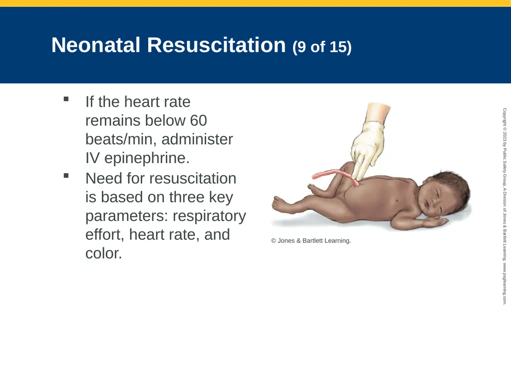
Neonatal Resuscitation (9 of 15) If the heart rate remains below 60 beats/min, administer IV epinephrine.
Need for resuscitation is based on three key parameters: respiratory effort, heart rate, and color. © Jones & Bartlett Learning.<br>
Need for resuscitation is based on three key parameters: respiratory effort, heart rate, and color. © Jones & Bartlett Learning.<br>
114
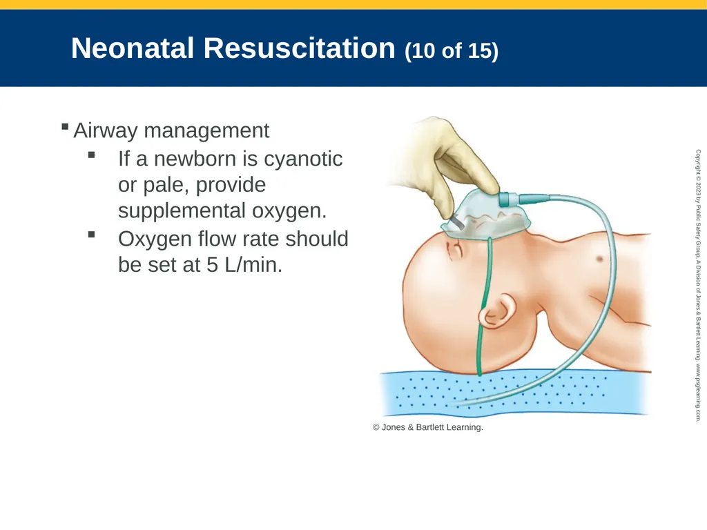
Neonatal Resuscitation (10 of 15) Airway management
If a newborn is cyanotic or pale, provide supplemental oxygen.
Oxygen flow rate should be set at 5 L/min. © Jones & Bartlett Learning.<br>
If a newborn is cyanotic or pale, provide supplemental oxygen.
Oxygen flow rate should be set at 5 L/min. © Jones & Bartlett Learning.<br>
115
Neonatal Resuscitation (11 of 15) Bag-mask ventilation
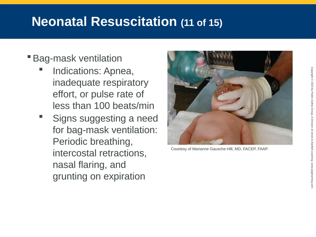
Indications: Apnea, inadequate respiratory effort, or pulse rate of less than 100 beats/min
Signs suggesting a need for bag-mask ventilation: Periodic breathing, intercostal retractions, nasal flaring, and grunting on expiration Courtesy of Marianne Gausche-Hill, MD, FACEP, FAAP.<br>
Indications: Apnea, inadequate respiratory effort, or pulse rate of less than 100 beats/min
Signs suggesting a need for bag-mask ventilation: Periodic breathing, intercostal retractions, nasal flaring, and grunting on expiration Courtesy of Marianne Gausche-Hill, MD, FACEP, FAAP.<br>
116
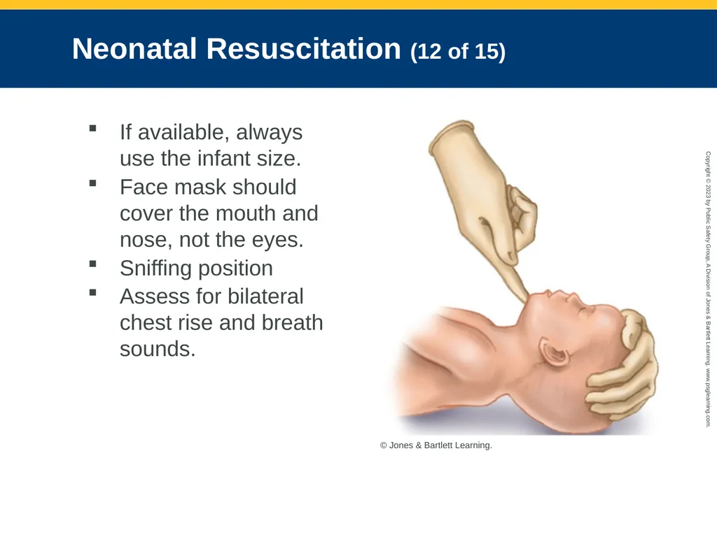
Neonatal Resuscitation (12 of 15) If available, always use the infant size.
Face mask should cover the mouth and nose, not the eyes.
Sniffing position
Assess for bilateral chest rise and breath sounds. © Jones & Bartlett Learning.<br>
Face mask should cover the mouth and nose, not the eyes.
Sniffing position
Assess for bilateral chest rise and breath sounds. © Jones & Bartlett Learning.<br>
117
Neonatal Resuscitation (13 of 15) Circulation
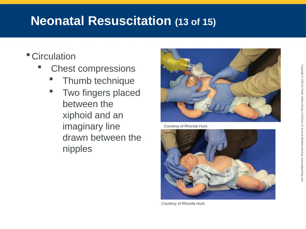
Chest compressions
Thumb technique
Two fingers placed between the xiphoid and an imaginary line drawn between the nipples Courtesy of Rhonda Hunt. Courtesy of Rhonda Hunt.<br>
Chest compressions
Thumb technique
Two fingers placed between the xiphoid and an imaginary line drawn between the nipples Courtesy of Rhonda Hunt. Courtesy of Rhonda Hunt.<br>
118
Neonatal Resuscitation (14 of 15) Bag-mask ventilation is performed during a pause after every third compression.
Adequate ventilation is absolutely critical to the successful resuscitation of the neonate.<br>
Adequate ventilation is absolutely critical to the successful resuscitation of the neonate.<br>
119
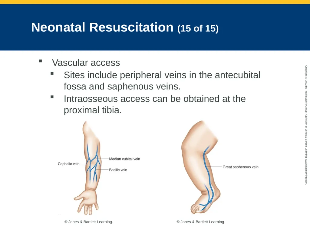
Neonatal Resuscitation (15 of 15) Vascular access
Sites include peripheral veins in the antecubital fossa and saphenous veins.
Intraosseous access can be obtained at the proximal tibia. © Jones & Bartlett Learning. © Jones & Bartlett Learning.<br>
Sites include peripheral veins in the antecubital fossa and saphenous veins.
Intraosseous access can be obtained at the proximal tibia. © Jones & Bartlett Learning. © Jones & Bartlett Learning.<br>
120
Pathophysiology, Assessment, and Management of Specific Conditions (1 of 7) Apnea
Common in newborns delivered before 32 weeks of gestation
If apnea does not respond to stimulation and further steps are not taken, hypoxemia and bradycardia can occur.<br>
Common in newborns delivered before 32 weeks of gestation
If apnea does not respond to stimulation and further steps are not taken, hypoxemia and bradycardia can occur.<br>
121
Pathophysiology, Assessment, and Management of Specific Conditions (2 of 7) Often follows a period of hypoxia or hypothermia
Other causes:
Maternal or infant narcotic exposure
Airway or respiratory muscle weakness
Septicemia
Prolonged or difficult labor and delivery
Gastroesophageal reflux
Central nervous system abnormalities<br>
Other causes:
Maternal or infant narcotic exposure
Airway or respiratory muscle weakness
Septicemia
Prolonged or difficult labor and delivery
Gastroesophageal reflux
Central nervous system abnormalities<br>
122
Pathophysiology, Assessment, and Management of Specific Conditions (3 of 7) Important to differentiate between primary apnea and secondary apnea.
If the newborn has experienced a relatively short period of hypoxia, drying and stimulation may cause resumption of breathing.
If hypoxia continues during primary apnea, the newborn will gasp and enter secondary apnea.
PPV by bag-mask device is required.<br>
If the newborn has experienced a relatively short period of hypoxia, drying and stimulation may cause resumption of breathing.
If hypoxia continues during primary apnea, the newborn will gasp and enter secondary apnea.
PPV by bag-mask device is required.<br>
123
Pathophysiology, Assessment, and Management of Specific Conditions (4 of 7) Bradycardia
Most frequently a result of inadequate ventilation and often responds to effective PPV.
Assess patency of the airway.
Begin chest compressions for a heart rate that is less than 60 beats/min despite at least 30 seconds of effective PPV.
For persistent bradycardia, call early for paramedic backup.<br>
Most frequently a result of inadequate ventilation and often responds to effective PPV.
Assess patency of the airway.
Begin chest compressions for a heart rate that is less than 60 beats/min despite at least 30 seconds of effective PPV.
For persistent bradycardia, call early for paramedic backup.<br>
124
Pathophysiology, Assessment, and Management of Specific Conditions (5 of 7) Hypoglycemia
Blood glucose level of less than 45 mg/dL
Most newborns remain asymptomatic until the glucose level is less than 20 mg/dL for a substantial length of time.
Symptoms include:
Cyanosis, apnea, irritability, poor sucking or feeding, hypotonia, irregular respirations, eye rolling, hypothermia, and decreased response to stimuli<br>
Blood glucose level of less than 45 mg/dL
Most newborns remain asymptomatic until the glucose level is less than 20 mg/dL for a substantial length of time.
Symptoms include:
Cyanosis, apnea, irritability, poor sucking or feeding, hypotonia, irregular respirations, eye rolling, hypothermia, and decreased response to stimuli<br>
125
Pathophysiology, Assessment, and Management of Specific Conditions (6 of 7) Check the blood glucose level in all sick newborns, and evaluate vital signs.
Establish good oxygenation, ventilation, and circulation before managing hypoglycemia.
Warm IV fluids can assist in rewarming the newborn who is hypothermic.<br>
Establish good oxygenation, ventilation, and circulation before managing hypoglycemia.
Warm IV fluids can assist in rewarming the newborn who is hypothermic.<br>
126
Pathophysiology, Assessment, and Management of Specific Conditions (7 of 7) Hypovolemia
Newborns appear pale and have weak pulses.
Persistent tachycardia or bradycardia
Administer 10 mL/kg of an isotonic crystalloid over 5 to 10 minutes.
Reassess and provide additional fluids as needed.<br>
Newborns appear pale and have weak pulses.
Persistent tachycardia or bradycardia
Administer 10 mL/kg of an isotonic crystalloid over 5 to 10 minutes.
Reassess and provide additional fluids as needed.<br>