ORTHO CURRICULUM Procedures: Arthrocentesis Indications Diagnosis of joint disease by synovial fluid analysis (gout, septic arthritis) Local instillation of medications into a joint for inflammatory disease Diagnosis of ligamentous or bony
"ORTHO CURRICULUM Procedures: Arthrocentesis" is the property of its rightful owner. Permission is granted to
download and print the materials on this website for personal, non-commercial use only, and to display it
on your personal computer provided you do not modify the materials and that you retain all copyright
notices contained in the materials. By downloading content from our website, you accept the terms of this
agreement.
Presentation Transcript
01
ORTHO CURRICULUM Procedures: Arthrocentesis<br>
02
Indications Diagnosis of joint disease by synovial fluid analysis (gout, septic arthritis)
Local instillation of medications into a joint for inflammatory disease
Diagnosis of ligamentous or bony injury by confirming presence of blood in the joint
Relief of painful hemarthrosis and effusion<br>
03
Contraindications Overlying skin infection
Known bacteremia
Caution with bleeding disorders<br>
04
Equipment 10-30cc syringe (depending on size of effusion)
18-22ga needle
1% lidocaine with 25-27ga needle and 10cc syringe
Chlorhexidine
Sterile gloves
Specimen cup
Culture bottles
Lavender tube
Light green tube<br>
05
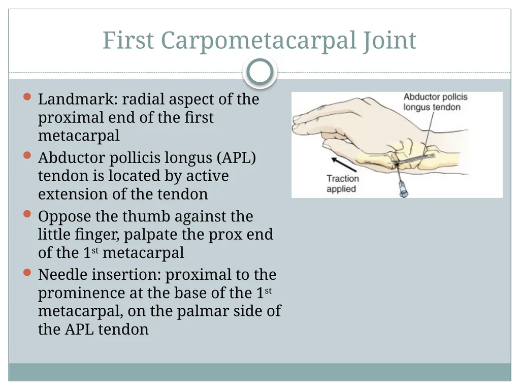
First Carpometacarpal Joint Landmark: radial aspect of the proximal end of the first metacarpal
Abductor pollicis longus (APL) tendon is located by active extension of the tendon
Oppose the thumb against the little finger, palpate the prox end of the 1st metacarpal
Needle insertion: proximal to the prominence at the base of the 1st metacarpal, on the palmar side of the APL tendon<br>
06
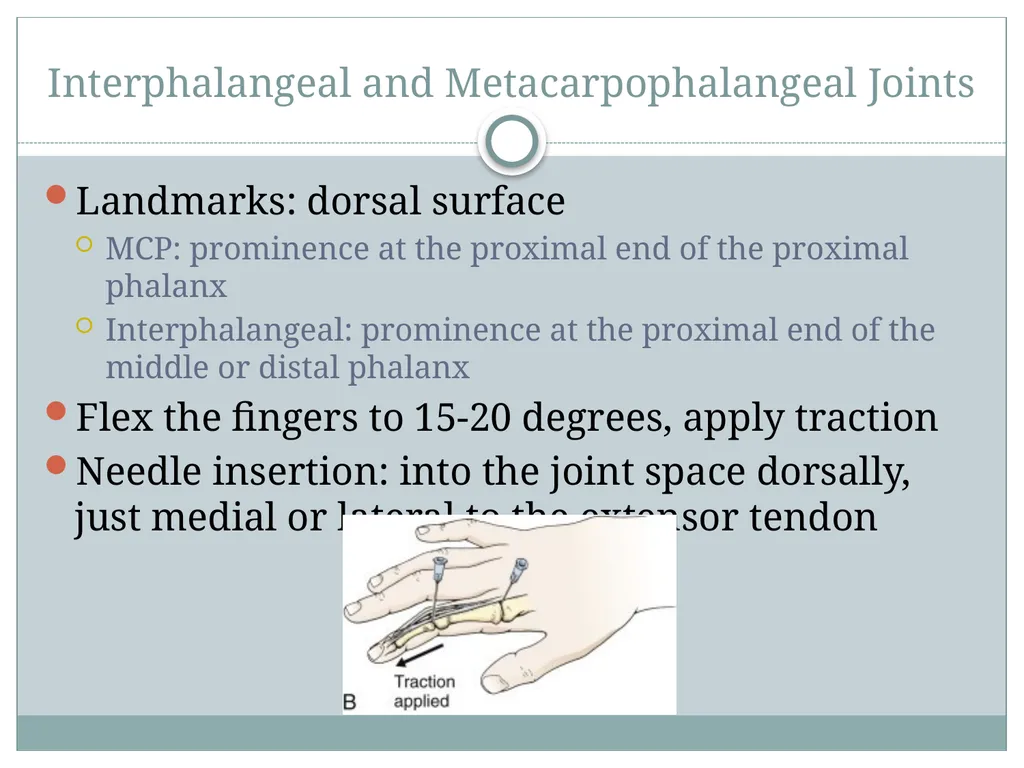
Interphalangeal and Metacarpophalangeal Joints Landmarks: dorsal surface
MCP: prominence at the proximal end of the proximal phalanx
Interphalangeal: prominence at the proximal end of the middle or distal phalanx
Flex the fingers to 15-20 degrees, apply traction
Needle insertion: into the joint space dorsally, just medial or lateral to the extensor tendon<br>
07
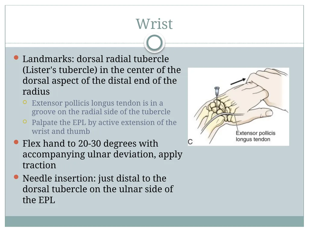
Wrist Landmarks: dorsal radial tubercle (Lister's tubercle) in the center of the dorsal aspect of the distal end of the radius
Extensor pollicis longus tendon is in a groove on the radial side of the tubercle
Palpate the EPL by active extension of the wrist and thumb
Flex hand to 20-30 degrees with accompanying ulnar deviation, apply traction
Needle insertion: just distal to the dorsal tubercle on the ulnar side of the EPL<br>
08
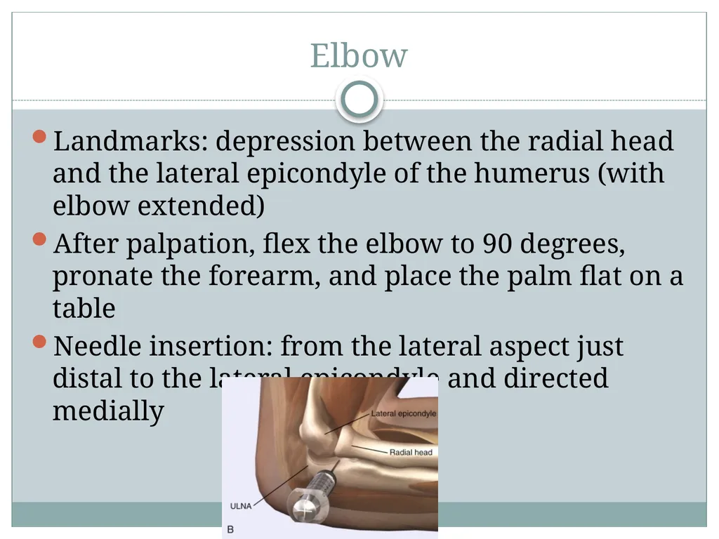
Elbow Landmarks: depression between the radial head and the lateral epicondyle of the humerus (with elbow extended)
After palpation, flex the elbow to 90 degrees, pronate the forearm, and place the palm flat on a table
Needle insertion: from the lateral aspect just distal to the lateral epicondyle and directed medially<br>
09
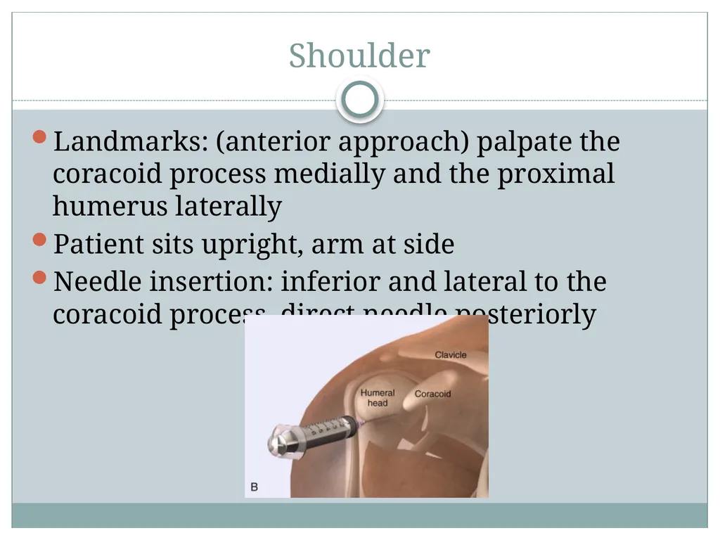
Shoulder Landmarks: (anterior approach) palpate the coracoid process medially and the proximal humerus laterally
Patient sits upright, arm at side
Needle insertion: inferior and lateral to the coracoid process, direct needle posteriorly<br>
10
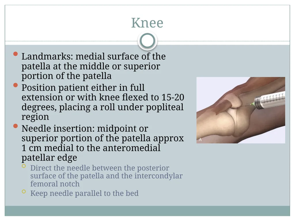
Knee Landmarks: medial surface of the patella at the middle or superior portion of the patella
Position patient either in full extension or with knee flexed to 15-20 degrees, placing a roll under popliteal region
Needle insertion: midpoint or superior portion of the patella approx 1 cm medial to the anteromedial patellar edge
Direct the needle between the posterior surface of the patella and the intercondylar femoral notch
Keep needle parallel to the bed<br>
11
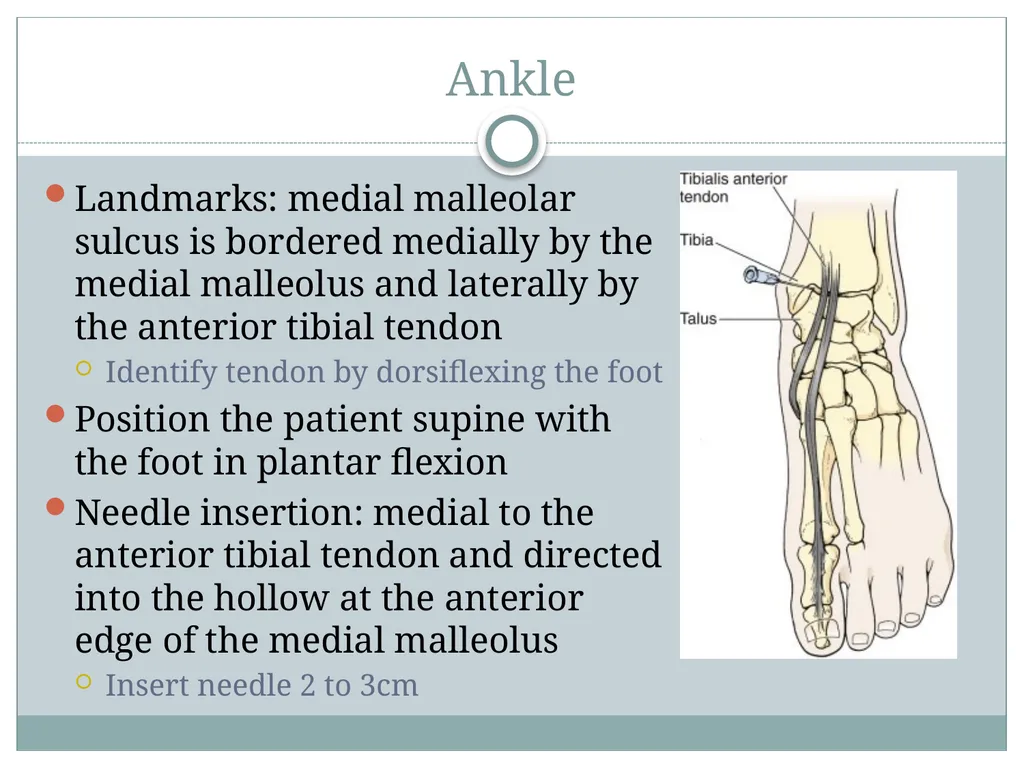
Ankle Landmarks: medial malleolar sulcus is bordered medially by the medial malleolus and laterally by the anterior tibial tendon
Identify tendon by dorsiflexing the foot
Position the patient supine with the foot in plantar flexion
Needle insertion: medial to the anterior tibial tendon and directed into the hollow at the anterior edge of the medial malleolus
Insert needle 2 to 3cm<br>
12
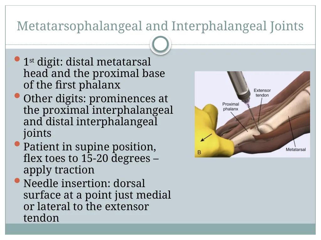
Metatarsophalangeal and Interphalangeal Joints 1st digit: distal metatarsal head and the proximal base of the first phalanx
Other digits: prominences at the proximal interphalangeal and distal interphalangeal joints
Patient in supine position, flex toes to 15-20 degrees – apply traction
Needle insertion: dorsal surface at a point just medial or lateral to the extensor tendon<br>