Papillomatosis (Warts) By Dr Marawan Elfky Definition Benign proliferative tumor of cutaneous and mucosal epithelia mainly in cattle. Ch. by formation of fibropapilloma outgrowths in form of fleshy lumps or large pendulous warts on
"Papillomatosis (Warts) By Dr/ Marawan Elfky" is the property of its rightful owner. Permission is granted to
download and print the materials on this website for personal, non-commercial use only, and to display it
on your personal computer provided you do not modify the materials and that you retain all copyright
notices contained in the materials. By downloading content from our website, you accept the terms of this
agreement.
Presentation Transcript
01
Papillomatosis (Warts)
By
Dr/ Marawan Elfky<br>
02
Definition
Benign proliferative tumor of cutaneous and mucosal epithelia mainly in cattle.
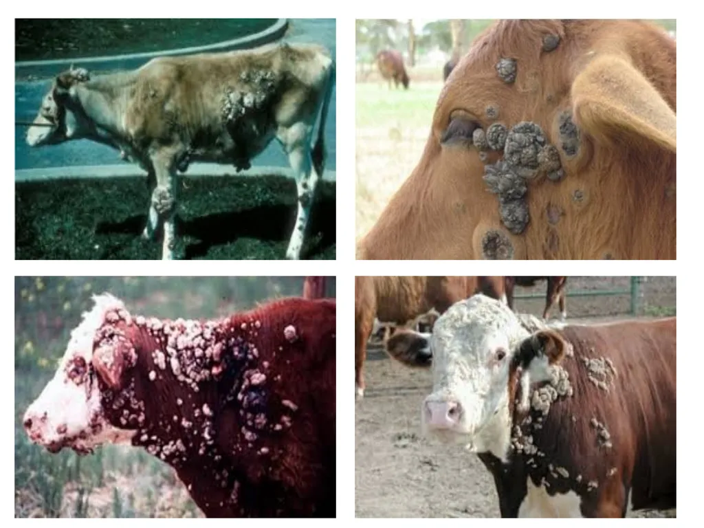
Ch. by formation of fibropapilloma outgrowths in form of fleshy lumps or large pendulous warts on different parts of the body.
Self-limiting disease.<br>
03
Etiology
Papovavirus or Papillomvirus, family Papovaviridae.
Small, non-enveloped, double stranded DNA virus (Strictly species-specific).
Six types (BPV-1 to BPV-6) which are divided into three broad subgroups:
Deltapapillomavirus or fibropapillomaviruses.
Xipapillomavirus or epitheliotropic BPVs.
Epsilonpapillomavirus.<br>
04
Deltapapillomavirus:
BPV-1 infects paragenital areas (penis, teats and udders)
BPV-2 infects skin, alimentary canal and urinary bladder
Xipapillomavirus:
BPV-3 infects skin
BPV-4 infects the upper alimentary tract
BPV-6 infects teats and udders
Epsilonpapillomavirus:
BPV-5 that infects teats and udders, and can cause both pure papillomas and fibropapillomas.
Predisposing factors: immune-suppressive factors (disease progression).<br>
05
Epidemiology
Distribution: Worldwide and recorded in Egypt.
Host rang:
All domestic animals, birds and fish can be infected.
In Cattle & buffaloes: mainly young animals up to two years of age; however, all ages can develop such lesions.
Seasonal incidence: no specific season for the diseases<br>
06
Transmission:
Source: Infected warts.
Mode:
Mainly horizontal transmission: direct contact with infected animals through skin abrasion, tattooing and dehorning instruments.
Flies and lice may be important in transmission.
Rare vertical intrauterine transmission.
Experimentally by I/d injection of warts tissue.<br>
07
5. Economic impact (minor):
Hides damage.
Decreases in body weight in animals with extensive lesions.
Interfere in the sales in pure bred
Secondary infection of traumatized warts.<br>
08
Pathogenesis
Upon infection, the virus infects basal cells of the epithelium causing extensive growth which is the characteristic of wart or tumor formation (fibropapilloma).<br>
09
Clinical signs
I.P from 3-8.w experimentally and more longer naturally.
Long course (3-18 m.), spontaneous recovery may be occurs after 1-8. m.
High morbidity rate (25%) and low mortality.<br>
10
Warts are solid outgrowths of epidermis: Pedunculated or cauliflower's like warts, gray or white and present in different parts of the body.
In young cattle present on the head especially around eyes and on the neck and spread to other parts of the body.
On teats (teat warts) may be round or flat-round or as an enlarged rice grain, it usually multiple and of diameter up to 2 cm.<br>
11
Genital warts: large size warts and bleed easily on the vulva and penis make matting is impracticable.
Interdigital fibropapilloma: round, flat and located in fleshy pad behind the pastern and above the heel bulbs (finger like projections with enlargement) these lesions are painful and animal may be recumbent and loss much of the condition occurs.<br>
12
Less common sites: urinary bladder, dorsal and lateral aspect of tongue, soft palate, oropharynx, esophagus, esophageal groove, rumen, and in the reticulum (chronic ruminal tympany) and in small intestine with possible of direct transmission of papilloma to carcinoma.
Persistent of fibropapillomas may be due to immunodeficiency in some animals.<br>
13
P/M lesions
As described in the signs. Diagnosis
1- Field diagnosis; depends on case history, clinical signs and P/M lesions.
2. Lab. Diagnosis;
Sample:
Biopsy of the lesions, slices from warts, whole blood and paired serum samples .<br>
14
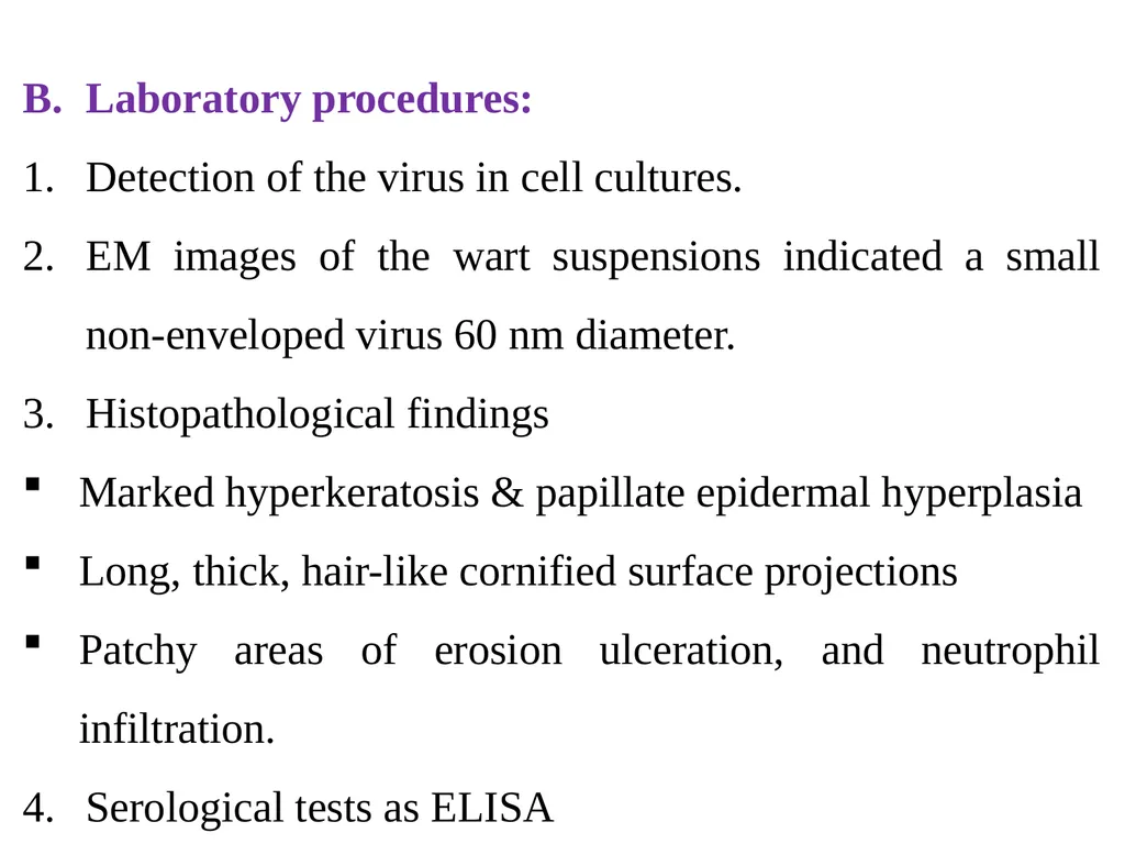
Laboratory procedures:
Detection of the virus in cell cultures.
EM images of the wart suspensions indicated a small non-enveloped virus 60 nm diameter.
Histopathological findings
Marked hyperkeratosis & papillate epidermal hyperplasia
Long, thick, hair-like cornified surface projections
Patchy areas of erosion ulceration, and neutrophil infiltration.
Serological tests as ELISA<br>
15
Differential Diagnosis
Atypical papillomas of cattle:
It affects all ages
lesions persist for long period
Discrete, low, flat & circular and often coalesce with each other to form large masses.
2. Ring worms and lumpy skin disease<br>
16
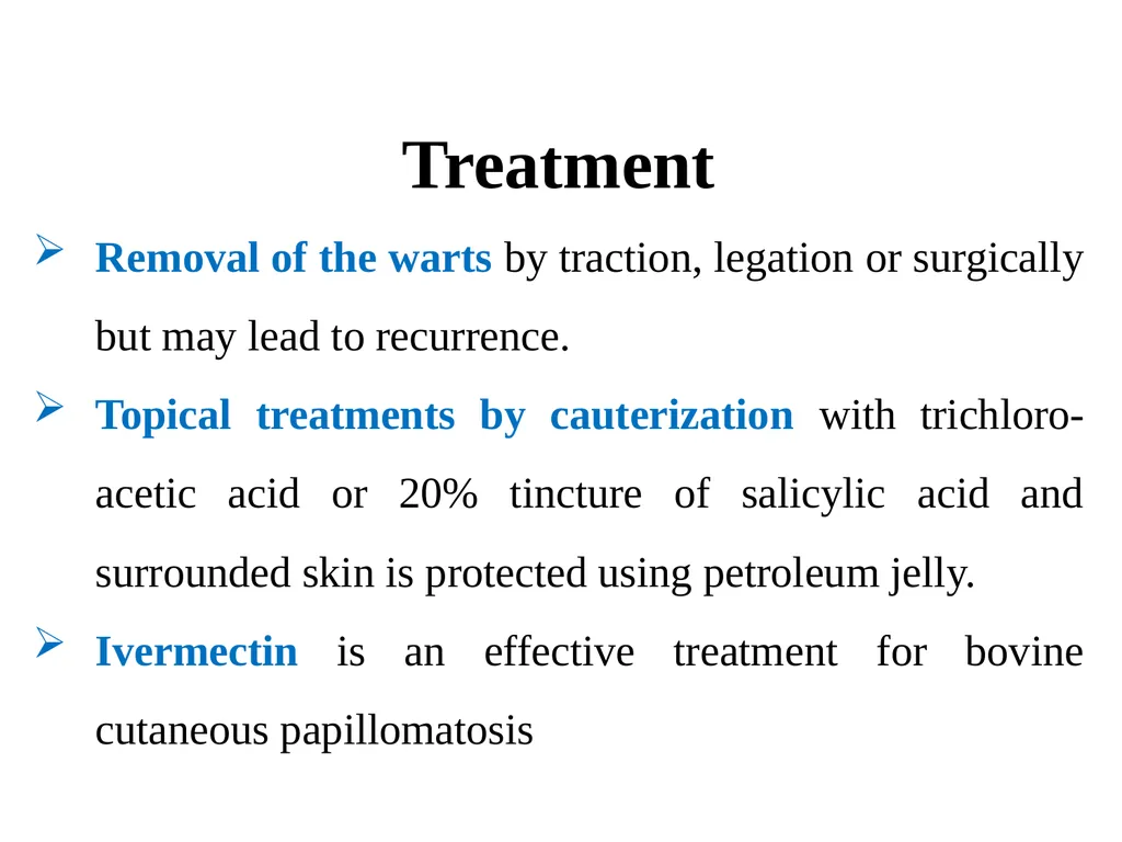
Treatment
Removal of the warts by traction, legation or surgically but may lead to recurrence.
Topical treatments by cauterization with trichloro-acetic acid or 20% tincture of salicylic acid and surrounded skin is protected using petroleum jelly.
Ivermectin is an effective treatment for bovine cutaneous papillomatosis<br>
17
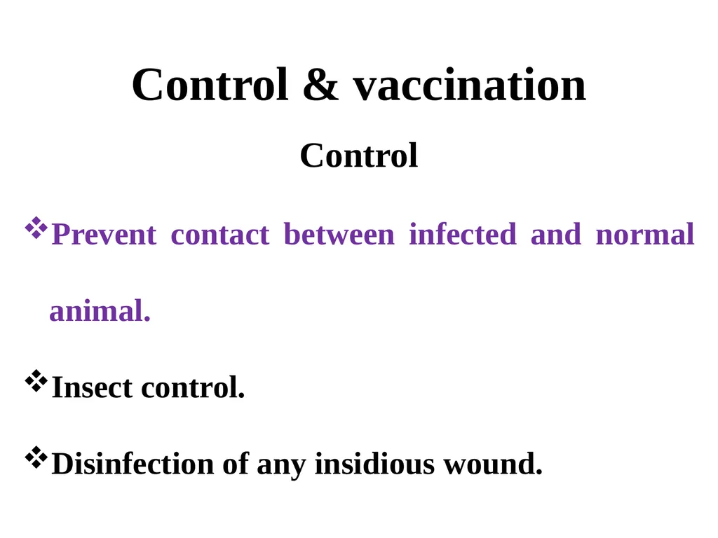
Control & vaccination
Control
Prevent contact between infected and normal animal.
Insect control.
Disinfection of any insidious wound.<br>
18
Prophylactic vaccination (of wart-free animals) with inactivated vaccine to calves as early as 4–6 weeks.
Therapeutic vaccination (of warts-existing animals) induces early regression of warts.
Autogenous vaccines from warts tissue, live or inactivated by formalin may be used as a therapy in two injections of 1-2. w apart, recovery after 3-6.w is produced in 80-85 % of cases.<br>