04
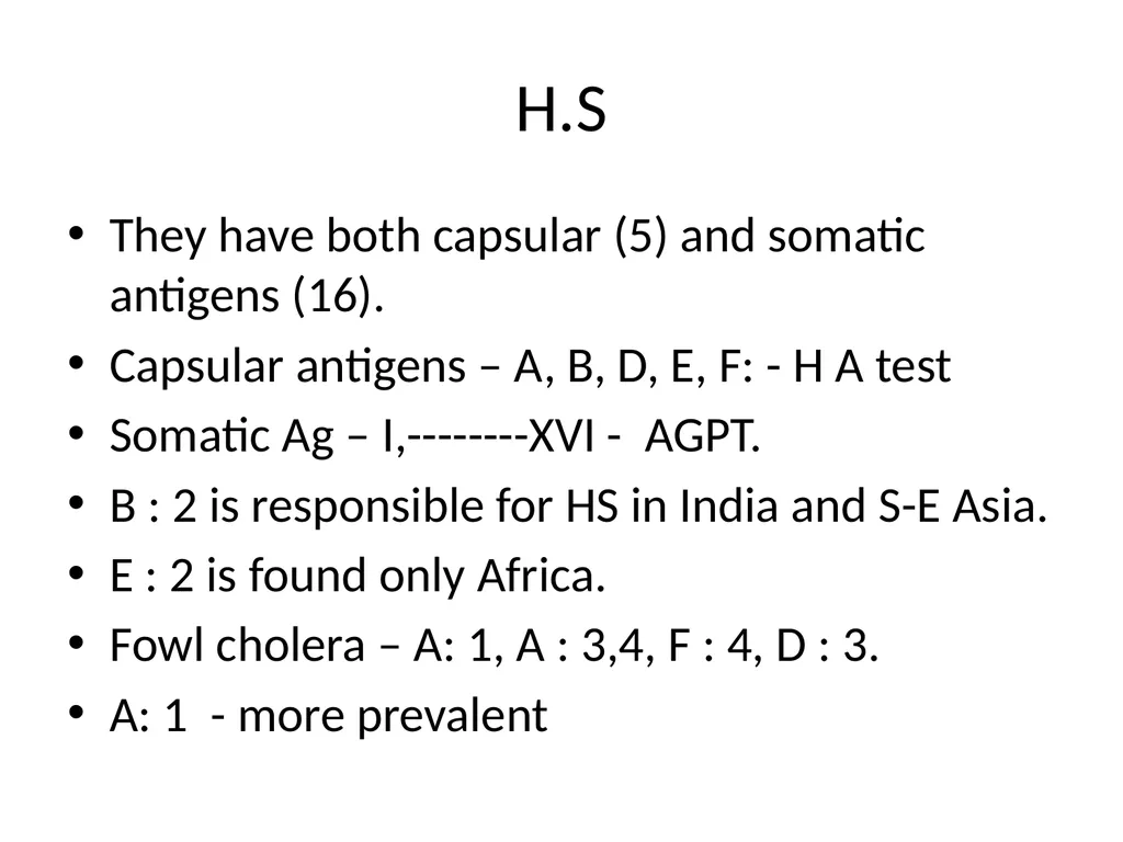
H.S They have both capsular (5) and somatic antigens (16).
Capsular antigens – A, B, D, E, F: - H A test
Somatic Ag – I,--------XVI - AGPT.
B : 2 is responsible for HS in India and S-E Asia.
E : 2 is found only Africa.
Fowl cholera – A: 1, A : 3,4, F : 4, D : 3.
A: 1 - more prevalent<br>
05
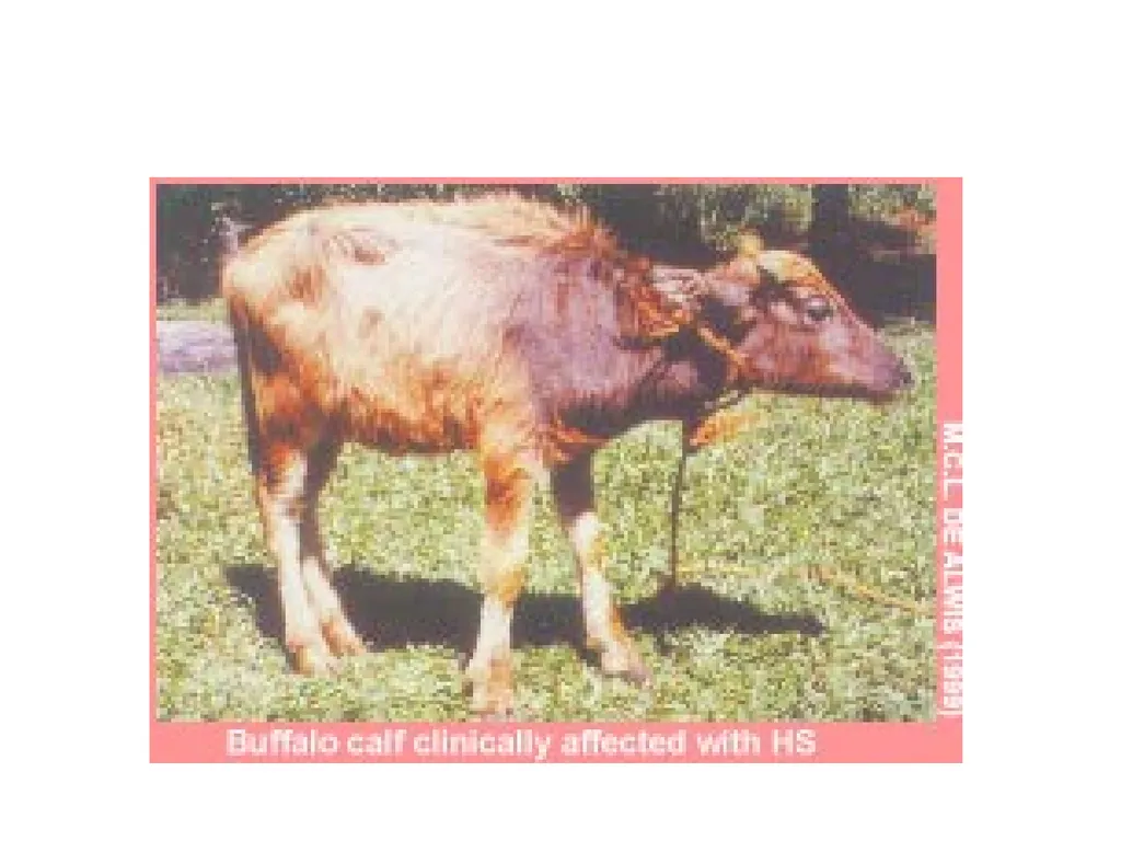
HAEMORRHAGIC SEPTICAEMIA Acute septacaemic disease with high fever, respiratory distress, diarrhea. In less acute case, oedema occurs at the brisket region. Absent in buffalo, they usually occurs with in 2-4 days.<br>
06
H.S Synonym: Stockyards disease , Pasteurellosis, Gala ghout.<br>
07
Incidence It is one of the important bacterial diseases of cattle and rarely buffaloes in India
Sheep and goat – Rarely affected
Swine and Fowls – May be affected<br>
08
H.S Transmission
Ingestion of contaminated feed, water etc.
The organism is found in the saliva of affected animals
Droplet infection may occur<br>
09
H.S Pathogenesis
Organisms on entry into the system reaches blood, proliferate and spread throughout the body due to its septicaemic nature and produces petechial and ecchymotic haemorrhjages on serous and mucous menbranes in different organs. Hence it is called as - “ HAEMORRHAGIC SEPTICAEMIA ”<br>
10
Pathogenesis The bacteria ( Bipolar) has capsule which prevent its opsonization and Phagocytosis.
The bacteria comes in lymph through pharyngeal tonsils and cause Bactermia.
The bacteria multiplies and produces septicemia and produce toxins.<br>
11
The toxins are –
A) Leukotoxin – it help in stabilizing bacteria & prevent its Phagocytosis by destroying monocyte, macrophages
B) Endotoxin – destroy endothelium of blood vessels and cause intravascular permeability, oedema & hemorrhage.
C) The toxin also activate macrophage to release Cytokines.<br>
12
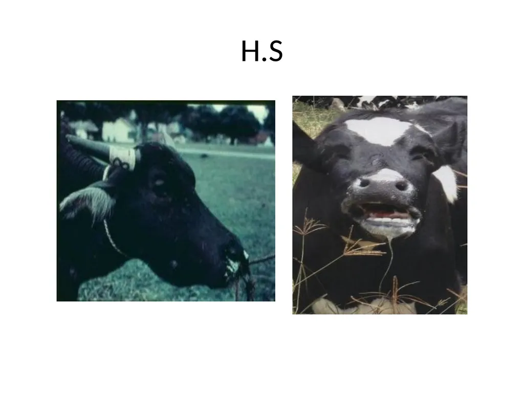
H.S Clinical signs
High temperature, dullness, dyspnoea, hot painful swelling – head, dewlap and neck with Inflammatory exudate in the subcutaneous tissues.<br>
14
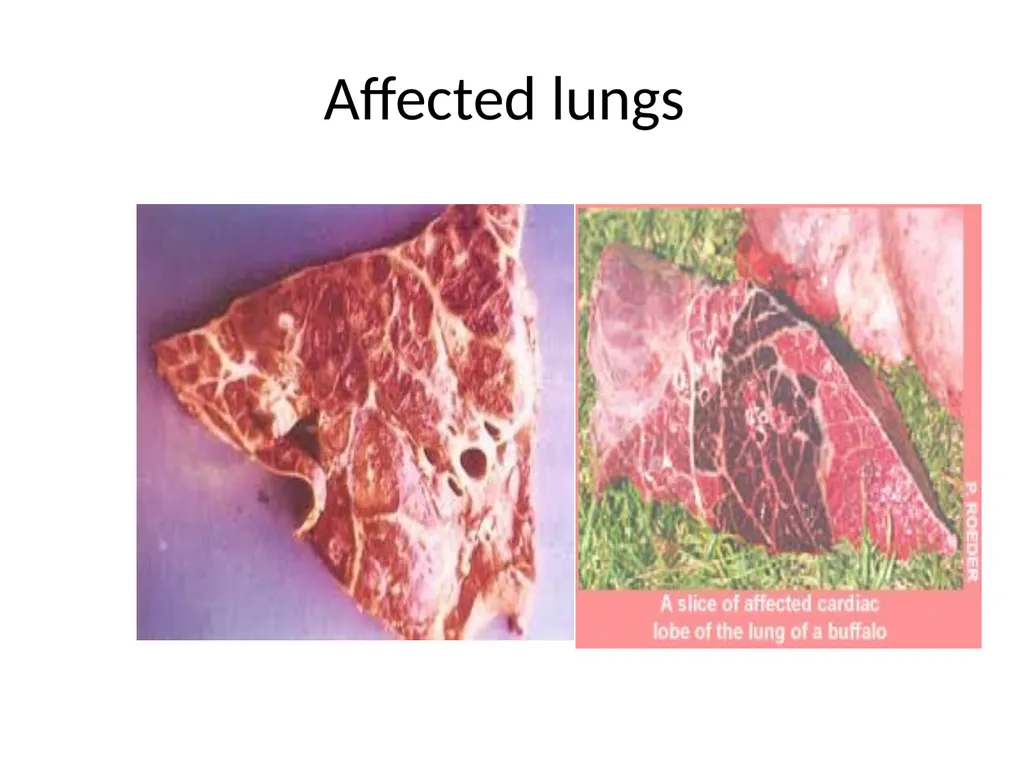
H.S Gross lesions
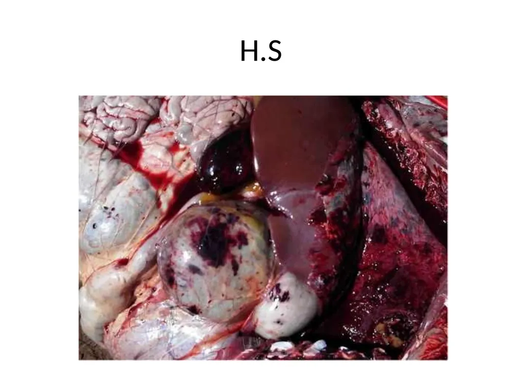
Petechiae on all serous and mucous membranes especially on epicardium, myocardium, pleura, peritoneum and Gastrointestinal tract.
Lymph glands- Swollen and haemorrhagic
G.I tract is severely inflammed with contents mixed with blood<br>
17
H.S Microscopic lesions
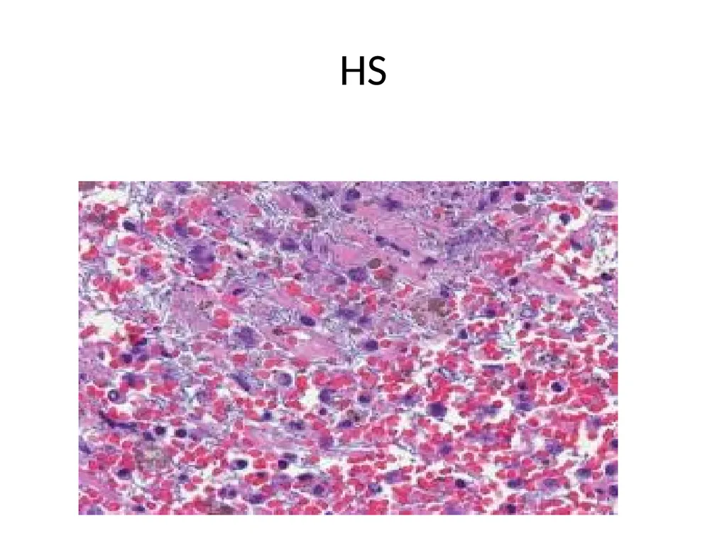
In acute and subacute cases, the predominant lesion is fibrinous bronchopneumonia
The organism is small ovoid rods the ends of rods are more deeply stained than central portion which gives them a bipolar appearance.
In rabbits – Haemorrhagic tracheitis.<br>
19
H.S Diagnosis
Examination of blood smear
A heart blood swab may be rubbed over the scarified abdomen of a rabbit. It will die within 24 to 40 hours showing haemorrhgaes, especially haemorrhagic tracheitis is pathognomonic lesion<br>
20
JOHNE'S DISEASE Synonym : Paratuberculosis
Chronic wasting illness of ruminants with prolonged course, recurrent diarrhoea, dehydration, emaciation and<br>
21
Aetiology
Mycobaterium paratuberculosis - Three strains:
Bovine strain
Ovine strain
Scottish strain<br>
22
Incidence
The disease is of worldwide distribution and constitutes an important economic problem in cattle
Susceptibility
Ruminants are usually affected. Horses and pigs may be affected very rarely
2-3 year-old female in high milk production most susceptible.
Cow – sub-clinical, organism abundantly in the milk – new born animals highly susceptible.
Gut is the main target organ
It is less frequently encountered sheep and goats<br>
23
Transmission
Infection is ingestion. Faeces containing the organisms serve as the primary source of infection
Incubation period : Long and protracted even upto 2 years<br>
24
Organisms enters the body through intestinal mucosa, sets up bacteremia and settles in the mucosa of intestine, mesenteric lymph nodes and tonsil produce a chronic granulomatous inflammation
Cell mediated immune response and followed by humoral immune response initiated by the release of bacteria by dying macrophages as the disease progresses.<br>
25
Immediate hypersensitivity – Mediates diarrhea.
Delayed hypersensitivity – mediates emaciation & anemia.
Failure of protein synthesis – mediates muscle atrophy.
Cytotoxic – muscle atrophy & renal damage.<br>
26
Clinical signs
Emaciation, hide and bound with dry coat
Normal appetite with increased thirst and diarrrhoea
Sheep and goat – Diarrhoea is not common, emaciation observed<br>
27
Gross lesions
Carcass – Emaciation with gelatinous fat
Terminal part of ileum – Wall thickened and oedematous
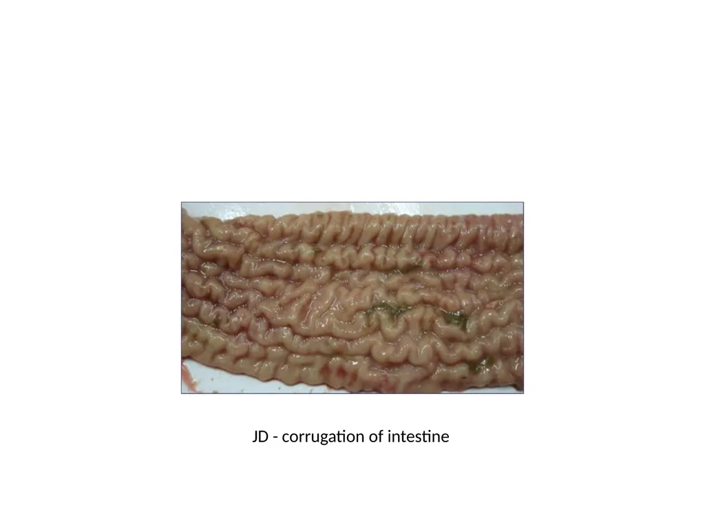
The mucosa is folded and showed Transverse corrugation or rugae (like cerebral convolution)<br>
28
JD - corrugation of intestine<br>
29
Microscopically – pronounced chronic granulomatous enteritis with heavy lymphocytic infiltration, typical Langerhans type gaint cells & scattered granulomatas.<br>