
PATHOLOGICAL PIGMENTATION Lecture by Assistant
DP
Published · 23 slides · 0 views
1 / 1


Description
PATHOLOGICAL PIGMENTATION Lecture by Assistant Professor Dr. Jihad A. Ahmed PATHOLOGICAL PIGMENTATION This is defined as deposition of inherently coloured substances(pigments) in aberrant locations or in excess in locations normally found.
Related Topics
Share
Embed code
Download this presentation From Below
"PATHOLOGICAL PIGMENTATION Lecture by Assistant" is the property of its rightful owner. Permission is granted to download and print the materials on this website for personal, non-commercial use only, and to display it on your personal computer provided you do not modify the materials and that you retain all copyright notices contained in the materials. By downloading content from our website, you accept the terms of this agreement.
Presentation Transcript
01
PATHOLOGICAL PIGMENTATION Lecture by
Assistant Professor
Dr. Jihad A. Ahmed<br>
Assistant Professor
Dr. Jihad A. Ahmed<br>
02
PATHOLOGICAL PIGMENTATION This is defined as deposition of inherently coloured substances(pigments) in aberrant locations or in excess in locations normally found.
Classification of pigments
Pigments are classified according to their origin as:
Exogenous pigments.
Endogenous pigments.
1. Exogenous pigments
Originate from outside the body, entering the body through the skin, lungs or intestines and being deposited in macrophages of regional lymph nodes
Some of the exogenous pigments undergo modification in the tissues e.g. mercury, which is converted to mercuric sulphide responsible for mercurial pigmentation<br>
Classification of pigments
Pigments are classified according to their origin as:
Exogenous pigments.
Endogenous pigments.
1. Exogenous pigments
Originate from outside the body, entering the body through the skin, lungs or intestines and being deposited in macrophages of regional lymph nodes
Some of the exogenous pigments undergo modification in the tissues e.g. mercury, which is converted to mercuric sulphide responsible for mercurial pigmentation<br>
03
Effects of exogenous pigments Some exogenous pigments like carbon dust cause little or no harm in the tissues
Others like silica, asbestos and iron produce tissue injury associated with extensive inflammatory reaction and healing by fibrosis
The general pathological term for such lesion in the lungs due to inhalation of irritant elements is pneumoconiosis
2. Endogenous pigments
Endogenous pigments are further classified into four types based on their source of formation in the body:<br>
Others like silica, asbestos and iron produce tissue injury associated with extensive inflammatory reaction and healing by fibrosis
The general pathological term for such lesion in the lungs due to inhalation of irritant elements is pneumoconiosis
2. Endogenous pigments
Endogenous pigments are further classified into four types based on their source of formation in the body:<br>
04
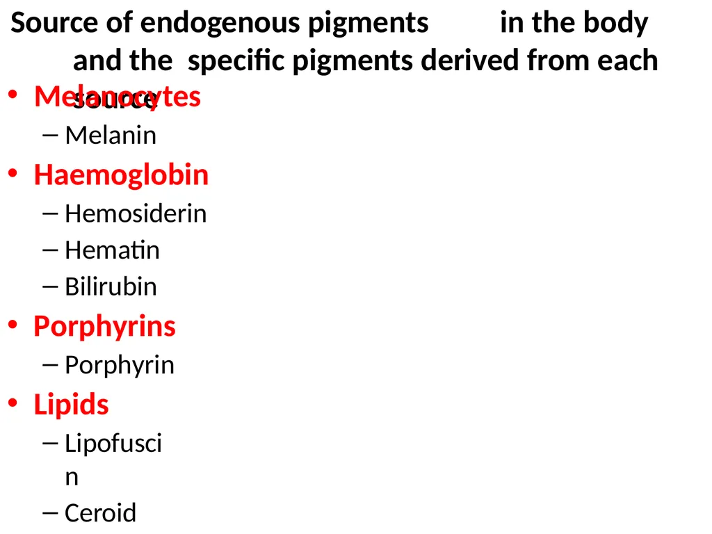
Source of endogenous pigments in the body and the specific pigments derived from each source Melanocytes
Melanin
Haemoglobin
Hemosiderin
Hematin
Bilirubin
Porphyrins
Porphyrin
Lipids
Lipofuscin
Ceroid<br>
Melanin
Haemoglobin
Hemosiderin
Hematin
Bilirubin
Porphyrins
Porphyrin
Lipids
Lipofuscin
Ceroid<br>
05
1. Melanin The pigment is produced in the skin by melanocytes located at the junction between the epidermis and the dermis
The melanocytes have branching processes through which melanin granules(melanosomes) are transferred to keratinocytes in the epidermis
The process of formation of melanin is called
melanization
The concentration of melanin in tissues determines the colour of the tissue<br>
The melanocytes have branching processes through which melanin granules(melanosomes) are transferred to keratinocytes in the epidermis
The process of formation of melanin is called
melanization
The concentration of melanin in tissues determines the colour of the tissue<br>
06
Melanin(cont.) In low concentrations, melanin appears yellow- brown, whereas in large quantities black
In other situation, its dispersion may be that it appears blue or green e.g. in the sexual skin of some baboons
Fish have additional reflecting cells called iridocytes, which contain guanin instead of melanin responsible for beautiful metallic sheen of many fish
There are two types of animal melanin: brown to black insoluble called eumelanin and yellow to reddish brown soluble called pheomelanin
The function of melanin is largely protective e.g. in chameleons, and in man against sunlight<br>
In other situation, its dispersion may be that it appears blue or green e.g. in the sexual skin of some baboons
Fish have additional reflecting cells called iridocytes, which contain guanin instead of melanin responsible for beautiful metallic sheen of many fish
There are two types of animal melanin: brown to black insoluble called eumelanin and yellow to reddish brown soluble called pheomelanin
The function of melanin is largely protective e.g. in chameleons, and in man against sunlight<br>
07
Melanization Melanoblasts in the skin differentiate into:
Melanocytes, which contain an amino acid
tyrosine
Tyrosine is oxidized to dioxyphenylalanine(DOPA) under the catalytic activity of enzyme tyrosinase
DOPA is changed into melanosomes
Melanosomes are sent to keratinocytes<br>
Melanocytes, which contain an amino acid
tyrosine
Tyrosine is oxidized to dioxyphenylalanine(DOPA) under the catalytic activity of enzyme tyrosinase
DOPA is changed into melanosomes
Melanosomes are sent to keratinocytes<br>
08
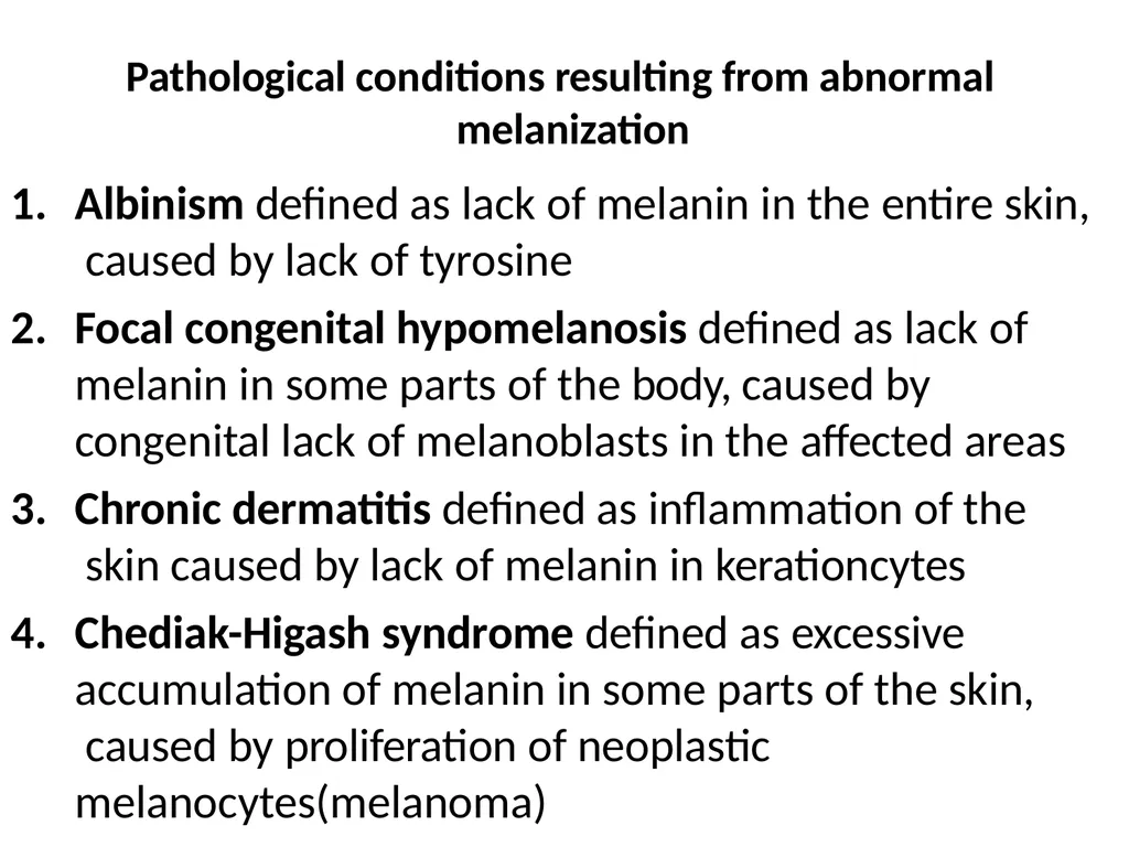
Pathological conditions resulting from abnormal melanization 1. Albinism defined as lack of melanin in the entire skin, caused by lack of tyrosine Focal congenital hypomelanosis defined as lack of melanin in some parts of the body, caused by congenital lack of melanoblasts in the affected areas
Chronic dermatitis defined as inflammation of the skin caused by lack of melanin in kerationcytes
Chediak-Higash syndrome defined as excessive accumulation of melanin in some parts of the skin, caused by proliferation of neoplastic melanocytes(melanoma)<br>
Chronic dermatitis defined as inflammation of the skin caused by lack of melanin in kerationcytes
Chediak-Higash syndrome defined as excessive accumulation of melanin in some parts of the skin, caused by proliferation of neoplastic melanocytes(melanoma)<br>
09
2. Hemosiderin:
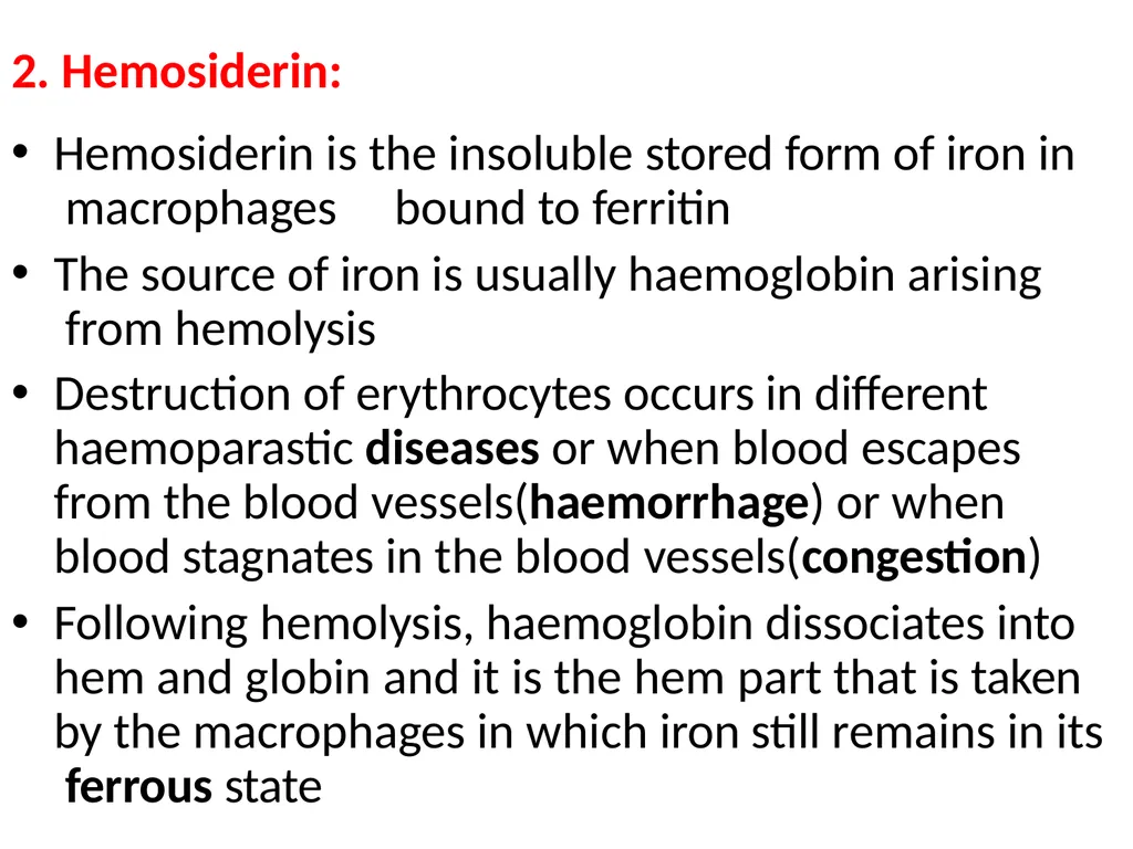
Hemosiderin is the insoluble stored form of iron in macrophages bound to ferritin
The source of iron is usually haemoglobin arising from hemolysis
Destruction of erythrocytes occurs in different haemoparastic diseases or when blood escapes from the blood vessels(haemorrhage) or when blood stagnates in the blood vessels(congestion)
Following hemolysis, haemoglobin dissociates into hem and globin and it is the hem part that is taken by the macrophages in which iron still remains in its ferrous state<br>
Hemosiderin is the insoluble stored form of iron in macrophages bound to ferritin
The source of iron is usually haemoglobin arising from hemolysis
Destruction of erythrocytes occurs in different haemoparastic diseases or when blood escapes from the blood vessels(haemorrhage) or when blood stagnates in the blood vessels(congestion)
Following hemolysis, haemoglobin dissociates into hem and globin and it is the hem part that is taken by the macrophages in which iron still remains in its ferrous state<br>
10
Hemosiderin(cont.)
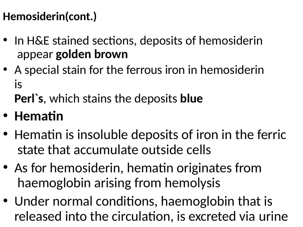
In H&E stained sections, deposits of hemosiderin appear golden brown
A special stain for the ferrous iron in hemosiderin is
Perl`s, which stains the deposits blue
Hematin
Hematin is insoluble deposits of iron in the ferric state that accumulate outside cells
As for hemosiderin, hematin originates from haemoglobin arising from hemolysis
Under normal conditions, haemoglobin that is released into the circulation, is excreted via urine<br>
In H&E stained sections, deposits of hemosiderin appear golden brown
A special stain for the ferrous iron in hemosiderin is
Perl`s, which stains the deposits blue
Hematin
Hematin is insoluble deposits of iron in the ferric state that accumulate outside cells
As for hemosiderin, hematin originates from haemoglobin arising from hemolysis
Under normal conditions, haemoglobin that is released into the circulation, is excreted via urine<br>
11
Hematin(cont.)
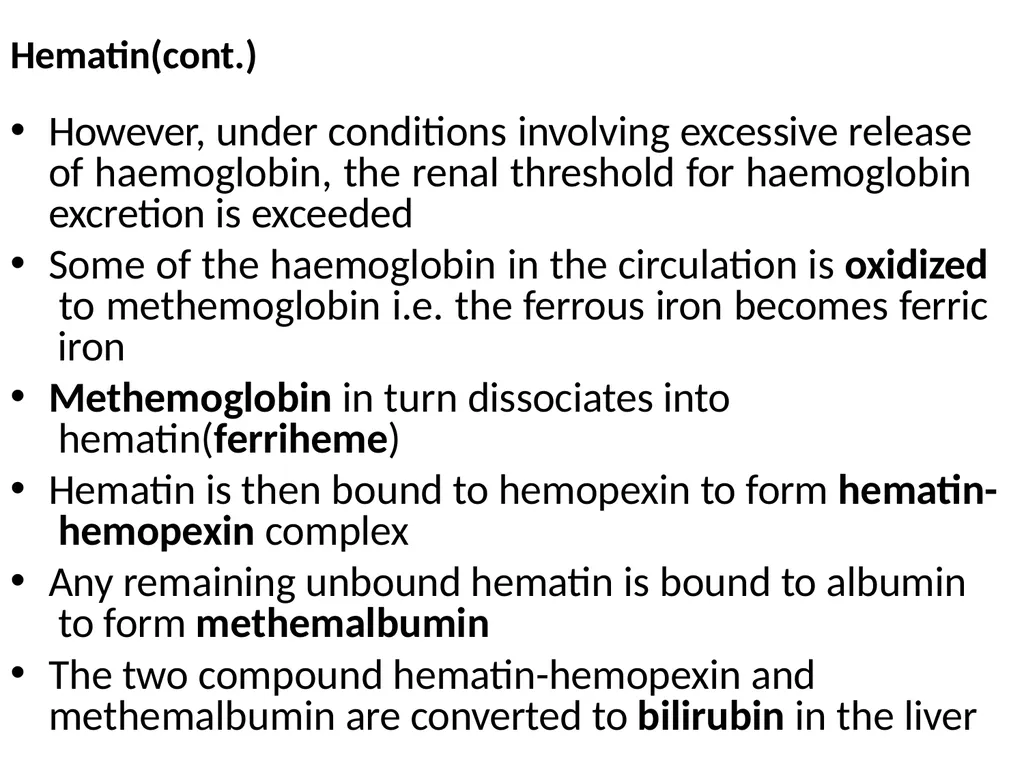
However, under conditions involving excessive release of haemoglobin, the renal threshold for haemoglobin excretion is exceeded
Some of the haemoglobin in the circulation is oxidized to methemoglobin i.e. the ferrous iron becomes ferric iron
Methemoglobin in turn dissociates into hematin(ferriheme)
Hematin is then bound to hemopexin to form hematin- hemopexin complex
Any remaining unbound hematin is bound to albumin to form methemalbumin
The two compound hematin-hemopexin and methemalbumin are converted to bilirubin in the liver<br>
However, under conditions involving excessive release of haemoglobin, the renal threshold for haemoglobin excretion is exceeded
Some of the haemoglobin in the circulation is oxidized to methemoglobin i.e. the ferrous iron becomes ferric iron
Methemoglobin in turn dissociates into hematin(ferriheme)
Hematin is then bound to hemopexin to form hematin- hemopexin complex
Any remaining unbound hematin is bound to albumin to form methemalbumin
The two compound hematin-hemopexin and methemalbumin are converted to bilirubin in the liver<br>
12
Identification of hematin In H&E stained sections, hematin appears brown, hence can be confused with hemosiderin
Distinction between hematin and hemosiderin in histological section is based on the facts that:;
Hematin is found outside cells
Hematin does not stain positive with Perl`s stain
NB haemorrhages and use of non-buffered formalin induce formation of hematin artifacts in tissues<br>
Distinction between hematin and hemosiderin in histological section is based on the facts that:;
Hematin is found outside cells
Hematin does not stain positive with Perl`s stain
NB haemorrhages and use of non-buffered formalin induce formation of hematin artifacts in tissues<br>
13
3. Bilirubin:
Bilirubin is the yellow bile pigment formed from haemoglobin
Following hemolysis of senescent erythrocytes, the released haemoglobin(Hb) is normally taken by macrophages of the reticuloendothelial system(RES)
In the RES, the Hb dissociates into heme and globin
The amino acids of globin are recycled, whereas the heme, a cyclic molecule made up of four pyrole rings and centrally placed ferrous iron is opened
Oxidation of the heme is catalysed by heme oxygenase and leads to its opening into a linear molecule called biliverdin and carbon monoxide<br>
Bilirubin is the yellow bile pigment formed from haemoglobin
Following hemolysis of senescent erythrocytes, the released haemoglobin(Hb) is normally taken by macrophages of the reticuloendothelial system(RES)
In the RES, the Hb dissociates into heme and globin
The amino acids of globin are recycled, whereas the heme, a cyclic molecule made up of four pyrole rings and centrally placed ferrous iron is opened
Oxidation of the heme is catalysed by heme oxygenase and leads to its opening into a linear molecule called biliverdin and carbon monoxide<br>
14
Bilirubin(cont.) In the next reaction, biliverdin is reduced to unconjugated bilirubin under the catalytic activity of biliverdin reductase
The unconjugated bilirubin is then released from the RES into the circulation, where it is bound to albumin
The albumin-bound unconjugated bilirubin is transferred into hepatocytes
The rate of transfer is dependent on:
The concentration of bilirubin in blood
The concentration of albumin in blood
The blood flow to the liver
The concentration of gamma-protein in hepatocytes
The concentration of unconjugated bilirubin on the gamma- protein<br>
The unconjugated bilirubin is then released from the RES into the circulation, where it is bound to albumin
The albumin-bound unconjugated bilirubin is transferred into hepatocytes
The rate of transfer is dependent on:
The concentration of bilirubin in blood
The concentration of albumin in blood
The blood flow to the liver
The concentration of gamma-protein in hepatocytes
The concentration of unconjugated bilirubin on the gamma- protein<br>
15
Bilirubin(cont.) The gamma-protein releases the unconjugated bilirubin at the smooth endoplasmic reticulum adjacent to bile canaliculi
At these sites, there is a conjugation enzyme called glucuronyl transferase, which activates two molecules of glucuronic acids, to enable them combine with the unconjugated bilirubin to produce conjugated bilirubin(bilirubin diglucuronide)
The conjugation enzyme is not fully developed until the new born is about one month, thus premature infants have impaired conjugation
Thus, abrupt hemolysis that occurs at the time of birth leads to uconjugated bilirubin load
When the levels exceed 200mg/100ml,the possibility of entry into the central nervous tissue(brain) and subsequent brain damage is quite high
Such brain damage resulting from unconjugated bilirubin is called
kernicterus<br>
At these sites, there is a conjugation enzyme called glucuronyl transferase, which activates two molecules of glucuronic acids, to enable them combine with the unconjugated bilirubin to produce conjugated bilirubin(bilirubin diglucuronide)
The conjugation enzyme is not fully developed until the new born is about one month, thus premature infants have impaired conjugation
Thus, abrupt hemolysis that occurs at the time of birth leads to uconjugated bilirubin load
When the levels exceed 200mg/100ml,the possibility of entry into the central nervous tissue(brain) and subsequent brain damage is quite high
Such brain damage resulting from unconjugated bilirubin is called
kernicterus<br>
16
Bilirubin(cont.)
The conjugated bilirubin then passes through the biliary tree to the gall bladder
Conjugated bilirubin is converted to urobilinogen in the large intestine by the action of bacterial glucuronidases
10% of urobilinogen are reabsorbed and passes unchanged in the liver and enters the circulation from where it is excreted through urine
Liver damage may lead to significant amounts of urobilinogen in the urine
90% of urobilinogen is reduced to give stercobilinogen that is finaly oxidized to stercobilin, which imparts the brown colour of feces<br>
The conjugated bilirubin then passes through the biliary tree to the gall bladder
Conjugated bilirubin is converted to urobilinogen in the large intestine by the action of bacterial glucuronidases
10% of urobilinogen are reabsorbed and passes unchanged in the liver and enters the circulation from where it is excreted through urine
Liver damage may lead to significant amounts of urobilinogen in the urine
90% of urobilinogen is reduced to give stercobilinogen that is finaly oxidized to stercobilin, which imparts the brown colour of feces<br>
17
Jaundice/Icterus These are synonymous terms, which mean yellow colouration of tissues due to hyperbilirubinaemia
Hyperbilirubinaemia may occur due to:
Excessive hemolysis
Damage of hepatocytes
Blockage of flow of bile/blokage of bile ducts
Types of jaundice
Pre-hepatic
Hepatic
Post-hepatic<br>
Hyperbilirubinaemia may occur due to:
Excessive hemolysis
Damage of hepatocytes
Blockage of flow of bile/blokage of bile ducts
Types of jaundice
Pre-hepatic
Hepatic
Post-hepatic<br>
18
3. Porphyrins Porphyrins are organic compounds made up of four pyrrole rings(pentagon-shaped rings of four carbon atoms with a nitrogen) and a centred bound metal ion
Porphyrins differ in terms of:
Metal ion e.g. Fe- heme, Mg – chlorophyll,
Eight side chains, two on each pyrrole ring
In the course of formation of heme, deficiencies in some enzymes have been reported to result into occurrence of abnormal intermediate metabolites of porphyrins<br>
Porphyrins differ in terms of:
Metal ion e.g. Fe- heme, Mg – chlorophyll,
Eight side chains, two on each pyrrole ring
In the course of formation of heme, deficiencies in some enzymes have been reported to result into occurrence of abnormal intermediate metabolites of porphyrins<br>
19
Porphyrins(cont.)
For example, lack of uroporphyrinogen decarboxylase(UPG decarboxylase), prevents uroporphyrinogen III to progress into the subsequent stage, hence being oxidized into a porphyrin isomer
Similarly, lack of corproporphyrinogen oxidase(CPG oxidase), prevents corproporphyrinogen III to progress to protoporphyrinogen III
Excess formation of abnormal porphyrin isomers leads to their deposition in tissues<br>
For example, lack of uroporphyrinogen decarboxylase(UPG decarboxylase), prevents uroporphyrinogen III to progress into the subsequent stage, hence being oxidized into a porphyrin isomer
Similarly, lack of corproporphyrinogen oxidase(CPG oxidase), prevents corproporphyrinogen III to progress to protoporphyrinogen III
Excess formation of abnormal porphyrin isomers leads to their deposition in tissues<br>
20
Effects of deposition of porphyrin isomers in tissues
Porphyria is a pathological state characterized by accumulation of abnormal porphyrins in tissues
The disease is also known as osteohemochromatosis due to reddish brown bone pigmentation or pink tooth due to colour of teeth
Deposition of abnormal porphyrins results into photodynamic dermatitis in cattle with sloughing of the affected areas
Photodynamic dermatitis occurs due to the fact that the porphyrin isomers are fluorescent, hence they become activated by sunrays and produce free radicals that induce photosensitization<br>
Porphyria is a pathological state characterized by accumulation of abnormal porphyrins in tissues
The disease is also known as osteohemochromatosis due to reddish brown bone pigmentation or pink tooth due to colour of teeth
Deposition of abnormal porphyrins results into photodynamic dermatitis in cattle with sloughing of the affected areas
Photodynamic dermatitis occurs due to the fact that the porphyrin isomers are fluorescent, hence they become activated by sunrays and produce free radicals that induce photosensitization<br>
21
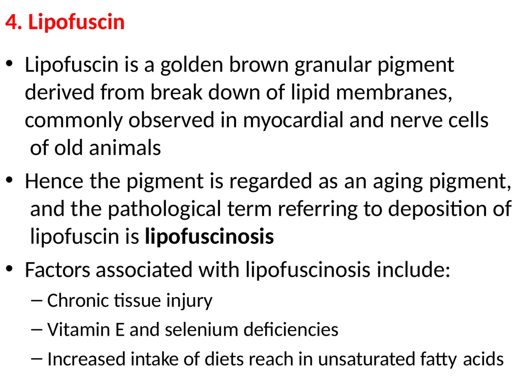
4. Lipofuscin Lipofuscin is a golden brown granular pigment derived from break down of lipid membranes, commonly observed in myocardial and nerve cells of old animals
Hence the pigment is regarded as an aging pigment, and the pathological term referring to deposition of lipofuscin is lipofuscinosis
Factors associated with lipofuscinosis include:
Chronic tissue injury
Vitamin E and selenium deficiencies
Increased intake of diets reach in unsaturated fatty acids<br>
Hence the pigment is regarded as an aging pigment, and the pathological term referring to deposition of lipofuscin is lipofuscinosis
Factors associated with lipofuscinosis include:
Chronic tissue injury
Vitamin E and selenium deficiencies
Increased intake of diets reach in unsaturated fatty acids<br>
22
Ceroid
Ceroid is a golden yellow brown pigment believed to be a variant of lipofuscin
It differs from lipofuscin by being acid-fast and
autofluorescent<br>
Ceroid is a golden yellow brown pigment believed to be a variant of lipofuscin
It differs from lipofuscin by being acid-fast and
autofluorescent<br>
23
Thank you Reference
All the scientific data cited from the lecture notes by Prof. Robert Mdoda Maselle
Dept. of veterinary pathology, faculty of veterinary medicine, Sokoine university of agriculture.<br>
All the scientific data cited from the lecture notes by Prof. Robert Mdoda Maselle
Dept. of veterinary pathology, faculty of veterinary medicine, Sokoine university of agriculture.<br>