
Physiologic Peculiarities of Specific
Description: Physiologic Peculiarities of Specific PulmonaryAbnormalities Chronic Pulmonary Emphysema :The term pulmonary emphysema literally means excess air in the lungs. However, this term is usually used to describe complex obstructive and
Related Topics
Download Presentation
"Physiologic Peculiarities of Specific" is the property of its rightful owner. Permission is granted to download and print the materials on this website for personal, non-commercial use only, and to display it on your personal computer provided you do not modify the materials and that you retain all copyright notices contained in the materials. By downloading content from our website, you accept the terms of this agreement.
Presentation Transcript
slide1. Physiologic Peculiarities of Specific PulmonaryAbnormalities Chronic Pulmonary Emphysema :The term pulmonary emphysema literally means excess air in the lungs. However, this term is usually used to describe complex obstructive and destructive process of the lungs caused by many years of smoking.
It results from the following major pathophysiologic
changes in the lungs:<br>
slide2. changes in the lungs:
Chronic infection
caused by inhaling smoke or other substances that irritate the bronchi and bronchioles.
The chronic infection seriously destroy the normal protective mechanisms of the airways, including partial paralysis of the cilia of the respiratory epithelium, an effect caused by nicotine.
As a result, mucus cannot be moved easily out of the passageways.
Also, stimulation of excess mucus secretion occurs, which further exacerbates the condition.
Too, inhibition of the alveolar macrophages occurs, so that they become less effective in combating infection.<br>
slide3. 2. The infection, excess mucus, and inflammatory
edema of the bronchiolar epithelium together cause chronic obstruction of many of the smaller airways.
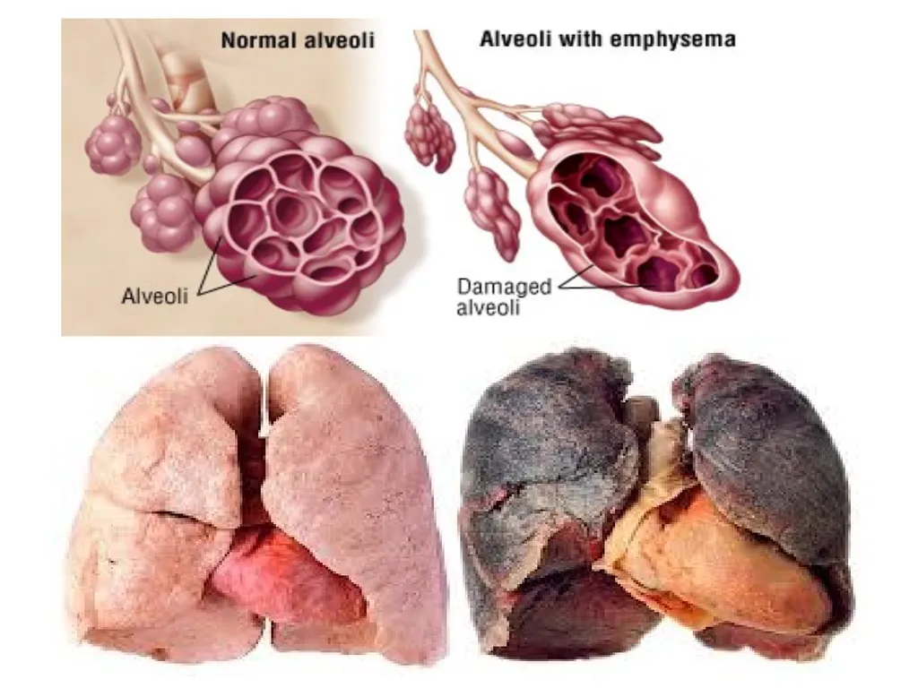
3. The obstruction of the airways makes it especially difficult to expire, thus causing entrapment of air in the alveoli and overstretching them. This, combined with the lung infection, causes marked destruction of as much as 50 to 80 percent of the alveolar walls. Therefore, the final picture of the emphysematous lung is that shown in Figures<br>
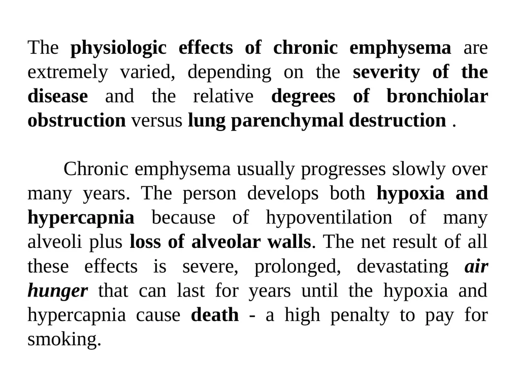
slide5. The physiologic effects of chronic emphysema are extremely varied, depending on the severity of the disease and the relative degrees of bronchiolar obstruction versus lung parenchymal destruction .
Chronic emphysema usually progresses slowly over many years. The person develops both hypoxia and hypercapnia because of hypoventilation of many alveoli plus loss of alveolar walls. The net result of all these effects is severe, prolonged, devastating air hunger that can last for years until the hypoxia and hypercapnia cause death - a high penalty to pay for smoking.<br>
slide6. Pneumonia
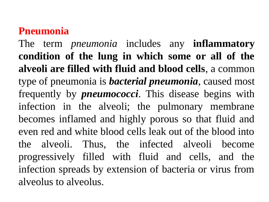
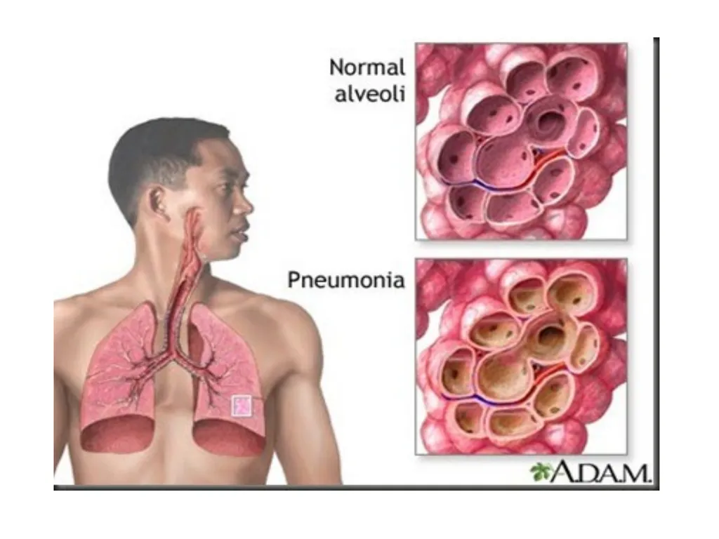
The term pneumonia includes any inflammatory condition of the lung in which some or all of the alveoli are filled with fluid and blood cells, a common type of pneumonia is bacterial pneumonia, caused most frequently by pneumococci. This disease begins with infection in the alveoli; the pulmonary membrane becomes inflamed and highly porous so that fluid and even red and white blood cells leak out of the blood into the alveoli. Thus, the infected alveoli become progressively filled with fluid and cells, and the infection spreads by extension of bacteria or virus from alveolus to alveolus.<br>
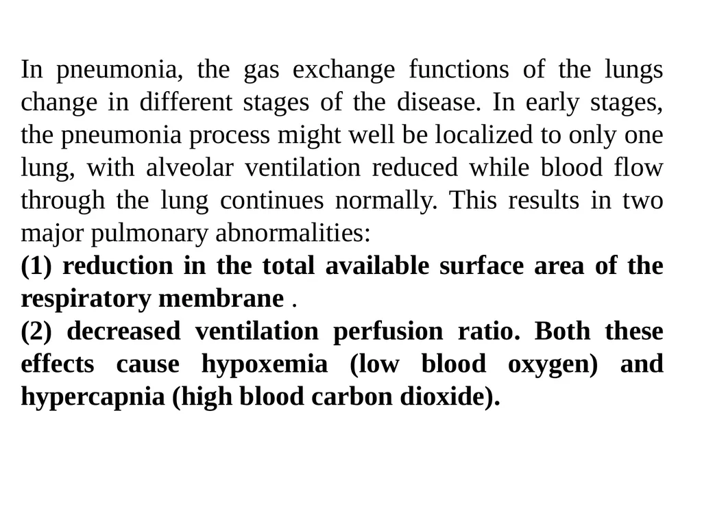
slide7. In pneumonia, the gas exchange functions of the lungs change in different stages of the disease. In early stages, the pneumonia process might well be localized to only one lung, with alveolar ventilation reduced while blood flow through the lung continues normally. This results in two major pulmonary abnormalities:
(1) reduction in the total available surface area of the respiratory membrane .
(2) decreased ventilation perfusion ratio. Both these effects cause hypoxemia (low blood oxygen) and hypercapnia (high blood carbon dioxide).<br>
slide9. Asthma:
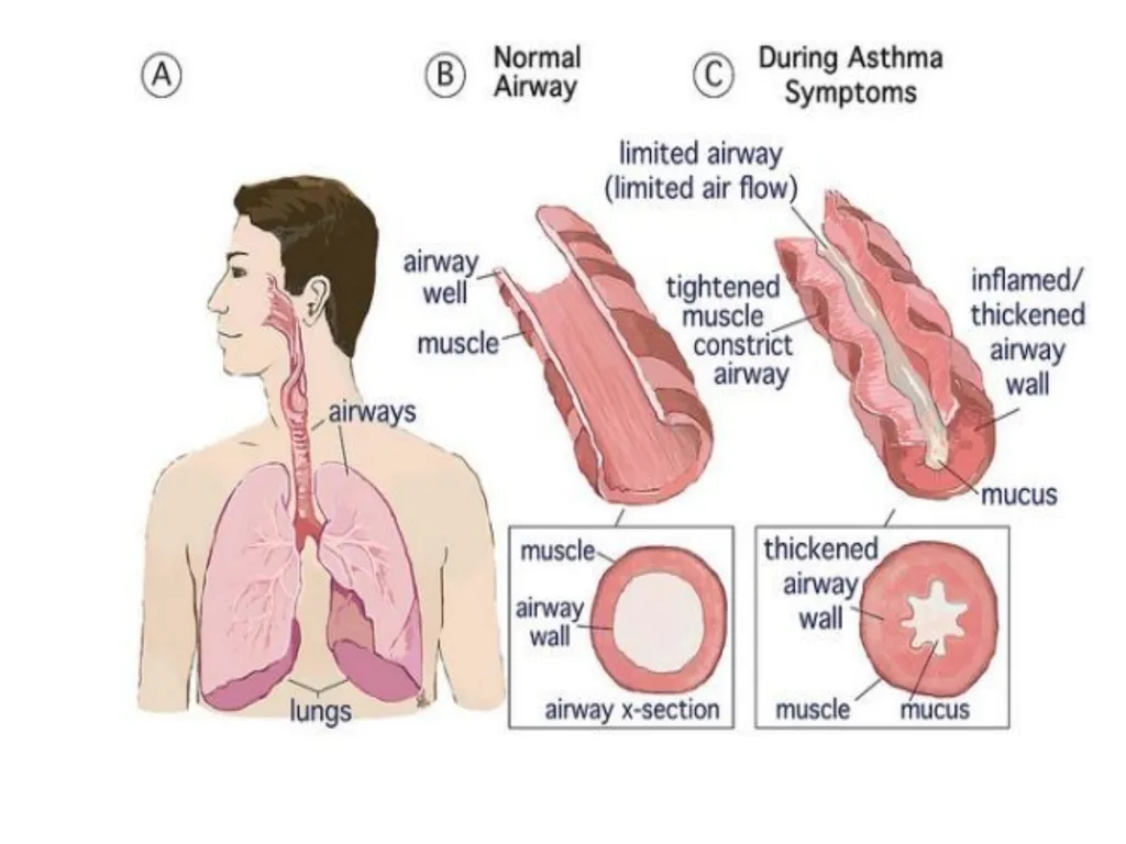
Asthma is characterized by spastic contraction of the smooth muscle in the bronchioles, which partially obstructs the bronchioles and causes extremely difficult breathing. It occurs in 3 to 5 percent of all people at some time in life.
The usual cause of asthma is contractile hypersensitivity
of the bronchioles in response to foreign substances in the air. In about 70 percent of patients younger than age 30 years, the asthma is caused by allergic hypersensitivity, especially sensitivity to plant pollens. In older people, the cause is almost always hypersensitivity to non allergenic types of irritants in the air, such as irritants in smog.<br>
slide10. The allergic reaction that occurs in the allergic type of asthma is believed to occur in the following ways: The typical allergic person has a tendency to form abnormally large amounts of IgE antibodies, and these antibodies cause allergic reactions when they react with the specific antigens that have caused them to develop in the first place, In asthma, these antibodies are mainly attached to mast cells that are present in the lung interstitium in close association with the bronchioles and small bronchi. When the asthmatic person breathes in pollen to which he or she is sensitive (that is, to which the person has developed IgE antibodies), the pollen reacts with the mast cell– attached antibodies and causes the mast cells to release several different substances.<br>
slide11. Among them are (a) histamine, (b) slow-reacting substance of anaphylaxis (which is a mixture of leukotrienes), (c) eosinophilic chemotactic factor, and (d) bradykinin. The combined effects of all these factors, especially the slow-reacting substance of anaphylaxis, are to produce (1) localized edema in the walls of the small bronchioles, as well as secretion of thick mucus into the bronchiolar lumens, and (2) spasm of the bronchiolar smooth muscle. Therefore, the airway resistance increases greatly. the asthmatic person often can inspire quite adequately but has great difficulty expiring.
Clinical measurements show (1) greatly reduced maximum expiratory rate and (2) reduced timed expiratory volume. Also, all of this together results in dyspnea, or “air hunger,”<br>
It results from the following major pathophysiologic
changes in the lungs:<br>
slide2. changes in the lungs:
Chronic infection
caused by inhaling smoke or other substances that irritate the bronchi and bronchioles.
The chronic infection seriously destroy the normal protective mechanisms of the airways, including partial paralysis of the cilia of the respiratory epithelium, an effect caused by nicotine.
As a result, mucus cannot be moved easily out of the passageways.
Also, stimulation of excess mucus secretion occurs, which further exacerbates the condition.
Too, inhibition of the alveolar macrophages occurs, so that they become less effective in combating infection.<br>
slide3. 2. The infection, excess mucus, and inflammatory
edema of the bronchiolar epithelium together cause chronic obstruction of many of the smaller airways.
3. The obstruction of the airways makes it especially difficult to expire, thus causing entrapment of air in the alveoli and overstretching them. This, combined with the lung infection, causes marked destruction of as much as 50 to 80 percent of the alveolar walls. Therefore, the final picture of the emphysematous lung is that shown in Figures<br>
slide5. The physiologic effects of chronic emphysema are extremely varied, depending on the severity of the disease and the relative degrees of bronchiolar obstruction versus lung parenchymal destruction .
Chronic emphysema usually progresses slowly over many years. The person develops both hypoxia and hypercapnia because of hypoventilation of many alveoli plus loss of alveolar walls. The net result of all these effects is severe, prolonged, devastating air hunger that can last for years until the hypoxia and hypercapnia cause death - a high penalty to pay for smoking.<br>
slide6. Pneumonia
The term pneumonia includes any inflammatory condition of the lung in which some or all of the alveoli are filled with fluid and blood cells, a common type of pneumonia is bacterial pneumonia, caused most frequently by pneumococci. This disease begins with infection in the alveoli; the pulmonary membrane becomes inflamed and highly porous so that fluid and even red and white blood cells leak out of the blood into the alveoli. Thus, the infected alveoli become progressively filled with fluid and cells, and the infection spreads by extension of bacteria or virus from alveolus to alveolus.<br>
slide7. In pneumonia, the gas exchange functions of the lungs change in different stages of the disease. In early stages, the pneumonia process might well be localized to only one lung, with alveolar ventilation reduced while blood flow through the lung continues normally. This results in two major pulmonary abnormalities:
(1) reduction in the total available surface area of the respiratory membrane .
(2) decreased ventilation perfusion ratio. Both these effects cause hypoxemia (low blood oxygen) and hypercapnia (high blood carbon dioxide).<br>
slide9. Asthma:
Asthma is characterized by spastic contraction of the smooth muscle in the bronchioles, which partially obstructs the bronchioles and causes extremely difficult breathing. It occurs in 3 to 5 percent of all people at some time in life.
The usual cause of asthma is contractile hypersensitivity
of the bronchioles in response to foreign substances in the air. In about 70 percent of patients younger than age 30 years, the asthma is caused by allergic hypersensitivity, especially sensitivity to plant pollens. In older people, the cause is almost always hypersensitivity to non allergenic types of irritants in the air, such as irritants in smog.<br>
slide10. The allergic reaction that occurs in the allergic type of asthma is believed to occur in the following ways: The typical allergic person has a tendency to form abnormally large amounts of IgE antibodies, and these antibodies cause allergic reactions when they react with the specific antigens that have caused them to develop in the first place, In asthma, these antibodies are mainly attached to mast cells that are present in the lung interstitium in close association with the bronchioles and small bronchi. When the asthmatic person breathes in pollen to which he or she is sensitive (that is, to which the person has developed IgE antibodies), the pollen reacts with the mast cell– attached antibodies and causes the mast cells to release several different substances.<br>
slide11. Among them are (a) histamine, (b) slow-reacting substance of anaphylaxis (which is a mixture of leukotrienes), (c) eosinophilic chemotactic factor, and (d) bradykinin. The combined effects of all these factors, especially the slow-reacting substance of anaphylaxis, are to produce (1) localized edema in the walls of the small bronchioles, as well as secretion of thick mucus into the bronchiolar lumens, and (2) spasm of the bronchiolar smooth muscle. Therefore, the airway resistance increases greatly. the asthmatic person often can inspire quite adequately but has great difficulty expiring.
Clinical measurements show (1) greatly reduced maximum expiratory rate and (2) reduced timed expiratory volume. Also, all of this together results in dyspnea, or “air hunger,”<br>