
Placenta & Membranes- Development & Anatomy Mrs.
AM
Published · 51 slides · 0 views
1 / 1




Description
Placenta Membranes- Development Anatomy Mrs. REENA MATHEW ASSO. PROFESSOR OBG DEPT JINSAR PURPOSE Placenta forms the link between mother and fetus. It acts as digestive, respiratory and excretory organ for the fetus. LEARNING
Related Topics
Share
Embed code
Download this presentation From Below
"Placenta & Membranes- Development & Anatomy Mrs." is the property of its rightful owner. Permission is granted to download and print the materials on this website for personal, non-commercial use only, and to display it on your personal computer provided you do not modify the materials and that you retain all copyright notices contained in the materials. By downloading content from our website, you accept the terms of this agreement.
Presentation Transcript
01
Placenta & Membranes- Development & Anatomy Mrs. REENA MATHEW
ASSO. PROFESSOR
OBG DEPT
JINSAR<br>
ASSO. PROFESSOR
OBG DEPT
JINSAR<br>
02
PURPOSE –
Placenta forms the link between mother
and fetus.
It acts as digestive, respiratory and
excretory organ for the fetus.<br>
Placenta forms the link between mother
and fetus.
It acts as digestive, respiratory and
excretory organ for the fetus.<br>
03
LEARNING OBJECTIVES –
Placenta
Development
Structures in placenta
Placental barrier
Placental circulation
Fetal membranes
Development of membranes
Embryonic development after implantation
Amniotic fluid<br>
Placenta
Development
Structures in placenta
Placental barrier
Placental circulation
Fetal membranes
Development of membranes
Embryonic development after implantation
Amniotic fluid<br>
04
Placenta Discoid – Shape
Haemochorial- Direct contact of chorion with Maternal Blood
Deciduate- Some maternal tissue is shed at Parturation.
Another Terminology -- Haemochorioendothelial<br>
Haemochorial- Direct contact of chorion with Maternal Blood
Deciduate- Some maternal tissue is shed at Parturation.
Another Terminology -- Haemochorioendothelial<br>
05
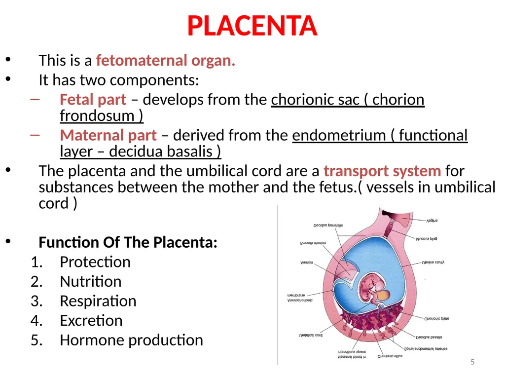
5 PLACENTA This is a fetomaternal organ.
It has two components:
Fetal part – develops from the chorionic sac ( chorion frondosum )
Maternal part – derived from the endometrium ( functional layer – decidua basalis )
The placenta and the umbilical cord are a transport system for substances between the mother and the fetus.( vessels in umbilical cord )
Function Of The Placenta:
Protection
Nutrition
Respiration
Excretion
Hormone production<br>
It has two components:
Fetal part – develops from the chorionic sac ( chorion frondosum )
Maternal part – derived from the endometrium ( functional layer – decidua basalis )
The placenta and the umbilical cord are a transport system for substances between the mother and the fetus.( vessels in umbilical cord )
Function Of The Placenta:
Protection
Nutrition
Respiration
Excretion
Hormone production<br>
06
Ovulation – Day 14
Fertilisation
Fertilisable life span of Oocyte- 12-24 hrs
Sperm-- 48-72 hrs
2,4,8 16 cell- Morula
Morula- 3 days in fallopian tube
Day 4- enters in uterus, called Blastocyst<br>
Fertilisation
Fertilisable life span of Oocyte- 12-24 hrs
Sperm-- 48-72 hrs
2,4,8 16 cell- Morula
Morula- 3 days in fallopian tube
Day 4- enters in uterus, called Blastocyst<br>
07
Implantation starts on day 6 (day 20 of M.C)
Interstitial implantation
Completes by day 10-11 (day 24-25 of M.C)<br>
Interstitial implantation
Completes by day 10-11 (day 24-25 of M.C)<br>
08
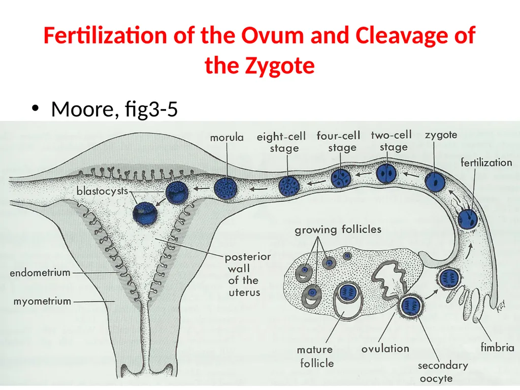
Fertilization of the Ovum and Cleavage of the Zygote Moore, fig3-5<br>
09
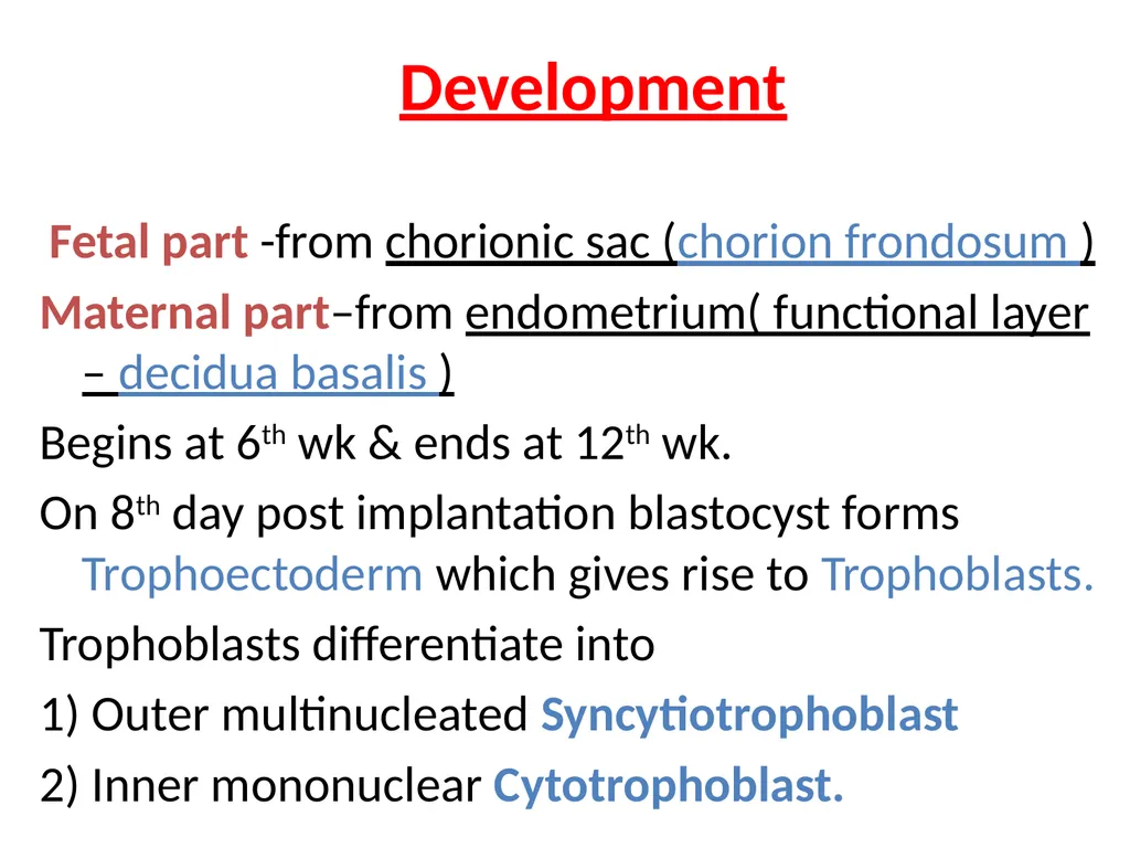
Development Fetal part -from chorionic sac (chorion frondosum )
Maternal part–from endometrium( functional layer – decidua basalis )
Begins at 6th wk & ends at 12th wk.
On 8th day post implantation blastocyst forms Trophoectoderm which gives rise to Trophoblasts.
Trophoblasts differentiate into
1) Outer multinucleated Syncytiotrophoblast
2) Inner mononuclear Cytotrophoblast.<br>
Maternal part–from endometrium( functional layer – decidua basalis )
Begins at 6th wk & ends at 12th wk.
On 8th day post implantation blastocyst forms Trophoectoderm which gives rise to Trophoblasts.
Trophoblasts differentiate into
1) Outer multinucleated Syncytiotrophoblast
2) Inner mononuclear Cytotrophoblast.<br>
10
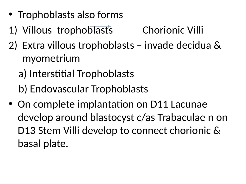
Trophoblasts also forms
Villous trophoblasts Chorionic Villi
Extra villous trophoblasts – invade decidua & myometrium
a) Interstitial Trophoblasts
b) Endovascular Trophoblasts
On complete implantation on D11 Lacunae develop around blastocyst c/as Trabaculae n on D13 Stem Villi develop to connect chorionic & basal plate.<br>
Villous trophoblasts Chorionic Villi
Extra villous trophoblasts – invade decidua & myometrium
a) Interstitial Trophoblasts
b) Endovascular Trophoblasts
On complete implantation on D11 Lacunae develop around blastocyst c/as Trabaculae n on D13 Stem Villi develop to connect chorionic & basal plate.<br>
11
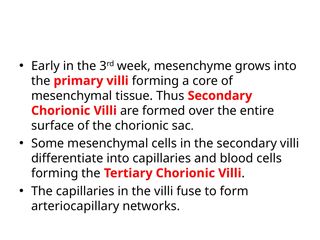
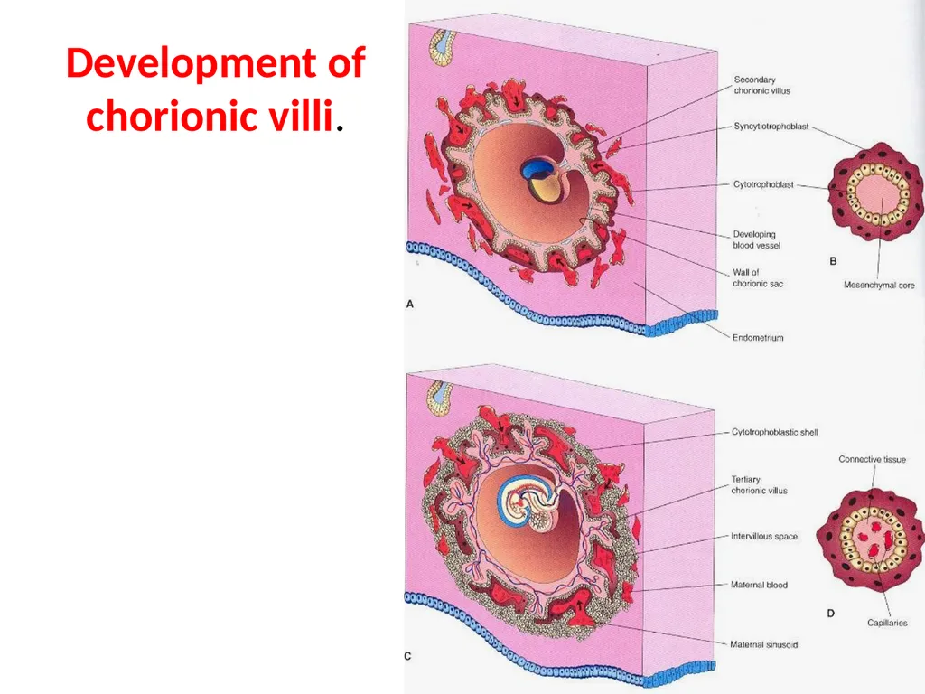
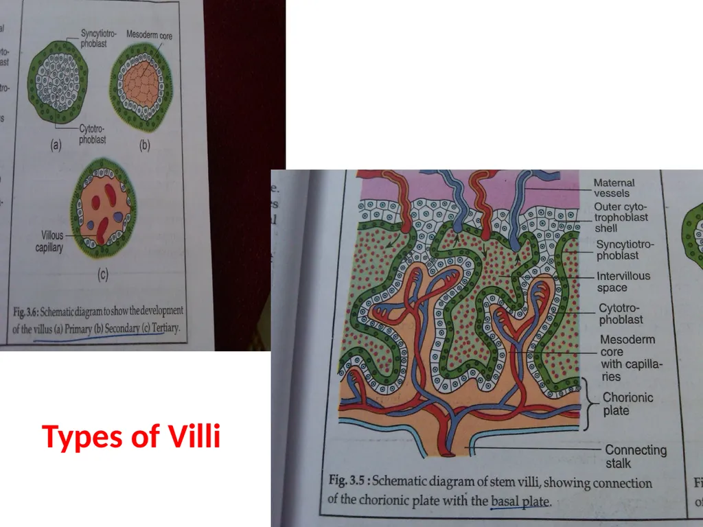
Early in the 3rd week, mesenchyme grows into the primary villi forming a core of mesenchymal tissue. Thus Secondary Chorionic Villi are formed over the entire surface of the chorionic sac.
Some mesenchymal cells in the secondary villi differentiate into capillaries and blood cells forming the Tertiary Chorionic Villi.
The capillaries in the villi fuse to form arteriocapillary networks.<br>
Some mesenchymal cells in the secondary villi differentiate into capillaries and blood cells forming the Tertiary Chorionic Villi.
The capillaries in the villi fuse to form arteriocapillary networks.<br>
12
Development of chorionic villi.<br>
13
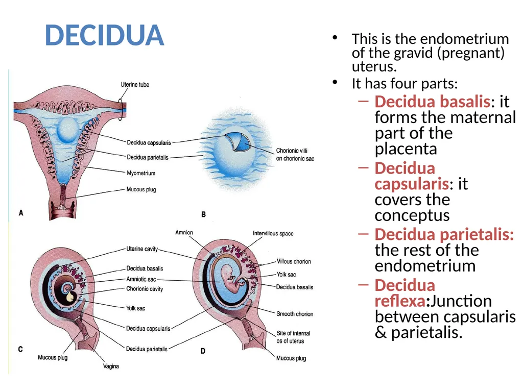
DECIDUA This is the endometrium of the gravid (pregnant) uterus.
It has four parts:
Decidua basalis: it forms the maternal part of the placenta
Decidua capsularis: it covers the conceptus
Decidua parietalis: the rest of the endometrium
Decidua reflexa:Junction between capsularis & parietalis.<br>
It has four parts:
Decidua basalis: it forms the maternal part of the placenta
Decidua capsularis: it covers the conceptus
Decidua parietalis: the rest of the endometrium
Decidua reflexa:Junction between capsularis & parietalis.<br>
14
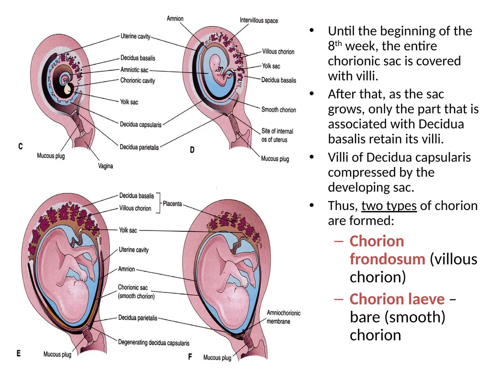
Until the beginning of the 8th week, the entire chorionic sac is covered with villi.
After that, as the sac grows, only the part that is associated with Decidua basalis retain its villi.
Villi of Decidua capsularis compressed by the developing sac.
Thus, two types of chorion are formed:
Chorion frondosum (villous chorion)
Chorion laeve – bare (smooth) chorion<br>
After that, as the sac grows, only the part that is associated with Decidua basalis retain its villi.
Villi of Decidua capsularis compressed by the developing sac.
Thus, two types of chorion are formed:
Chorion frondosum (villous chorion)
Chorion laeve – bare (smooth) chorion<br>
15
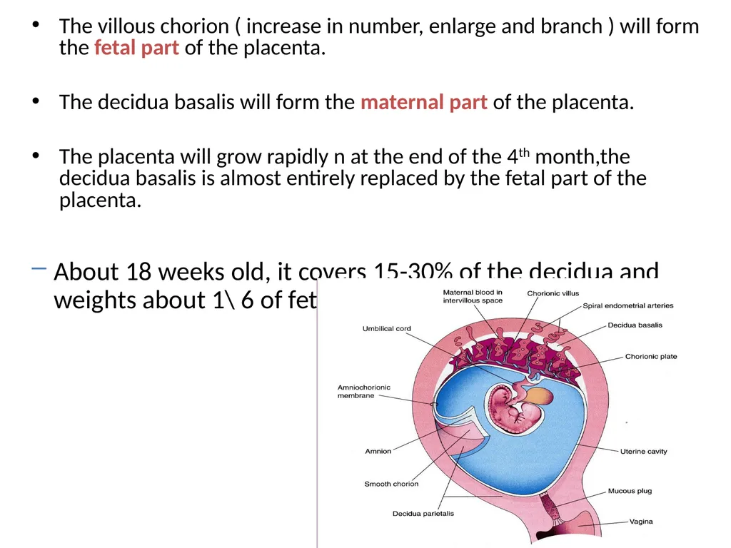
The villous chorion ( increase in number, enlarge and branch ) will form the fetal part of the placenta.
The decidua basalis will form the maternal part of the placenta.
The placenta will grow rapidly n at the end of the 4th month,the decidua basalis is almost entirely replaced by the fetal part of the placenta.
About 18 weeks old, it covers 15-30% of the decidua and weights about 1\ 6 of fetus<br>
The decidua basalis will form the maternal part of the placenta.
The placenta will grow rapidly n at the end of the 4th month,the decidua basalis is almost entirely replaced by the fetal part of the placenta.
About 18 weeks old, it covers 15-30% of the decidua and weights about 1\ 6 of fetus<br>
16
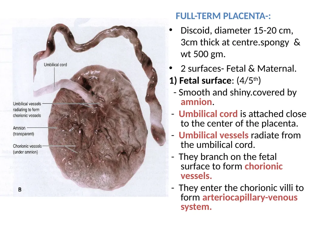
FULL-TERM PLACENTA-:
Discoid, diameter 15-20 cm, 3cm thick at centre.spongy & wt 500 gm.
2 surfaces- Fetal & Maternal.
1) Fetal surface: (4/5th)
- Smooth and shiny.covered by amnion.
- Umbilical cord is attached close to the center of the placenta.
- Umbilical vessels radiate from the umbilical cord.
- They branch on the fetal surface to form chorionic vessels.
- They enter the chorionic villi to form arteriocapillary-venous system.<br>
Discoid, diameter 15-20 cm, 3cm thick at centre.spongy & wt 500 gm.
2 surfaces- Fetal & Maternal.
1) Fetal surface: (4/5th)
- Smooth and shiny.covered by amnion.
- Umbilical cord is attached close to the center of the placenta.
- Umbilical vessels radiate from the umbilical cord.
- They branch on the fetal surface to form chorionic vessels.
- They enter the chorionic villi to form arteriocapillary-venous system.<br>
17
2) Maternal Surface- (1/5th)
Rough & spongy, dull red,
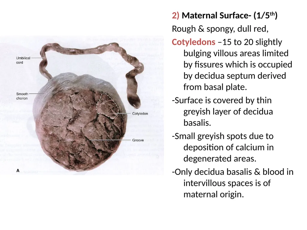
Cotyledons –15 to 20 slightly bulging villous areas limited by fissures which is occupied by decidua septum derived from basal plate.
-Surface is covered by thin greyish layer of decidua basalis.
-Small greyish spots due to deposition of calcium in degenerated areas.
-Only decidua basalis & blood in intervillous spaces is of maternal origin.<br>
Rough & spongy, dull red,
Cotyledons –15 to 20 slightly bulging villous areas limited by fissures which is occupied by decidua septum derived from basal plate.
-Surface is covered by thin greyish layer of decidua basalis.
-Small greyish spots due to deposition of calcium in degenerated areas.
-Only decidua basalis & blood in intervillous spaces is of maternal origin.<br>
18
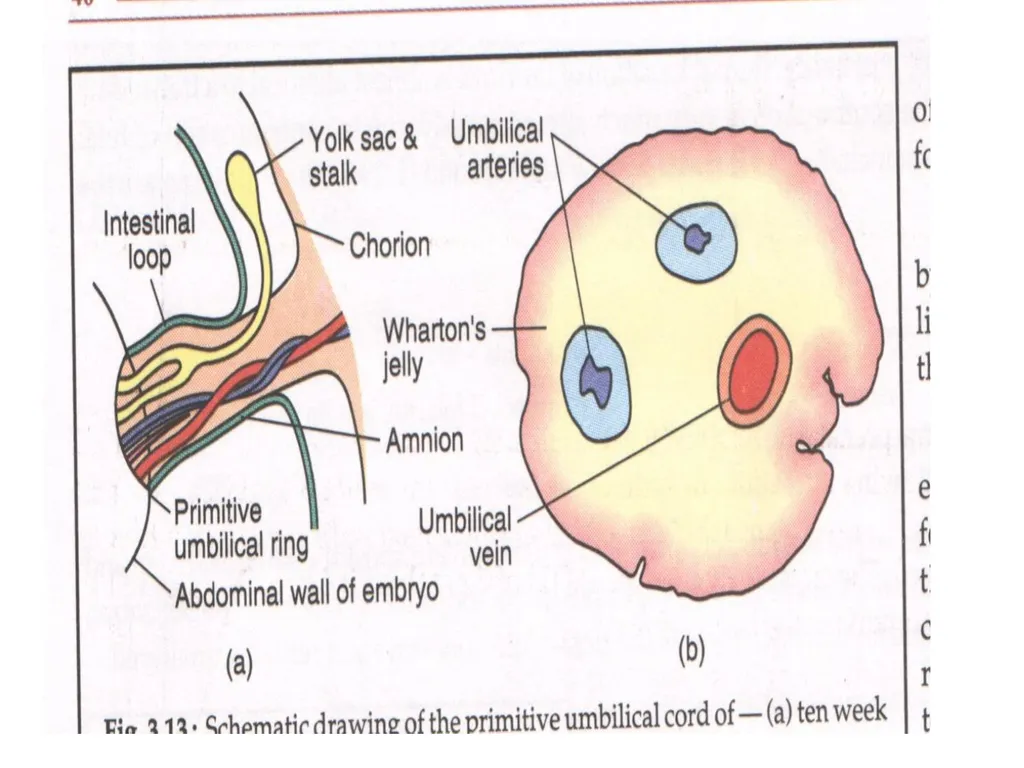
Development of Umbilical Cord Connecting Stalk or umbilical cord develops from Extra-embryonic Mesoderm
Connecting Link between embryo & fetus
Contents- 1) Vitello-intestinal duct & remnant of yolk sac
2)Extraembryonic mesoderm– Gelatinous sub K/ as Wharton’s Jelly
3) Blood Vessels
4)Extraembryonic coelom
5) Covering epithelium
6) Allantois<br>
Connecting Link between embryo & fetus
Contents- 1) Vitello-intestinal duct & remnant of yolk sac
2)Extraembryonic mesoderm– Gelatinous sub K/ as Wharton’s Jelly
3) Blood Vessels
4)Extraembryonic coelom
5) Covering epithelium
6) Allantois<br>
19
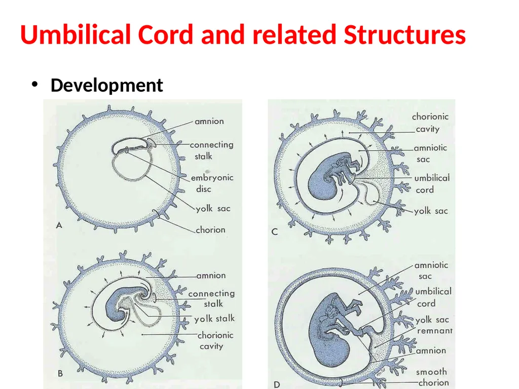
Umbilical Cord and related Structures Development<br>
20
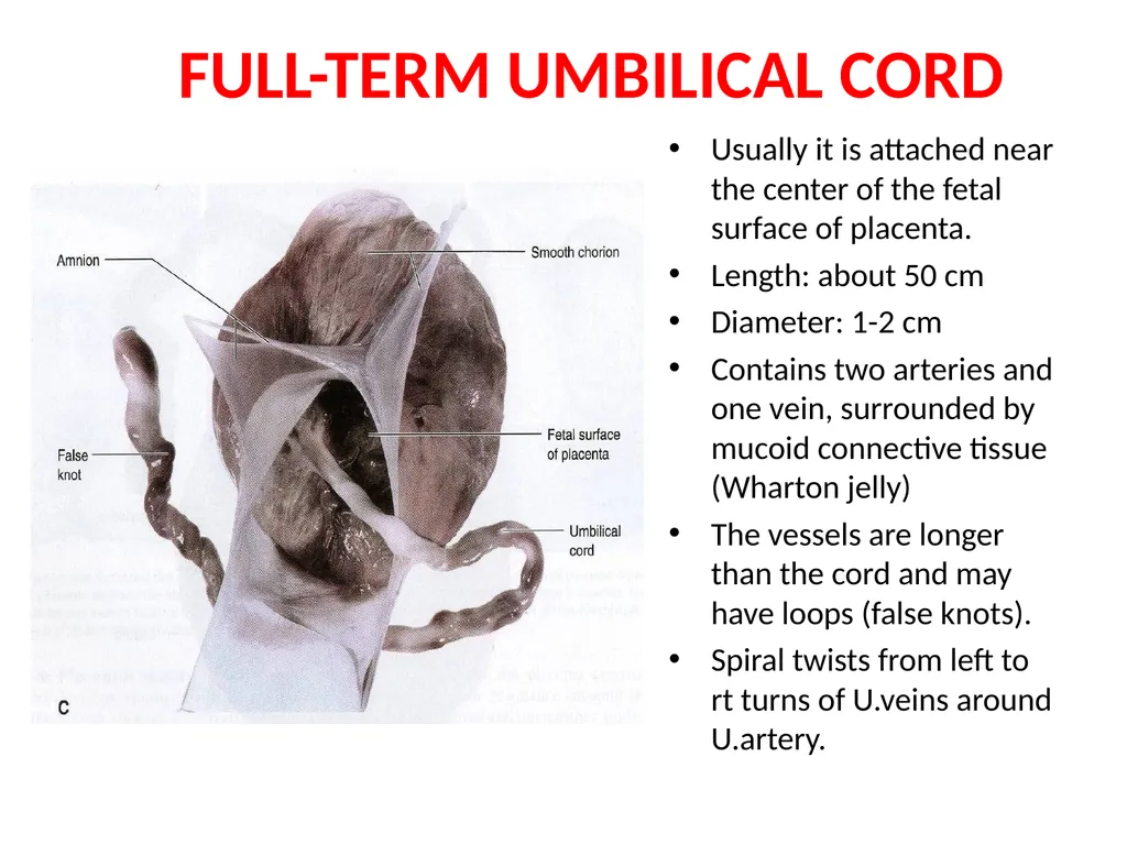
FULL-TERM UMBILICAL CORD Usually it is attached near the center of the fetal surface of placenta.
Length: about 50 cm
Diameter: 1-2 cm
Contains two arteries and one vein, surrounded by mucoid connective tissue (Wharton jelly)
The vessels are longer than the cord and may have loops (false knots).
Spiral twists from left to rt turns of U.veins around U.artery.<br>
Length: about 50 cm
Diameter: 1-2 cm
Contains two arteries and one vein, surrounded by mucoid connective tissue (Wharton jelly)
The vessels are longer than the cord and may have loops (false knots).
Spiral twists from left to rt turns of U.veins around U.artery.<br>
21
STRUCTURES In Placenta 1)Amniotic Membrane-Single layer of cubical epithilium loosely lining chorionic plate internally.
2 ) 2 Plates- Chorionic & Basal.
Chorionic Plate- Forms inner boundry of choriodesidual space.
-Stem Villi & Umbilical cord arise from this plate.
-From outwards consists of-
Primitive mesenchymal tissue containing branches of umbilical vessel.
Layer of Cytotrophoblasts.
Syncitiotrophoblasts.<br>
2 ) 2 Plates- Chorionic & Basal.
Chorionic Plate- Forms inner boundry of choriodesidual space.
-Stem Villi & Umbilical cord arise from this plate.
-From outwards consists of-
Primitive mesenchymal tissue containing branches of umbilical vessel.
Layer of Cytotrophoblasts.
Syncitiotrophoblasts.<br>
22
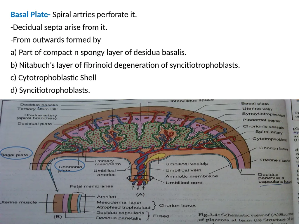
Basal Plate- Spiral artries perforate it.
-Decidual septa arise from it.
-From outwards formed by
a) Part of compact n spongy layer of desidua basalis.
b) Nitabuch’s layer of fibrinoid degeneration of syncitiotrophoblasts.
c) Cytotrophoblastic Shell
d) Syncitiotrophoblasts.<br>
-Decidual septa arise from it.
-From outwards formed by
a) Part of compact n spongy layer of desidua basalis.
b) Nitabuch’s layer of fibrinoid degeneration of syncitiotrophoblasts.
c) Cytotrophoblastic Shell
d) Syncitiotrophoblasts.<br>
23
3)Intervillous space- bounded by Chorionic & Basal plate.
-Lined internally all around by syncitiotrophoblasts.
-Filled with Maternal Blood
-Numerous branches form stem villi project into it.
4)Stem villi & their branches-(primary,secondary,tertiary)
-Stem villi supplies Cotyledon(15-20)-functional unit of placenta.
-Tertiary villi supplies Lobule-functional subunit of placenta.
-Each cotyledon contain 3-4 major stem villi
-Total villi surface-10-14 sq km, capillary system in villi 50km long.
-Function-1) Anchoring Placenta to desidua
2) Nutritive function.
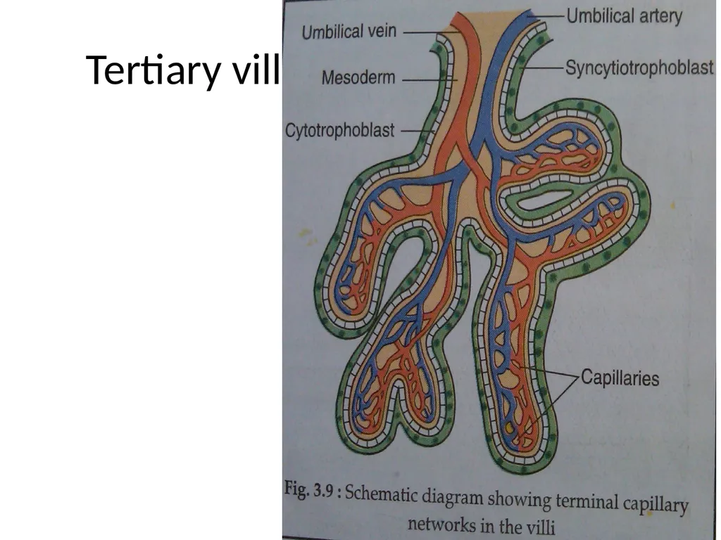
Tertiary Villi-: Composed Outside in of-
-outer syncitiotrophoblasts, Cytotrophoblasts, basement Membrane,
Central stroma-fetal capillaries,mesenchymal cells,connective tissue & Hofbauer phagocytic cells.<br>
-Lined internally all around by syncitiotrophoblasts.
-Filled with Maternal Blood
-Numerous branches form stem villi project into it.
4)Stem villi & their branches-(primary,secondary,tertiary)
-Stem villi supplies Cotyledon(15-20)-functional unit of placenta.
-Tertiary villi supplies Lobule-functional subunit of placenta.
-Each cotyledon contain 3-4 major stem villi
-Total villi surface-10-14 sq km, capillary system in villi 50km long.
-Function-1) Anchoring Placenta to desidua
2) Nutritive function.
Tertiary Villi-: Composed Outside in of-
-outer syncitiotrophoblasts, Cytotrophoblasts, basement Membrane,
Central stroma-fetal capillaries,mesenchymal cells,connective tissue & Hofbauer phagocytic cells.<br>
24
Types of Villi<br>
25
Tertiary villi<br>
26
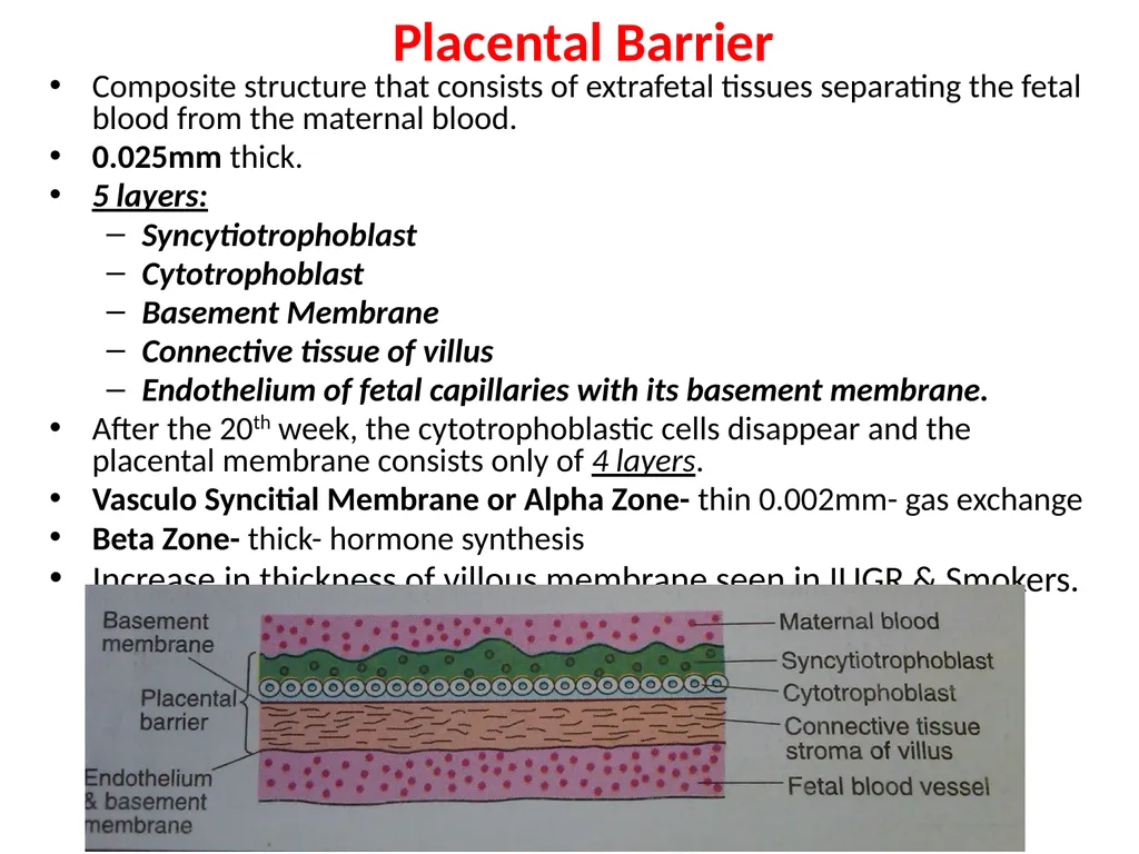
Placental Barrier Composite structure that consists of extrafetal tissues separating the fetal blood from the maternal blood.
0.025mm thick.
5 layers:
Syncytiotrophoblast
Cytotrophoblast
Basement Membrane
Connective tissue of villus
Endothelium of fetal capillaries with its basement membrane.
After the 20th week, the cytotrophoblastic cells disappear and the placental membrane consists only of 4 layers.
Vasculo Syncitial Membrane or Alpha Zone- thin 0.002mm- gas exchange
Beta Zone- thick- hormone synthesis
Increase in thickness of villous membrane seen in IUGR & Smokers.<br>
0.025mm thick.
5 layers:
Syncytiotrophoblast
Cytotrophoblast
Basement Membrane
Connective tissue of villus
Endothelium of fetal capillaries with its basement membrane.
After the 20th week, the cytotrophoblastic cells disappear and the placental membrane consists only of 4 layers.
Vasculo Syncitial Membrane or Alpha Zone- thin 0.002mm- gas exchange
Beta Zone- thick- hormone synthesis
Increase in thickness of villous membrane seen in IUGR & Smokers.<br>
27
Placental Circulation Utero-Placental Circulation-:
-100-200 spiral atreries open in intervillous space.
-Volume of blood in mature placenta- 500ml
-Volume of blood in intervillous spaces- 150 ml
-Blood flow in intervillous spaces-500-600ml/min
-Pressure in intervillous spaces- ut contraction- 30-50mmHg
- ut relaxation- 10-15mmHg
-Pressure in supplying Uterine artery- 70-80 mmHg
-Pressure in draining Uterine Vein- 8mmHg
-Blood in intervillous space replaced every 3-4 mins.
-Endovascular extravillous trophoblasts invade spiral artries upto myometrium causing their dialatation & failure of this invasion leads to PIH, IUGR etc.<br>
-100-200 spiral atreries open in intervillous space.
-Volume of blood in mature placenta- 500ml
-Volume of blood in intervillous spaces- 150 ml
-Blood flow in intervillous spaces-500-600ml/min
-Pressure in intervillous spaces- ut contraction- 30-50mmHg
- ut relaxation- 10-15mmHg
-Pressure in supplying Uterine artery- 70-80 mmHg
-Pressure in draining Uterine Vein- 8mmHg
-Blood in intervillous space replaced every 3-4 mins.
-Endovascular extravillous trophoblasts invade spiral artries upto myometrium causing their dialatation & failure of this invasion leads to PIH, IUGR etc.<br>
28
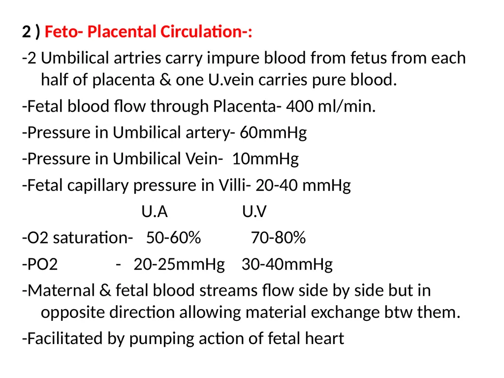
2 ) Feto- Placental Circulation-:
-2 Umbilical artries carry impure blood from fetus from each half of placenta & one U.vein carries pure blood.
-Fetal blood flow through Placenta- 400 ml/min.
-Pressure in Umbilical artery- 60mmHg
-Pressure in Umbilical Vein- 10mmHg
-Fetal capillary pressure in Villi- 20-40 mmHg
U.A U.V
-O2 saturation- 50-60% 70-80%
-PO2 - 20-25mmHg 30-40mmHg
-Maternal & fetal blood streams flow side by side but in opposite direction allowing material exchange btw them.
-Facilitated by pumping action of fetal heart<br>
-2 Umbilical artries carry impure blood from fetus from each half of placenta & one U.vein carries pure blood.
-Fetal blood flow through Placenta- 400 ml/min.
-Pressure in Umbilical artery- 60mmHg
-Pressure in Umbilical Vein- 10mmHg
-Fetal capillary pressure in Villi- 20-40 mmHg
U.A U.V
-O2 saturation- 50-60% 70-80%
-PO2 - 20-25mmHg 30-40mmHg
-Maternal & fetal blood streams flow side by side but in opposite direction allowing material exchange btw them.
-Facilitated by pumping action of fetal heart<br>
29
Fetal Membranes Outer Chorion & Inner Amnion
Chorion-
Renmnant of chorion leave
Thicker than amnion, friable & shaggy
No vessels & nerves.<br>
Chorion-
Renmnant of chorion leave
Thicker than amnion, friable & shaggy
No vessels & nerves.<br>
30
Amnion-
Inner layer of fetal membranes
Smooth & shiny & contact with AF
Can be peeled off from fetal surface except at placenta<br>
Inner layer of fetal membranes
Smooth & shiny & contact with AF
Can be peeled off from fetal surface except at placenta<br>
31
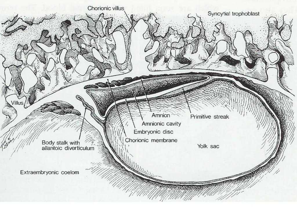
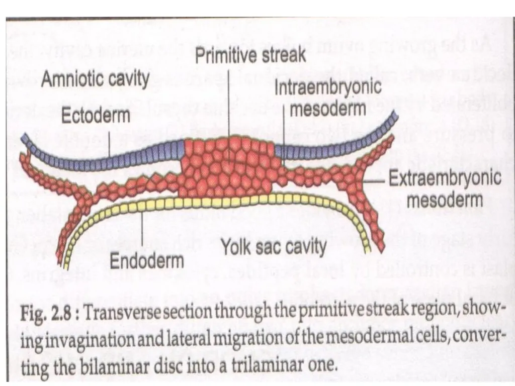
Development of Membranes Along with Trophoblast on day 8, Embryoblast differentiates into Bilaminar germ disc.
Bilaminar germ disc – Dorsal ectodermal layer
Ventral endodermal layer
Two cavities appear on each side of disc.
Day 12- fluid filled space appears between ectodermal layer & cytotrphoblast- Amniotic Cavity<br>
Bilaminar germ disc – Dorsal ectodermal layer
Ventral endodermal layer
Two cavities appear on each side of disc.
Day 12- fluid filled space appears between ectodermal layer & cytotrphoblast- Amniotic Cavity<br>
32
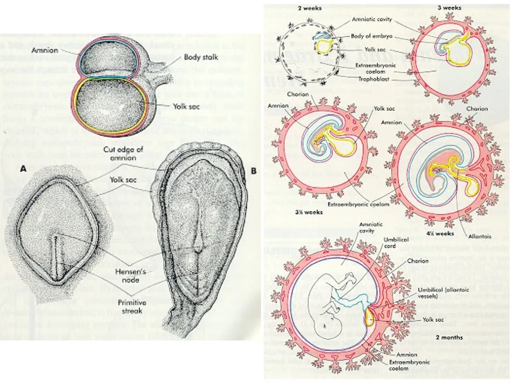
Ventral side yolk sac appears.
Amniotic Cavity-
Initially fluid accumulates slowly ,later large & obliterates chorionic cavity. Initially located dorsally but after formation of head tail & lateral folds surround fetus.<br>
Amniotic Cavity-
Initially fluid accumulates slowly ,later large & obliterates chorionic cavity. Initially located dorsally but after formation of head tail & lateral folds surround fetus.<br>
33
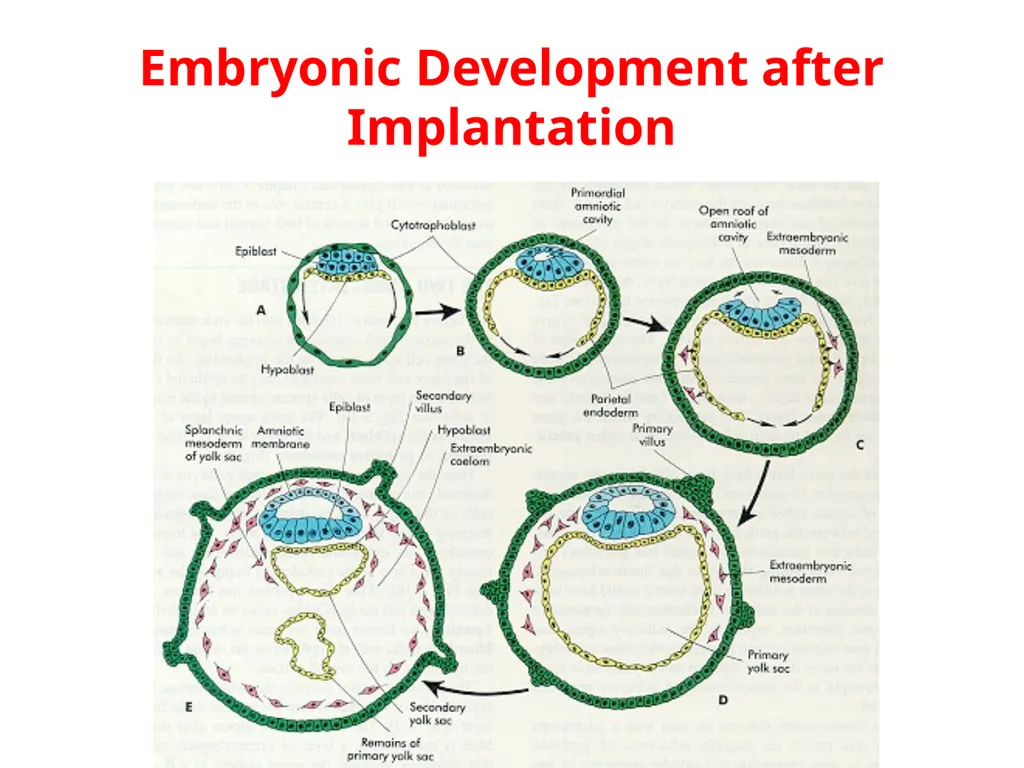
Embryonic Development after Implantation<br>
34
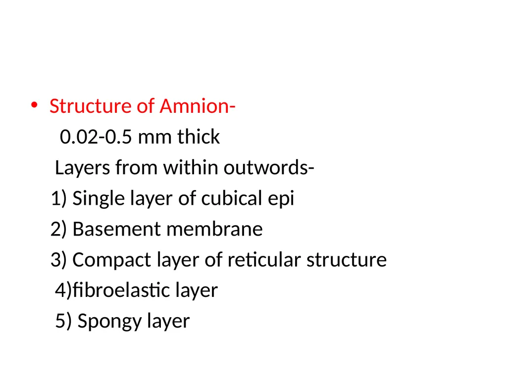
Structure of Amnion-
0.02-0.5 mm thick
Layers from within outwords-
1) Single layer of cubical epi
2) Basement membrane
3) Compact layer of reticular structure
4)fibroelastic layer
5) Spongy layer<br>
0.02-0.5 mm thick
Layers from within outwords-
1) Single layer of cubical epi
2) Basement membrane
3) Compact layer of reticular structure
4)fibroelastic layer
5) Spongy layer<br>
35
Amnion- No blood, nerve & lymphatic supply
Functions-
1)Formation of amniotic fluid
2) Prevent infection
3) Facilitates cervical dilatation in labor
4)Enzymatic activity
5) Rich source of glycerophospholipids- Precursor of PG’S<br>
Functions-
1)Formation of amniotic fluid
2) Prevent infection
3) Facilitates cervical dilatation in labor
4)Enzymatic activity
5) Rich source of glycerophospholipids- Precursor of PG’S<br>
36
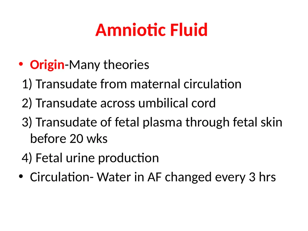
Amniotic Fluid Origin-Many theories
1) Transudate from maternal circulation
2) Transudate across umbilical cord
3) Transudate of fetal plasma through fetal skin before 20 wks
4) Fetal urine production
Circulation- Water in AF changed every 3 hrs<br>
1) Transudate from maternal circulation
2) Transudate across umbilical cord
3) Transudate of fetal plasma through fetal skin before 20 wks
4) Fetal urine production
Circulation- Water in AF changed every 3 hrs<br>
37
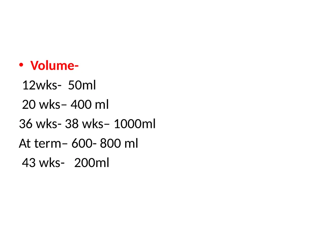
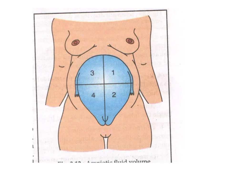
Volume-
12wks- 50ml
20 wks– 400 ml
36 wks- 38 wks– 1000ml
At term– 600- 800 ml
43 wks- 200ml<br>
12wks- 50ml
20 wks– 400 ml
36 wks- 38 wks– 1000ml
At term– 600- 800 ml
43 wks- 200ml<br>
38
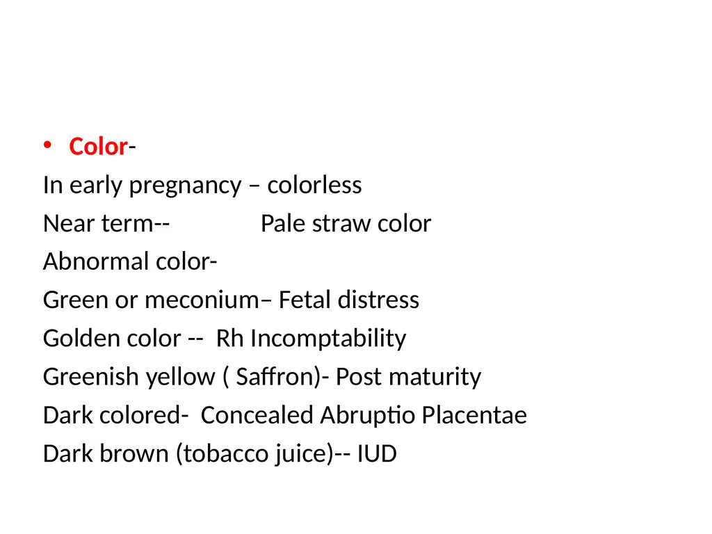
Color-
In early pregnancy – colorless
Near term-- Pale straw color
Abnormal color-
Green or meconium– Fetal distress
Golden color -- Rh Incomptability
Greenish yellow ( Saffron)- Post maturity
Dark colored- Concealed Abruptio Placentae
Dark brown (tobacco juice)-- IUD<br>
In early pregnancy – colorless
Near term-- Pale straw color
Abnormal color-
Green or meconium– Fetal distress
Golden color -- Rh Incomptability
Greenish yellow ( Saffron)- Post maturity
Dark colored- Concealed Abruptio Placentae
Dark brown (tobacco juice)-- IUD<br>
39
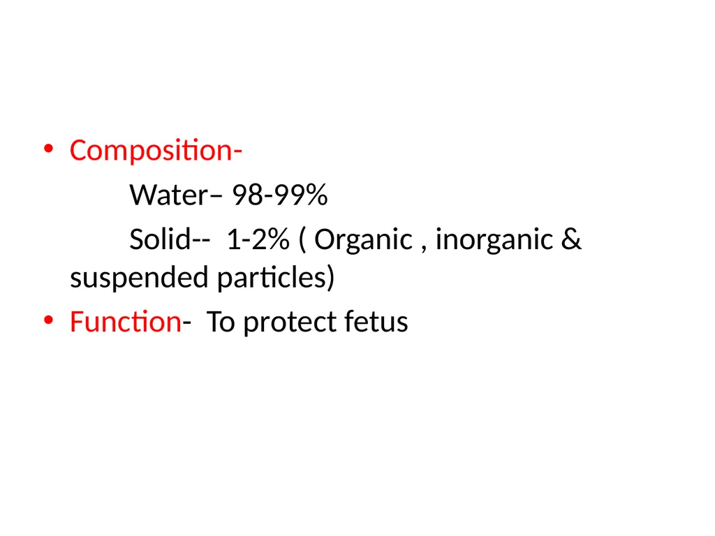
Composition-
Water– 98-99%
Solid-- 1-2% ( Organic , inorganic & suspended particles)
Function- To protect fetus<br>
Water– 98-99%
Solid-- 1-2% ( Organic , inorganic & suspended particles)
Function- To protect fetus<br>
40
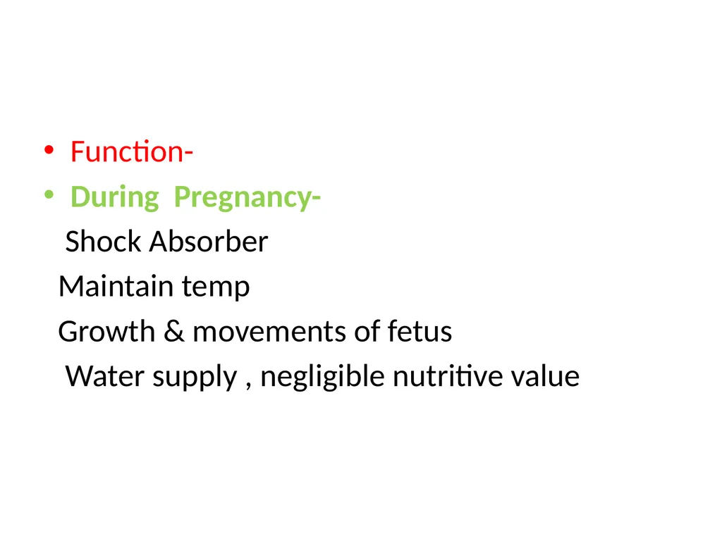
Function-
During Pregnancy-
Shock Absorber
Maintain temp
Growth & movements of fetus
Water supply , negligible nutritive value<br>
During Pregnancy-
Shock Absorber
Maintain temp
Growth & movements of fetus
Water supply , negligible nutritive value<br>
41
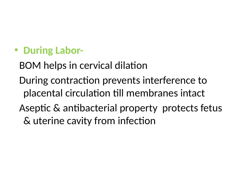
During Labor-
BOM helps in cervical dilation
During contraction prevents interference to placental circulation till membranes intact
Aseptic & antibacterial property protects fetus & uterine cavity from infection<br>
BOM helps in cervical dilation
During contraction prevents interference to placental circulation till membranes intact
Aseptic & antibacterial property protects fetus & uterine cavity from infection<br>
42
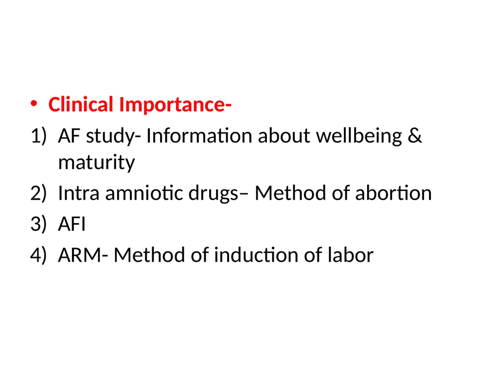
Clinical Importance-
AF study- Information about wellbeing & maturity
Intra amniotic drugs– Method of abortion
AFI
ARM- Method of induction of labor<br>
AF study- Information about wellbeing & maturity
Intra amniotic drugs– Method of abortion
AFI
ARM- Method of induction of labor<br>
43
Summary:
We have studied about the development and main functions of placenta and amniotic fluids<br>
We have studied about the development and main functions of placenta and amniotic fluids<br>
44
REFERENCES –
D.C. Dutta’s Textbook Of Obstetrics
Williams Textbook Of Obstetrics<br>
D.C. Dutta’s Textbook Of Obstetrics
Williams Textbook Of Obstetrics<br>
45
Exam Appeared Questions –
SAQ – 4 marks
Q)Describe gross structure of placenta. Enumerate the
functions of placenta.<br>
SAQ – 4 marks
Q)Describe gross structure of placenta. Enumerate the
functions of placenta.<br>
46
THANK YOU…<br>